
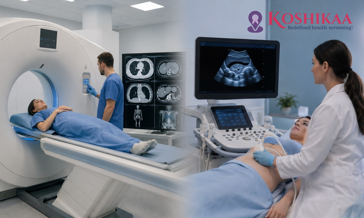
Your doctor has just told you that you need a scan, but you may be wondering: should it be a CT scan or an ultrasound? Both are widely used forms of diagnostic imaging, and both allow doctors to examine structures inside the body without surgery. However, they use different technologies and are better suited to different medical situations. At a reliable Health Screening Centre in Bangalore, the choice of scan is generally based on what your doctor needs to evaluate.
This guide, wIll help you compare CT scan vs ultrasound, explain how each works, where they are commonly used, and the key differences in preparation, safety, and imaging capabilities. This can help you understand what to expect before booking a CT Scan in Bangalore or undergoing an ultrasound.
Medical Disclaimer: This article is intended for general educational and awareness purposes only. It explains the typical differences, uses, and safety profiles of CT scans and ultrasound imaging, but it is not a substitute for professional medical advice. The right test for you depends on your specific symptoms, medical history, and your doctor’s clinical judgment. Always follow your doctor’s recommendation regarding which scan to undergo.
How Each Test Actually Works
How a CT Scan Works
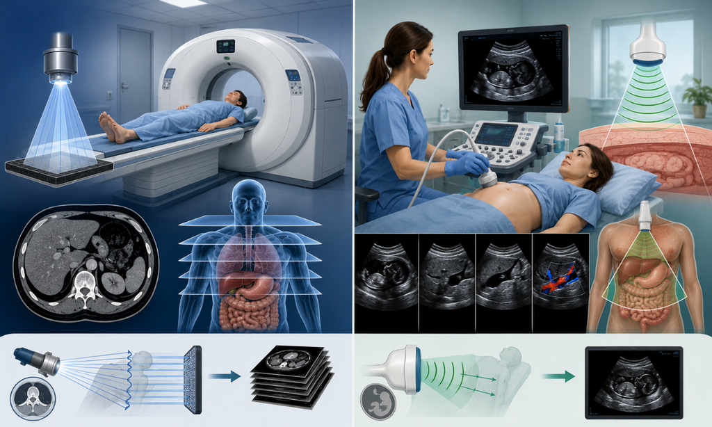
A CT (Computed Tomography) scan uses X-rays taken from multiple angles around the body, which a computer then combines into detailed cross-sectional images of your internal anatomy. It’s especially good at capturing bone, lung tissue, and complex internal structures in high resolution, and an entire scan usually takes just a few minutes.
How Ultrasound Imaging Works
Ultrasound imaging (sonography) uses high-frequency sound waves instead of radiation. A handheld probe called a transducer is moved across the skin, sending sound waves into the body; as these waves bounce off organs and tissues, they create echoes that are converted into real-time, moving images on a screen. This real-time quality is one of ultrasound’s biggest strengths doctors can watch blood flow, a beating heart, or a moving fetus as it happens.
CT Scan vs Ultrasound: Key Differences at a Glance
| Factor | CT Scan | Ultrasound |
| Technology | X-rays (ionizing radiation) | Sound waves (no radiation) |
| Image type | Detailed, static cross-sections | Real-time, moving images |
| Best for | Bone, lungs, complex trauma, internal bleeding | Soft tissue, organs, blood flow, pregnancy |
| Speed | Very fast (minutes) | Slightly longer, operator-dependent |
| Radiation exposure | Small but measurable dose | None |
| Safe in pregnancy | Generally avoided, especially early pregnancy | Yes, safe throughout pregnancy |
| Typical cost | Higher | Generally more affordable |
| Preparation | Sometimes needs fasting or contrast dye | Often needs fasting for abdominal scans |
Neither test is universally “more accurate” than the other; each is simply built to answer different clinical questions. A radiologist or treating doctor picks the best option based on which one is most likely to show what they’re looking for.
When Doctors Recommend a CT Scan
CT scans are usually the first choice when speed, detail, or the ability to see through bone and air-filled structures like the lungs matters most. Common situations include:
- Trauma and accidents: CT is the fastest way to check for internal bleeding, fractures, or organ damage after a serious injury.
- Suspected appendicitis: CT has notably higher accuracy than ultrasound for confirming appendicitis in adults.
- Chest and lung conditions: Persistent cough, unexplained breathlessness, or suspected infections are often evaluated with a chest CT.
- Brain injuries or suspected stroke: A CT scan can quickly identify bleeding or a blood clot in the brain you can read more in our detailed guide on diagnosing a blood clot in the brain via CT scan.
- Cancer staging and treatment planning: CT helps doctors see the size, location, and spread of tumours clearly.
- Heart and blood vessel evaluation: A cardiac CT scan can assess blockages and structural heart issues. Our article on what a cardiac CT scan reveals explains this in more detail.
When Doctors Recommend an Ultrasound
Ultrasound imaging is typically the first-line test whenever radiation should be avoided, when real-time movement needs to be observed, or when soft tissues and organs close to the skin’s surface need evaluation. Common situations include:
- Pregnancy monitoring: Ultrasound is the standard, safe imaging method throughout all trimesters.
- Abdominal pain or suspected gallstones: Ultrasound is usually the first test ordered for gallbladder and liver-related symptoms.
- Thyroid nodules or neck lumps: Ultrasound gives a clear, detailed view of the thyroid gland without radiation.
- Reproductive health concerns: Conditions like PCOS are commonly evaluated using pelvic ultrasound. Our guide on how ultrasound helps diagnose PCOS covers this in depth.
- Blood flow and vascular issues: A specialised Doppler ultrasound can assess blood flow in veins and arteries in real time.
- Guided procedures: Ultrasound is often used to safely guide needle biopsies or fluid drainage.
Abdominal CT Scan vs Abdominal Ultrasound
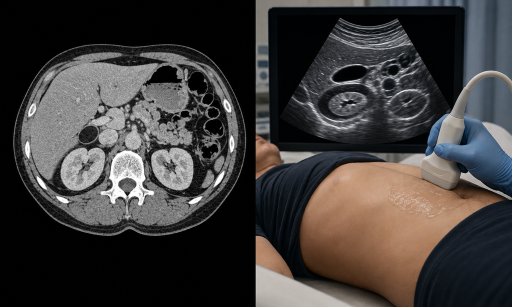
Abdominal complaints are one of the most common reasons doctors have to choose between these two tests, so it’s worth looking at this comparison specifically. An abdominal ultrasound is usually tried first for symptoms like suspected gallstones, liver conditions, kidney issues, or general abdominal discomfort, largely because it’s radiation-free, affordable, and very effective at visualising soft organs like the liver, gallbladder, and kidneys.
An abdominal CT scan, on the other hand, is generally reserved for situations needing more detail or a wider field of view such as suspected diverticulitis, internal bleeding, complex abdominal pain that ultrasound couldn’t fully explain, or cancer staging. It’s also frequently used in emergency settings, since it provides fast, comprehensive answers when time matters.
It’s common and completely normal for a doctor to start with an ultrasound and then order a CT scan if the findings need further clarification. This step-by-step approach helps limit unnecessary radiation exposure while still ensuring an accurate diagnosis.
Safety Considerations: Radiation, Pregnancy, and Children
One of the most important differences in the CT scan vs ultrasound decision is safety, particularly around radiation exposure:
- CT scans use a small dose of ionizing radiation. A single scan carries a low individual risk, but doctors avoid unnecessary repeat CT scans, especially in children, and generally avoid CT scans during pregnancy unless it’s a genuine emergency.
- Ultrasound uses no ionizing radiation at all, which is why it’s considered safe for pregnant women in every trimester, for children, and for patients who may need repeated imaging over time to monitor a condition.
- Radiologists follow the principle of using the lowest radiation dose necessary to get a clear diagnosis, choosing ultrasound first whenever it can reasonably answer the clinical question.
If you have any concerns about radiation exposure for yourself or a family member it’s always worth discussing this directly with your doctor, who can weigh the diagnostic benefit against the (typically small) risk.
Cost and Convenience
Ultrasound scans are generally more affordable and quicker to schedule than CT scans, largely because the equipment and preparation involved are simpler. CT scans, while typically costing more, are often unavoidable when the clinical situation calls for the level of detail only CT can provide such as trauma cases or cancer staging. At Koshikaa, CT scan packages start at an affordable ₹4,000, with transparent pricing and no hidden charges, so cost never has to be a barrier to timely diagnosis.
How Doctors Actually Decide
In practice, the choice isn’t always CT or ultrasound; many patients end up needing both at different stages of diagnosis. A doctor typically considers:
- What symptom or organ needs to be evaluated: soft tissue and real-time movement favour ultrasound; bone, lungs, and complex trauma favour CT.
- How urgent the situation is: CT is often faster and more definitive in emergencies.
- Radiation sensitivity: pregnancy, young age, or the need for repeated monitoring usually tips the decision toward ultrasound.
- Whether an earlier test needs further clarification: sometimes an ultrasound finding needs a CT scan to confirm or characterise it in more detail.
This is also where advances in radiology are making a difference. AI-assisted image analysis is increasingly helping radiologists read both CT and ultrasound images with greater speed and consistency, a trend we cover in our article on how AI is improving diagnostic imaging accuracy.
Why Choose Koshikaa for Your Diagnostic Imaging
As a trusted Health Screening Centre in Bangalore, Koshikaa offers both CT scan and ultrasound imaging under one roof, so your doctor’s recommendation can be acted on quickly, without running between multiple centres. Here’s what sets us apart:
- Both modalities available in-house, no need to visit separate facilities for CT scans and ultrasound imaging.
- Experienced radiologists who interpret every scan with precision and explain results in clear, simple language.
- Fast reporting, with most results ready within 24 – 48 hours.
- Radiation-conscious practices; our team always uses the appropriate imaging method for your specific condition, minimising unnecessary radiation exposure.
- Transparent, affordable pricing on every CT scan in Bangalore and ultrasound imaging package we offer.
Conclusion
There is no single “better” option when comparing a CT scan vs ultrasound. The right test depends on what your doctor needs to examine and the clinical question that needs to be answered. CT scans can provide detailed and rapid images of bones, lungs, and complex internal structures, while ultrasound uses sound waves and is particularly useful for many soft-tissue examinations, real-time imaging, and pregnancy-related care.
Understanding the differences can help you feel more informed when your doctor recommends diagnostic imaging. If you need a reliable facility for your imaging needs, Koshikaa offers a range of diagnostic services with a focus on patient care and accurate reporting.
Book your diagnostic scan with Koshikaa and take the next step towards informed healthcare.
Frequently Asked Questions
1. Is a CT scan more accurate than an ultrasound?
Not universally, each is more accurate for different purposes. CT is generally better for bone, lungs, and complex trauma, while ultrasound is often more effective for soft tissue, real-time movement, and pregnancy monitoring.
2. Is ultrasound always the first test doctors order?
Not always, but for many abdominal, pelvic, and pregnancy-related concerns, ultrasound is typically tried first since it’s radiation-free and more affordable. In emergencies or when more detail is needed, a CT scan may be ordered directly.
3. Is CT scan radiation dangerous?
A single CT scan carries a small, generally low individual risk. Doctors avoid unnecessary repeat scans, especially in children, and weigh the diagnostic benefit against this risk on a case-by-case basis.
4. Can I have an ultrasound and a CT scan for the same condition?
Yes, this is common. Doctors often start with an ultrasound and follow up with a CT scan if the findings need further clarification or a more detailed view.
5. Which is more affordable, a CT scan or an ultrasound?
Ultrasound scans are generally more affordable than CT scans, largely due to simpler equipment and preparation requirements.
6. Is a CT scan safe during pregnancy?
CT scans are generally avoided during pregnancy, especially in the first trimester, unless it’s a genuine emergency. Ultrasound is considered the safe imaging option throughout pregnancy.