Health Screening

Welcome to KOSHIKAA Health Screening, your comprehensive guide to maintaining your well-being through regular health screenings. We understand the importance of early detection and prevention in ensuring long and healthy life. Our cutting-edge health screening services are designed to identify potential health risks and provide you with personalized insights to take control of your health.
Health Screening Centre

How Health Screening Can Save Lives ?

At Koshikaa, we are driven by a bold vision for the future of healthcare. We envision a world where every individual has the opportunity to thrive in good health, where medical care is not just a reactive measure but a proactive journey towards holistic well-being. Our vision encompasses not only the treatment of ailments but also the prevention of illnesses through education, empowerment, and personalized care. We strive to pioneer this transformative shift in healthcare by delivering innovative solutions that prioritize preventive measures, empower patients, and foster communities of wellness. Together, let’s build a future where health is not just a privilege but a fundamental right for all.

Health Packages In Bengaluru

Take the First Step Towards Optimal Health

Regular health screenings are essential for maintaining a proactive approach to your well-being. By detecting health issues at an early stage, you can significantly increase the chances of successful treatment and better long-term outcomes. Don’t wait until symptoms manifest; prioritize your health today and schedule a health screening with KOSHIKAA Health Screening.

Why Choose Our Health Screening Services?

Regular health screenings are essential for maintaining a proactive approach to your well-being. By detecting health issues at an early stage, you can significantly increase the chances of successful treatment and better long-term outcomes. Don’t wait until symptoms manifest; prioritize your health today and schedule a health screening with KOSHIKAA Health Screening.
Comprehensive Health Assessment

Our extensive range of health screening packages covers a wide spectrum of medical conditions, ensuring that no potential health issue goes undetected. From cardiovascular diseases and diabetes to cancer and hormonal imbalances, our screenings are tailored to suit your unique needs.

Advanced Diagnostic Tools

Our clinics are equipped with advanced medical equipment, enabling us to conduct thorough examinations and deliver precise results. We utilize cutting-edge imaging technology, such as MRI and CT scans, to provide detailed insights into your body’s overall health.

Expert Medical Team

Our team of experienced doctors and specialists are dedicated to providing exceptional care. They will guide you through the entire screening process, answer any questions you may have, and offer professional advice based on your individual health profile

Personalized Health Reports

Upon completion of your health screening, you will receive a comprehensive health report that highlights any potential areas of concern. Our reports are easy to understand, providing actionable recommendations to improve your well-being and prevent future health complications.

Convenience and Efficiency

We value your time and strive to make your health screening experience as convenient as possible. With multiple locations and flexible appointment scheduling, we ensure minimal disruption to your daily routine.

Remember, prevention is always better than cure. Invest in your health now to enjoy a lifetime of vitality and wellness. Contact us today to book your health screening appointment. Let us be your trusted partner in safeguarding your most precious asset your health

Frequently Asked Questions

How does early detection through cancer screening save lives?

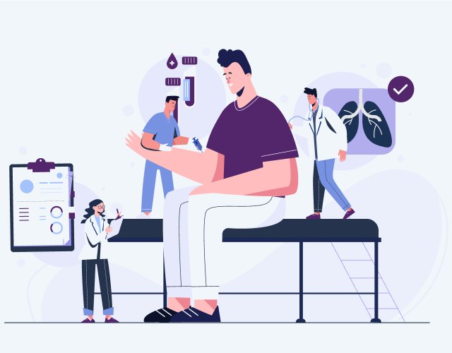
Cancer is a formidable disease that affects millions of lives worldwide. However, the good news is that many cancers can be treated successfully when detected early. That’s why cancer screening plays a crucial role in saving lives and improving treatment outcomes. In this segment, we will explore the importance of cancer screening and how it can positively impact your health.

Cancer screening is a proactive approach to preserving your health and increasing your chances of successful treatment. By detecting cancer in its early stages, you provide yourself with a fighting chance against this disease. Don’t delay; prioritize your well-being by scheduling regular cancer screenings. Remember, early detection saves lives.

What are the benefits of cancer screening and detecting cancer in its early stages?
  • Early Detection : Cancer screening allows for the identification of cancerous cells or tumors before symptoms manifest. Early-stage cancer is often easier to treat and has higher survival rates.

  • Increased Treatment Options : By catching cancer at an early stage, a wider range of treatment options is available, including less invasive procedures and targeted therapies.

  • Improved Survival Rates : Studies consistently show that regular cancer screening leads to higher survival rates as it enables early intervention and appropriate treatment plans.

What are the different types of cancer screening, and how are tailored approaches used for different types of cancer?
  • Breast Cancer Screening : Mammograms and clinical breast exams are vital tools for the early detection of breast cancer, particularly for women aged 40 and above.

  • Colorectal Cancer Screening : Tests such as colonoscopies, fecal occult blood tests (FOBT), and stool DNA tests help identify precancerous polyps or early-stage colorectal cancer.

  • Cervical Cancer Screening : The Pap test and HPV DNA test aid in the detection of abnormal cells in the cervix, allowing for timely treatment and prevention of cervical cancer.

  • Prostate Cancer Screening : Prostate-specific antigen (PSA) tests and digital rectal exams (DRE) are utilized to detect prostate cancer in its early stages, assisting in prompt intervention.

What are the consequences of skipping cancer screening and the risks of neglecting cancer detection?
  • Delayed Diagnosis : Without regular cancer screening, cancers may go undetected until they have advanced, resulting in more aggressive treatment and reduced chances of successful outcomes

  • Limited Treatment Options : Advanced-stage cancers may limit the available treatment options, making it more challenging to achieve long-term remission or cure.

  • Higher Mortality Rates : Studies have shown that individuals who skip cancer screening have higher mortality rates compared to those who undergo regular screenings.

MRI of the uterus is primarily used for diagnosing and evaluating conditions such as uterine fibroids, adenomyosis, and malignancies. It is particularly useful in the planning of surgical procedures and assessing the extent of gynecological diseases. This imaging test is valuable for its ability to deliver detailed cross-sectional images of the uterus and surrounding structures.
An MRI of the spleen can help diagnose various disorders affecting the spleen including splenomegaly (enlarged spleen), tumors, and other abnormalities. It provides clear, detailed images that are crucial for assessing the health of the spleen and planning potential surgical interventions.
Prostate MRI is a non-invasive imaging technique that provides detailed pictures of the prostate gland and surrounding tissues. It helps in the detection, staging, and monitoring of prostate cancer. Prostate MRIs are increasingly used to guide biopsy procedures and assist in the planning of treatment strategies for prostate health.
An MRI of the pancreas is used to identify issues such as pancreatitis, pancreatic cancer, and other pancreatic disorders. It offers high-resolution images that help in detecting small lesions and assessing the pancreatic ducts. This imaging technique is valuable for guiding biopsies and planning surgeries involving the pancreas.
MRI scans of the liver, kidney, and bile ducts provide detailed images that help diagnose various conditions, such as tumors, cysts, and blockages in the bile ducts. They are crucial for assessing organ damage, staging cancer, and planning liver or kidney-related surgeries. This type of MRI can also evaluate the health and function of these organs comprehensively.
Breast MRI is highly sensitive for detecting breast cancer, especially in individuals with dense breast tissue or a high risk of cancer. It is often used in conjunction with mammography to provide a more comprehensive evaluation. Breast MRIs can also monitor the response to breast cancer treatments and provide detailed pre-surgical mapping.
Brain and spine MRIs are critical tools for diagnosing and monitoring neurological conditions, including tumors, spinal cord injuries, and multiple sclerosis. These MRIs can reveal abnormalities in brain tissue and spinal cord structure that might not be visible with other imaging methods. They are essential for planning surgeries and assessing the effectiveness of treatments in neurology.
PET-CT scans of transplant organs like the liver, kidney, or pancreas are performed to assess metabolic activity, detect complications, and monitor post-transplant function. These scans play a vital role in the early detection of issues such as organ rejection, recurrence of disease, or infection, guiding timely interventions and optimizing transplant outcomes for patients.
PET-CT examinations of organs such as the liver, kidney, or prostate are crucial for assessing metabolic activity, detecting abnormalities, and monitoring disease progression. These scans help diagnose conditions such as liver metastases, renal cell carcinoma, or prostate cancer, guiding treatment decisions and monitoring response to therapy over time.
PET-CT scans of soft-tissue masses are utilized to assess metabolic activity and detect malignant tumors or other abnormalities. By combining PET imaging with CT imaging, these scans provide detailed information about tumor location, size, and metabolic characteristics, aiding in accurate diagnosis and treatment planning for soft-tissue cancers.
PET-CT scans are valuable for detecting cysts or fluid collections within the body, including cysts in the liver, kidneys, or other organs. These scans help characterize the nature and metabolic activity of cysts, guiding diagnosis and treatment planning for conditions such as cystic liver disease, renal cysts, or pancreatic cysts.
PET-CT scans of muscles are performed to assess metabolic activity and detect abnormalities such as inflammation, infection, or tumors. These scans provide valuable information about muscle function and pathology, aiding in the diagnosis and management of conditions like myositis, muscular dystrophy, or soft-tissue sarcomas.
PET-CT scans of the brain are utilized to evaluate brain metabolism and detect abnormalities such as tumors, epilepsy, or neurodegenerative diseases. By combining functional PET imaging with anatomical CT imaging, these scans offer comprehensive information about brain function and structure, guiding neurosurgical interventions and treatment decisions for neurological disorders
PET-CT scans of bones are commonly used to detect bone metastases in cancer patients, assess bone health in conditions like osteoporosis, and evaluate response to treatment. By combining PET and CT imaging, these scans provide detailed information about metabolic activity and anatomical changes in bone structures, aiding in accurate diagnosis and treatment planning for various bone-related conditions.
CT scans of the spine are conducted to assess the vertebrae, spinal cord, and surrounding soft tissues for conditions like herniated discs, spinal fractures, or spinal stenosis, aiding in the diagnosis and management of spinal disorders with detailed imaging essential for surgical planning and intervention.
CT scans of the pelvis are utilized to visualize the pelvic bones, reproductive organs, bladder, and surrounding structures for conditions such as fractures, tumors, or pelvic inflammatory disease, guiding treatment plans and surgical interventions with precise anatomical information.
CT scans of the neck are performed to evaluate structures such as the throat, thyroid gland, lymph nodes, and blood vessels for abnormalities like tumors, infections, or injuries, assisting in diagnosis and management of head and neck disorders with detailed imaging.
CT scans of the extremities are employed to assess bones, joints, and soft tissues in the arms or legs, aiding in the diagnosis of fractures, arthritis, or other musculoskeletal conditions, guiding orthopedic treatment and rehabilitation plans for optimal recovery.
CT scans of the chest are utilized to examine the lungs, heart, blood vessels, and surrounding structures for conditions such as lung nodules, pneumonia, or pulmonary embolism, facilitating accurate diagnosis and management of respiratory and cardiovascular diseases.
CT scans of the brain are conducted to assess brain structures, identify abnormalities like tumors, hemorrhages, or strokes, and aid in diagnosing neurological conditions, guiding neurosurgical interventions and treatment strategies with precise imaging of the brain’s anatomy.
CT scans of the abdomen are performed to visualize and evaluate the internal organs such as the liver, kidneys, pancreas, and intestines for conditions like tumors, infections, or injuries, providing detailed images essential for diagnosis and treatment planning.
Transplant organ ultrasound scans, including those of the liver, kidney, or pancreas, are performed to assess organ viability, detect complications, and monitor post-transplant function. These scans play a vital role in the early detection of issues such as organ rejection, vascular complications, or fluid collections, guiding timely interventions and optimizing transplant outcomes for patients.
Ultrasound examinations of organs such as the liver, kidney, or prostate are crucial for assessing organ structure, detecting abnormalities, and monitoring disease progression. These scans help diagnose conditions such as liver cirrhosis, kidney stones, or prostate enlargement, guiding treatment decisions and monitoring response to therapy over time.
Soft-tissue masses can be evaluated using ultrasound to determine their size, location, and characteristics. Ultrasound imaging helps differentiate between benign and malignant masses, guiding further diagnostic evaluation and treatment planning. By providing real-time imaging, ultrasound assists in the precise localization of soft-tissue masses for biopsy or surgical removal if needed.
Ultrasound scans are valuable for detecting cysts or fluid collections within the body, including cysts in the liver, kidneys, or other organs. These scans help characterize the nature and size of cysts, aiding in diagnosis and treatment planning for conditions such as cystic liver disease or renal cysts, and guiding interventions such as cyst drainage or sclerotherapy.
Musculoskeletal ultrasound scans are used to assess the health and integrity of muscles, identifying issues such as strains, tears, or muscle inflammation. These scans provide detailed images of muscle structures, allowing healthcare providers to diagnose muscle injuries accurately and develop personalized rehabilitation plans to promote recovery and restore function.
Ultrasound examinations of joints are essential for evaluating joint health, detecting abnormalities such as inflammation, fluid accumulation, or cartilage damage. By visualizing the joint structures in real-time, ultrasound helps diagnose conditions like arthritis, bursitis, or ligament injuries, guiding treatment decisions and monitoring disease progression over time.
Ultrasound scans of tendons are performed to assess for injuries, inflammation, or tears, aiding in the diagnosis and management of conditions such as tendonitis or tendon tears. These scans provide detailed images of tendon structures, helping healthcare providers determine the extent of damage and plan appropriate treatment strategies, including physical therapy or surgical intervention if necessary.
Pelvis X-rays play a crucial role in diagnosing injuries, diseases, and developmental conditions affecting the pelvic region. They provide detailed images of the hip bones, sacrum, and coccyx, aiding in the assessment of fractures, dislocations, or signs of arthritis. Pelvic X-rays are instrumental in guiding surgical interventions, monitoring healing progress, and evaluating conditions such as osteoporosis or tumors. By providing precise anatomical information, these imaging studies enable healthcare professionals to deliver personalized care and improve patient outcomes.
Teeth X-rays, known as dental radiographs, are indispensable tools for dentists to assess oral health. They aid in diagnosing dental decay, gum disease, and abnormalities in tooth structure or alignment. These images provide valuable insights into hidden dental issues, allowing for early intervention and prevention of complications. Additionally, dental X-rays are essential for planning treatments such as orthodontics, root canals, or dental implants, ensuring optimal outcomes and patient satisfaction.
Spine X-rays are pivotal in diagnosing spinal fractures, degenerative conditions, and spinal misalignments, providing critical information for treatment planning and monitoring spinal health. They offer detailed images of the vertebrae and surrounding structures, enabling healthcare professionals to identify issues such as scoliosis, disc herniation, or spinal stenosis
Skull X-rays are valuable for detecting fractures, assessing head trauma, and evaluating the alignment of facial bones, crucial in diagnosing conditions like fractures, tumors, or developmental abnormalities in the skull. They provide detailed views of the cranial structure, aiding clinicians in determining appropriate treatment strategies and ensuring patient safety.
For the heart, X-ray solutions can aid in the detection of various cardiac problems, ranging from mild to severe, including – Heart failure, Cardiac congestion and other heart-related conditions. In addition to cardiac assessments, X-ray solutions are also used for breast examinations to identify tumors or signs of breast cancer.
Bone X-rays are essential for diagnosing fractures and assessing bone alignment, aiding doctors in developing treatment plans and monitoring healing progress. They provide detailed images of the skeletal system, helping identify abnormalities or injuries that may not be visible through other imaging techniques
Abdominal X-rays are performed when patients experience symptoms such as significant nausea, ongoing vomiting, abdominal pain, flank or back pain, or abdominal swelling. These X-rays can help diagnose various conditions.
Search Bar

Frequently Booked Tests

Frequently Booked Checkups