Looking for a budget-friendly, trustworthy ultrasound scan in Bangalore with quick, accurate reports and caring staff at a clean, reliable place like Koshikaa Health Screening Centre.
Pregnancy – to check the baby’s development and health.
Abdomen – to examine organs like the liver, kidneys, gallbladder, or bladder.
Pelvis – to assess the uterus, ovaries, or prostate.
Heart (echocardiogram) – to evaluate heart function.
Blood vessels (Doppler ultrasound) – to detect blockages or clots.
When it comes to accurate diagnostics Koshikaa considered as a best for ultrasound scan in Bangalore and patient-first care, Koshikaa stands out as one of Bangalore’s most trusted imaging centers. Here’s why patients and doctors alike recommend us:


Breast ultrasound is a non-invasive test that identifies breast lumps and cysts. Moreover, it is often recommended after an abnormal mammogram to further assess breast abnormalities.
A pelvic ultrasound examines the organs in the pelvic area between the lower abdomen (belly) and legs. It assesses organs such as the bladder, prostate, rectum, ovaries, uterus, and vagina. Moreover, you can consider an ultrasound scan in Bangalore for a comprehensive examination of your pelvic organs.
In a transvaginal ultrasound, a probe is inserted into the vaginal canal to evaluate reproductive tissues like the uterus and ovaries. Moreover, it is also referred to as a pelvic ultrasound because it assesses structures within the pelvis. If you’re looking for a transvaginal ultrasound scan near you, reach out to us.
Thyroid ultrasound is used to evaluate the thyroid gland, which is located in the neck. Additionally, it allows providers to measure the size of the thyroid and identify any nodules or lesions within the gland.
Transrectal ultrasound involves inserting an ultrasound probe into the rectum to evaluate the rectum or nearby tissues, such as the prostate in individuals assigned male at birth. Moreover, while the exact cost may vary, searching for “Ultrasound Scan Price in Bangalore” can help you find a clinic that fits your budget.
At Koshikaa, we do Ultrasound KUB scans to check your kidneys, ureters, and bladder. This scan helps find problems like kidney stones, infections, or blockages. Moreover, it is safe and painless. Before the scan, you only need to drink water to keep your bladder full. The test takes about 15 to 20 minutes. Additionally, there is no radiation, and our team ensures you are comfortable and get clear, accurate results.
Using an ultrasound scan helps doctors clearly see different parts of the body. Moreover, it helps detect various conditions effectively.
Booking an Affordable ultrasound at Koshikaa Diagnostic scan center bangalore is easy and takes less than a minute. Just follow these 4 simple steps:
Please enter your name, mobile number, and email. This way, we can contact you quickly and, moreover, confirm your appointment without any delays.

Our team will call you to confirm your appointment. Moreover, they will guide you on any instructions for the scan.

Once confirmed, you’ll receive the appointment details. Additionally, the center address and reporting time will be sent directly to your phone or inbox

Visit the center at your scheduled time. Furthermore, our expert team will ensure a smooth, comfortable experience
Abdominal scan: You may need to fast for 6–8 hours.
Pelvic scan: Drink 3–4 glasses of water before the test and do not empty your bladder.
Pregnancy ultrasound: No fasting needed for most routine checks.
Doppler scan: Usually no special prep, but follow the doctor’s advice.
The scan is done in 15–30 minutes and results are usually available the same day or within 24 hours.
For pelvic ultrasounds, you may need to fill your bladder by drinking water before the test. This is because a full bladder provides better visualization of the pelvic organs. Moreover, your healthcare provider will give you specific instructions on how much water to drink and when to do so before the ultrasound.


I was thoroughly impressed with my thyroid ultrasound here. The staff was friendly, the technician skilled, and results were promptly provided with clear explanations
Koshikaa for breast ultrasounds surpassed my expectations. The staff was professional and caring, the technician thorough, and the facility clean and comfortable. Highly recommended.
My pelvic ultrasound experience here was seamless. The knowledgeable technician answered all my questions, and results were promptly delivered with follow up to ensure understanding.
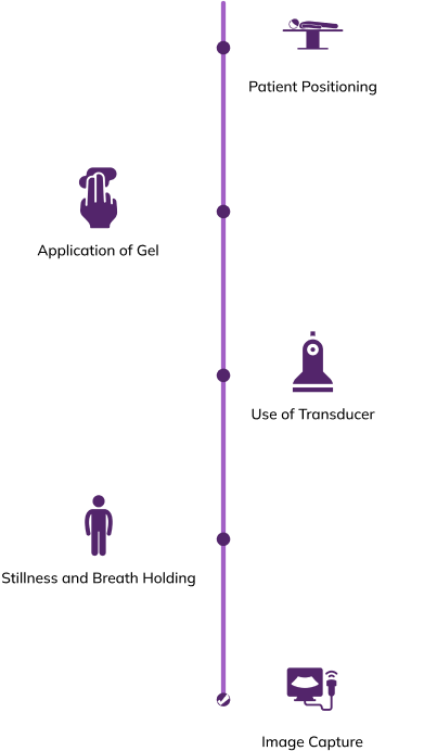
An ultrasound scan in Bangalore is a painless procedure where you just have to lie down on a low table while the technician is applying gel on your skin. The doctors then slide a transducer across the body part to capture images utilizing sound waves. Believe it or not, you may be required to take deep breaths and then stop breathing for some time in the process of taking pictures.
Ultrasound also known as USG is a diagnostic tool used to look deep inside the body tissues, which include the tendons, muscles, joints, cysts, and other internal organs such as the liver and kidneys among others. This is especially helpful in cancers involving soft tissues and in the differentiation between cystic lesions and solid masses. The test is preferred over a CT scan because it is faster, and does not require exposure to ionizing radiation.
It depends. For abdominal scans, fasting is usually needed. For others like obstetric or pelvic, no fasting is required.
Preparation for an ultrasound scan in Bangalore varies depending on the area being scanned. Some of the preparations for the pelvic ultrasounds include you may be required to drink water to fill your bladder. Especially, before undergoing abdominal ultrasonography you might be advised to observe a few hours of fasting. However, make sure that you follow your healthcare provider’s instructions if you are to get the right results.
An ultrasound scan in Bangalore effectively detects cancer in soft tissues and is often the first step in diagnosis. It is fast, inexpensive, and noninvasive and allows for locating parts of the body that need additional assessment. Ultrasound is used in the first instance for assessment but may not be as clear as CT or MRI scans hence requiring further procedures such as biopsies for a cancer confirmation.
Non-invasive imaging through an ultrasound scan in Bangalore is safe and comfortable for all ages. Our advanced technology allows for exceptional clarity in viewing internal organs, aiding accurate diagnoses. Detailed images depict proper treatment approaches to different diseases since patients require proper diagnosis to gain the correct treatment. Ultrasound has limitations too including poor transmission, it cannot image through gas or fatty tissue-bearing organs or structures which are hidden by bones such as the lungs and skull. To enhance assessment, an ultrasound scan in Bangalore can be combined with other imaging tests for comprehensive cancer evaluations.
An ultrasound procedure usually takes around 20 to 30 minutes to complete. However, the actual duration can vary depending on the type of examination being performed and the complexity of finding any changes or abnormalities in the organs being studied.
Not at all. It’s non-invasive and typically feels like a warm gel on your skin.
yes we give preliminary results quickly. A full radiology report may take a few hours.

Bone X-rays are essential for diagnosing fractures and assessing bone alignment, aiding doctors in developing treatment plans and monitoring healing progress. They provide detailed images of the skeletal system, helping identify abnormalities or injuries that may not be visible through other imaging techniques