
Getting an MRI report can feel like reading a foreign language. With long medical terms, unfamiliar abbreviations, and detailed descriptions, it is understandable that many patients skip straight to the last section, hoping to see the word “normal.” However, an MRI scan report follows a specific structure, and understanding a few common terms can make it much easier to follow. At a reliable Health Screening Centre in Bangalore, patients can also discuss their results with the appropriate healthcare professionals after an MRI Scan in Bangalore.
In this guide, we’ll explain the main sections of an MRI report, what common terms generally mean, why the Impression section matters, and what you should do after receiving your results all in simple, easy-to-understand language.
Why MRI Reports Can Be Confusing
MRI reports aren’t written for patients. They’re created by radiologists for other doctors. That’s why they use precise medical language instead of everyday words. For example, “mild degenerative changes” might sound alarming, but it often just means normal wear and tear that most people have by their 40s or 50s. This gap in language is why many patients feel anxious when they open their report. Once you learn the basic structure and some common terms, that anxiety can fade.
The Basic Structure of an MRI Scan Report
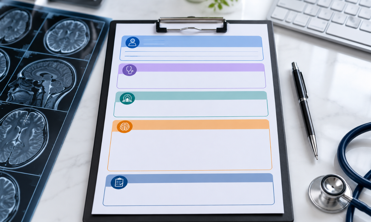
Most radiology reports have a similar layout, regardless of the body part scanned. Here’s what each section means:
1. Patient and Study Details
This section includes your name, age, the scan date, and which body part was examined, like “MRI Brain” or “MRI Lumbar Spine.” It’s just identifying information.
2. Clinical History
This is a brief note on why the scan was done, such as “persistent lower back pain for 3 months” or “follow-up for known disc bulge.” It gives context to the radiologist, helping them focus on what matters for your symptoms.
3. Technique
This section describes how the scan was performed, the type of machine used, and whether contrast dye was involved. Most patients can skip this; it’s mainly for other doctors.
4. Findings
This is the main part of the report. It details what the radiologist observed, organ by organ or region by region. It can seem intimidating since it lists normal and abnormal observations.
5. Impression (or Conclusion)
This is the most important part of the report. It summarizes the key findings in order of importance. If you read only one section, read this one, alongside your doctor’s explanation.
Common Terms in MRI Findings
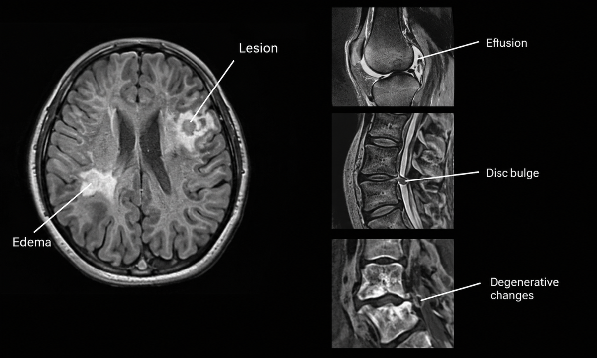
Radiology reports use a specific vocabulary, and a lot of patient anxiety comes down to simply not knowing what these words mean. Here’s a simple glossary of terms you’ll often come across in MRI findings:
| Term You Might See | What It Usually Means |
|---|---|
| Unremarkable / Within normal limits | No abnormality was found in that area |
| Lesion | A general term for any area that looks different from surrounding tissue, not necessarily cancer or anything serious |
| Mass | A lump or growth; the report will usually describe its size and characteristics for further evaluation |
| Signal intensity | How bright or dark a structure appears on the scan, which helps identify tissue type or fluid |
| Enhancement | An area that becomes more visible after contrast dye is used, often to check blood flow or inflammation |
| Degenerative changes | Wear-and-tear changes, most often age-related, especially common in spine and joint MRIs |
| Effusion | A build-up of fluid, often seen around a joint |
| Herniation / Bulge | Displacement of disc material in the spine, ranging from mild to significant |
| Edema | Swelling caused by excess fluid in the tissue |
| Incidental finding | Something noticed on the scan that is unrelated to the original reason for the MRI |
Seeing these words in your MRI scan report doesn’t mean something is seriously wrong. Many describe minor or normal variations. Always consider the context, which is why the Impression section and your doctor’s explanation are crucial
How to Read the Impression Section

The Impression section answers the question: “What does this mean for the patient?” It usually lists the most significant findings first, often numbered, and may suggest next steps, like “clinical correlation recommended” or “follow-up imaging in 6 months.”
Read this section as a summary, not a diagnosis. Radiologists describe what they see; your doctor combines that with your symptoms and other test results to make a diagnosis and treatment plan. This is why the same MRI finding can mean different things for different patients.
What Families Should Avoid with an MRI Report
It’s natural to want quick answers, especially when a loved one is unwell, but some habits can cause unnecessary worry:
- Searching terms online without context. A word like “lesion” can lead to frightening results that don’t apply to your case.
- Focusing only on the Findings section. The Findings detail every observation, including harmless ones. The Impression tells you what matters.
- Comparing your report to someone else’s. MRI findings are unique to each individual based on age, symptoms, and medical history.
- Making decisions before speaking with the doctor. Reports are meant to be explained, not self-diagnosed.
If you’re unsure about anything, always ask your doctor to walk you through the report.
When to Discuss the Report With Your Doctor
Once your MRI report is ready, book a follow-up appointment instead of trying to interpret it alone. It helps to prepare a short list of questions, like “What does this finding mean for my symptoms?” or “Do I need follow-up scans?” If you’re new to MRIs and want to know what to expect, our guide on what to expect during an MRI scan can help.
Why Choose Koshikaa for Your MRI Scan and Report Support
At Koshikaa Health Screening Centre in Bangalore, we believe an MRI shouldn’t end with confusion. Here’s how we make the process easier:
- Experienced radiologists prepare clear, accurate reports for every scan.
- Simple explanations at handover: our team helps you understand your findings before you leave.
- Fast turnaround, with most reports ready in 24 – 48 hours.
- A calm, patient-first environment, whether it’s your first MRI or a follow-up. If you’re interested in our imaging for the brain and spine, our guide on MRI for the brain and spine is a great next read.
- Transparent pricing, so there are no surprises when booking your MRI scan with us.
Conclusion
Understanding MRI scan reports doesn’t require a medical degree. Familiarising yourself with the basic structure and common terms in a radiology report can help you have a more informed conversation with your doctor. Remember, the Impression section is an important starting point, but MRI interpretation should always be considered alongside your symptoms, medical history, and other tests rather than used for self-diagnosis.
If you or a family member has been advised to undergo an MRI Scan in Bangalore, Koshikaa provides diagnostic imaging services with a focus on patient care and reliable reporting. Get in touch with Koshikaa to learn more or schedule your scan.
Frequently Asked Questions
1. How long does it take to get an MRI scan report?
Most MRI reports are ready within 24 – 48 hours, though complex scans or those requiring a specialist’s second opinion may take a little longer.
2. Can I understand my MRI report without a doctor’s help?
You can understand the general structure and common terms using guides like this one, but the report should always be interpreted by your doctor, since they can connect the findings to your specific symptoms and history.
3. What does “clinical correlation recommended” mean in an MRI report?
It simply means the radiologist is asking your treating doctor to match the imaging findings with your actual symptoms and examination before drawing a final conclusion. It’s a routine phrase, not a red flag by itself.
4. Does the word “mass” in my MRI findings always mean cancer?
No. “Mass” is a general descriptive term for any lump or growth seen on the scan. Many masses are benign (non-cancerous), and further evaluation is usually needed to determine what they actually are.
5. Why does my MRI report mention things unrelated to why I got the scan?
These are called incidental findings: observations noticed while scanning the targeted area that aren’t related to your original symptoms. They’re common and often harmless, but your doctor will let you know if any need follow-up.
6. Can I ask the radiologist directly to explain my report?
At most centres, including Koshikaa, you can request a consultation with the reporting doctor or your treating physician to go through the report in detail and ask any questions you have.