
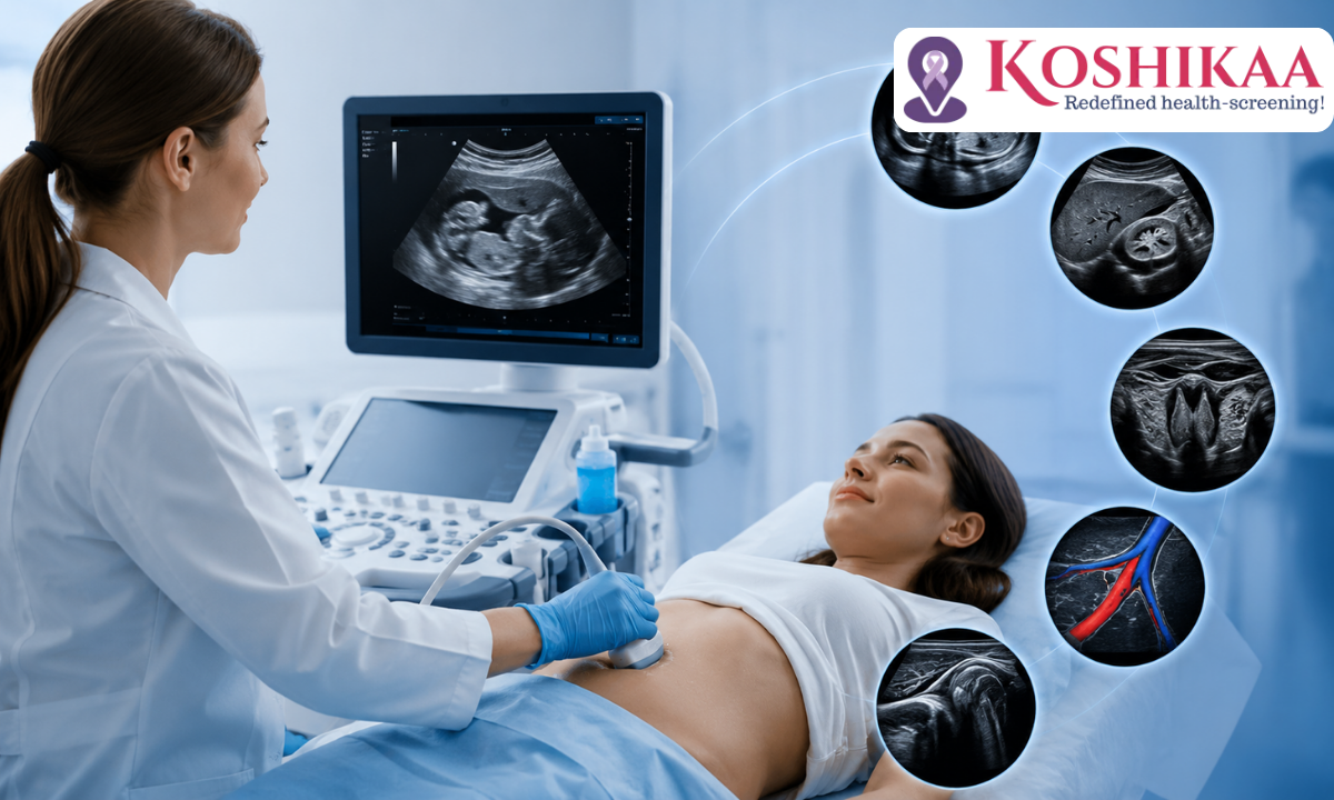
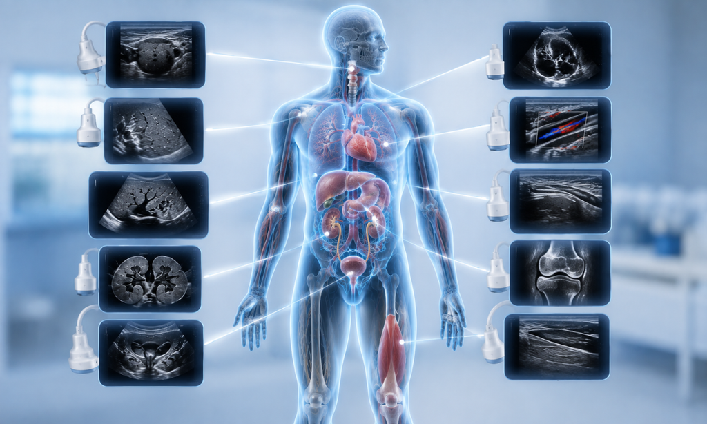
Ultrasound is one of the most commonly used diagnostic imaging tools in modern healthcare, with applications that go far beyond what many people realise. The range of ultrasound diagnostic uses covers different parts of the body, including the liver, kidneys, thyroid, heart, blood vessels, muscles, and joints. Since ultrasound uses sound waves instead of ionising radiation and can provide real-time images, it is a useful option for evaluating many health conditions.
If your doctor has recommended an Ultrasound Scan in Bangalore, understanding why it may be used can help you feel more informed.
In this guide, we’ll explore the common medical conditions ultrasound can help detect and how this imaging method supports diagnosis and evaluation.
Medical Disclaimer
This article is intended for general educational and awareness purposes only. It is not a substitute for professional medical advice, diagnosis, or treatment. Whether an ultrasound or another imaging test is appropriate for your specific symptoms should always be determined by a qualified doctor. Please consult your physician for any health concerns.
What Is an Ultrasound?
An ultrasound is a non-invasive imaging test that uses high-frequency sound waves to create images of structures inside the body. Unlike X-rays and CT scans, ultrasound uses sound waves instead of ionising radiation
How Ultrasound Actually Works
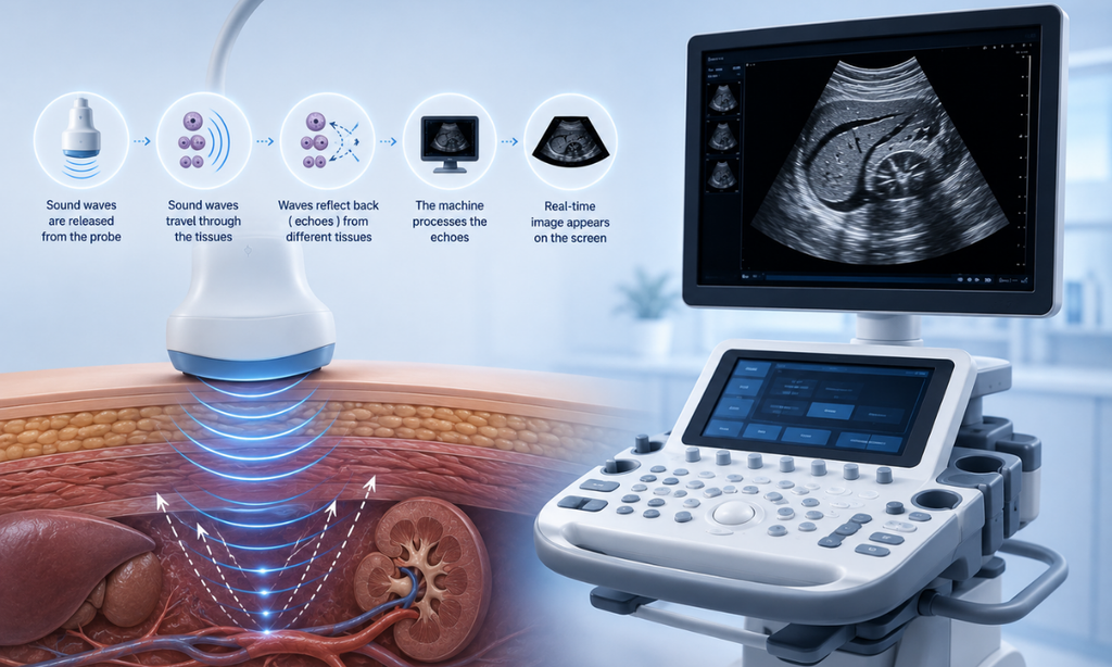
The process is fairly simple:
Probe → Sound waves enter the body → Echoes return from tissues → Computer processes the echoes → Real-time images appear on the screen
This allows doctors to examine organs, tissues, and blood flow while they are being observed in real time.
Why Is Ultrasound Commonly Used?
- No ionising radiation
- Non-invasive and generally painless
- Provides real-time imaging
- Can be used to examine many organs and soft tissues
- Can be repeated when clinically necessary
This makes ultrasound particularly useful in evaluating many other conditions involving the abdomen, pelvis, thyroid, blood vessels, muscles, and joints.
Note: Although ultrasound is generally considered safe, the type of scan and whether it is appropriate depends on the patient’s condition and the doctor’s recommendation.
What Can Ultrasound Diagnose?
1. Abdominal Conditions (Liver, Gallbladder, Pancreas, Spleen)
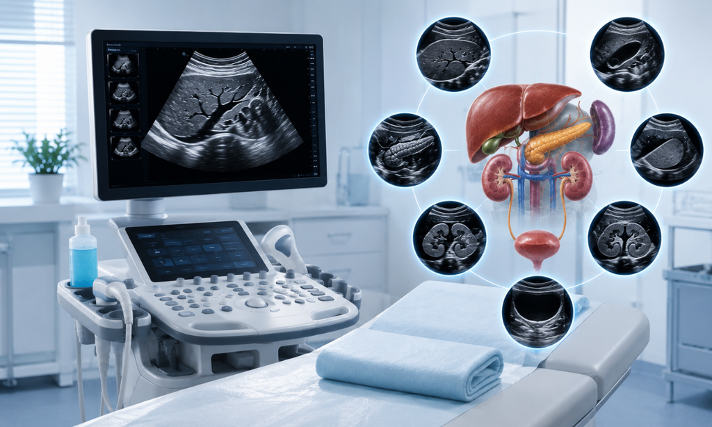
An abdominal ultrasound is commonly used to investigate unexplained abdominal pain, digestive symptoms, or suspected problems involving the abdominal organs. It can provide images of organs such as the liver, gallbladder, spleen, and parts of the pancreas, without exposing the patient to ionising radiation.
A liver ultrasound can help identify fatty changes, cysts, enlargement, and other structural abnormalities. It can also help doctors evaluate the liver when there is concern about chronic liver disease. Ultrasound can detect gallstones, gallbladder inflammation, bile duct abnormalities, and some pancreatic changes as well. However, not every abdominal condition can be diagnosed through ultrasound alone, and additional imaging may sometimes be recommended.
If you’d like to understand fatty liver findings in more depth, our guide on how fatty liver disease is diagnosed is a helpful next read.
2. Kidney and Urinary Tract Conditions
A kidney ultrasound, often performed as part of a KUB (Kidneys, Ureters, Bladder) scan, is a useful non-invasive way to examine the kidneys and urinary system. It can help identify kidney cysts, swelling of the kidneys (hydronephrosis), changes in kidney size, and certain structural abnormalities.
Ultrasound may also detect some kidney stones, although very small stones or stones located in certain parts of the urinary tract may not be clearly visible. A bladder ultrasound can assess bladder emptying and may help investigate symptoms such as difficulty urinating or recurrent urinary problems.
In children and other patients where radiation exposure is a concern, ultrasound is often a useful first-line imaging option for evaluating the urinary tract. Our detailed guides on KUB ultrasound for first-time patients, when doctors recommend a renal ultrasound, and ultrasound’s role in diagnosing UTIs explain these applications in more detail.
3. Pelvic and Reproductive Health
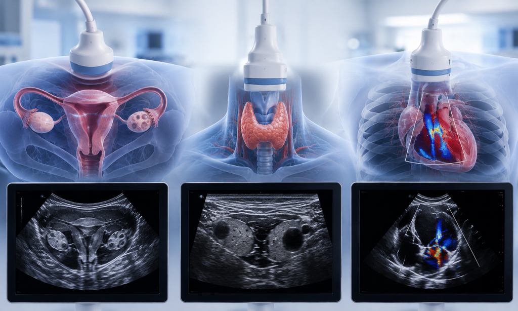
A pelvic ultrasound is an important imaging test used to examine the reproductive and pelvic organs in both women and men. In women, it provides detailed images of the uterus, ovaries, cervix, and surrounding pelvic structures, helping doctors investigate symptoms such as pelvic pain, irregular periods, heavy menstrual bleeding, or unusual bleeding.
It can help identify findings such as ovarian cysts, uterine fibroids, endometrial thickening, and other structural changes. Depending on the reason for the scan, an abdominal or transvaginal ultrasound may be recommended. A transvaginal scan uses a specialised probe to obtain closer, more detailed images of the uterus and ovaries and may be particularly useful when evaluating ovarian or uterine abnormalities. You can learn more in our article on transvaginal ultrasound for ovarian cyst detection.
Pelvic ultrasound may also be used as part of the evaluation of PCOS (Polycystic Ovary Syndrome). However, PCOS is not diagnosed through ultrasound alone. Doctors consider the scan findings alongside symptoms, medical history, and other clinical or laboratory findings. Our guide on how ultrasound helps diagnose PCOS explains what doctors may look for during the examination.
4. Thyroid and Neck Abnormalities
Ultrasound is commonly used to evaluate thyroid nodules, enlarged thyroid glands, and unexplained neck swellings. It can provide detailed information about the size, shape, and internal characteristics of a thyroid nodule and help doctors determine whether further evaluation is appropriate.
For example, ultrasound can help distinguish between fluid-filled cystic areas and solid nodules and identify certain features that may require closer assessment. If a nodule needs tissue testing, ultrasound can also be used to guide a fine-needle biopsy accurately.
Because ultrasound does not use ionising radiation, it is particularly useful for repeated monitoring of known thyroid nodules. However, an ultrasound appearance alone does not confirm whether a nodule is cancerous; doctors may recommend additional testing based on the findings.
5. Heart and Blood Vessel Conditions
Ultrasound is also an important tool for examining the heart and circulatory system. An echocardiogram, or cardiac ultrasound, produces moving images of the heart and can help doctors assess the size of its chambers, heart muscle movement, valve function, and pumping ability.
Another technique called Doppler ultrasound evaluates the movement of blood through blood vessels. It can help investigate reduced blood flow, narrowing or blockages in certain arteries, and blood clots such as deep vein thrombosis (DVT).
Because Doppler imaging can show blood flow in real time, it is particularly useful when a doctor needs to understand not only the structure of a blood vessel but also how blood is moving through it.
6. Musculoskeletal and Soft Tissue Issues
Ultrasound isn’t limited to internal organs. It can also examine muscles, tendons, ligaments, joints, and other soft tissues, making it useful for investigating sports injuries, persistent pain, swelling, and soft-tissue abnormalities.
Depending on the area being examined, ultrasound can help identify findings such as tendon tears, inflammation, fluid collections, muscle injuries, and some soft-tissue masses. One of its advantages is that imaging can be performed while the patient moves the affected body part.
This real-time imaging can help doctors assess how a tendon or other structure behaves during movement. Ultrasound can also be used to guide certain procedures, such as injections or fluid drainage, when clinically appropriate.
7. Cancer and Tumour Evaluation
Ultrasound can play an important role in evaluating lumps, masses, and other abnormalities, including those involving the breast, thyroid, liver, and reproductive organs. It can help doctors understand the location, size, and characteristics of an abnormal area and may help distinguish between a fluid-filled cyst and a solid mass.
However, it is important to understand that ultrasound alone cannot confirm cancer. If an ultrasound shows a suspicious finding, the doctor may recommend additional imaging, follow-up examinations, or a biopsy to determine what the abnormality represents.
Ultrasound can therefore be an important part of the diagnostic pathway, helping doctors decide what further evaluation may be appropriate. Our detailed article on
the role of ultrasound scans in early detection of cancer explores this topic further.
8. Emergency and Trauma Situations
Ultrasound can also be valuable in emergency settings, where doctors sometimes need answers quickly. A FAST scan (Focused Assessment with Sonography in Trauma) is a bedside ultrasound examination used in certain trauma situations to look for signs of internal bleeding, particularly around organs and within the abdomen or chest.
Because the examination can be performed quickly at the patient’s bedside and does not involve radiation, it can provide useful information while emergency teams assess the patient’s condition.
Ultrasound is not a replacement for CT or other imaging when more detailed evaluation is required. Instead, in appropriate emergency situations, it can provide rapid initial information that helps doctors decide what should happen next.
Quick Reference: Ultrasound Diagnostic Uses by Body Area
| Body Area | Common Conditions Diagnosed | Type of Ultrasound |
| Liver, gallbladder, pancreas, spleen | Fatty liver, gallstones, cysts | Abdominal ultrasound |
| Kidneys, ureters, bladder | Kidney stones, hydronephrosis, UTIs | Renal / KUB ultrasound |
| Uterus, ovaries | Fibroids, ovarian cysts, PCOS | Pelvic / transvaginal ultrasound |
| Thyroid gland | Nodules, cysts, swelling | Thyroid ultrasound |
| Heart | Valve issues, pumping efficiency | Echocardiogram |
| Veins and arteries | Blood clots, blockages | Doppler ultrasound |
| Tendons, joints, muscles | Tears, fluid buildup, inflammation | Musculoskeletal ultrasound |
| Breast, liver, thyroid, ovaries | Early tumour or mass evaluation | Diagnostic sonography |
| Abdomen/chest (emergency) | Internal bleeding, organ injury | FAST / emergency ultrasound |
Why Ultrasound Is Often the First Test Doctors Choose
Beyond its safety profile, ultrasound has a few practical advantages that make it a frequent first step in diagnosis:
- No radiation, making it repeatable and safe even for children and patients
- Real-time imaging, allowing doctors to observe movement blood flow, a beating heart, or a joint in motion
- Faster and more affordable than CT or MRI scans, in most cases
- No special preparation for many scan types, and results are available quickly
- Widely accessible, making it a practical first-line test before deciding if more advanced imaging is needed
Why Choose Koshikaa for Ultrasound Scans in Bangalore
As a trusted Health Screening Centre in Bangalore, Koshikaa offers a full range of ultrasound diagnostic uses under one roof, supported by experienced radiologists and modern imaging equipment:
- Comprehensive ultrasound services abdominal, pelvic, thyroid, renal, cardiac, Doppler, and more
- Skilled sonographers and radiologists who ensure accurate, detailed imaging and reporting
- Fast turnaround, with most reports delivered within 4 to 6 hours
- Transparent, affordable pricing, with ultrasound scans starting at ₹850
- Emergency ultrasound availability for time-critical situations, alongside routine and specialised scans
Conclusion
Ultrasound has become an important part of modern diagnostic care, From evaluating abdominal and urinary conditions to examining the thyroid, heart, blood vessels, and soft tissues, its wide range of uses makes it a valuable tool for investigating many health concerns.
If your doctor has recommended an Ultrasound Scan in Bangalore, understanding why the scan is being performed can help you feel more informed and prepared. The appropriate imaging test will always depend on your symptoms and clinical needs.
For reliable diagnostic imaging and patient-focused care, Koshikaa offers ultrasound services to support accurate evaluation and timely medical guidance.