Early cancer detection has become crucial in modern healthcare as it allows for timely intervention, leading to improved patient outcomes. Among the various diagnostic tools available, ultrasound scans stand out for their versatility, non-invasive nature, and widespread accessibility. Ultrasound scans utilize high-frequency sound waves to produce images of internal organs, offering valuable insights into possible abnormalities. They play a vital role in diagnosing various types of cancer, including breast, liver, thyroid, and more. As a reliable imaging tool, ultrasound is widely used for early cancer detection in cities like Bangalore, where access to advanced healthcare facilities is more common.
In this article, we explore how ultrasound scans contribute to cancer detection. The following subtopics delve into the workings of ultrasound technology, its applications in detecting specific cancers, and how the service is available in urban centers like Bangalore.
How Ultrasound Scans Work
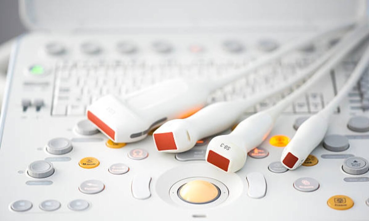
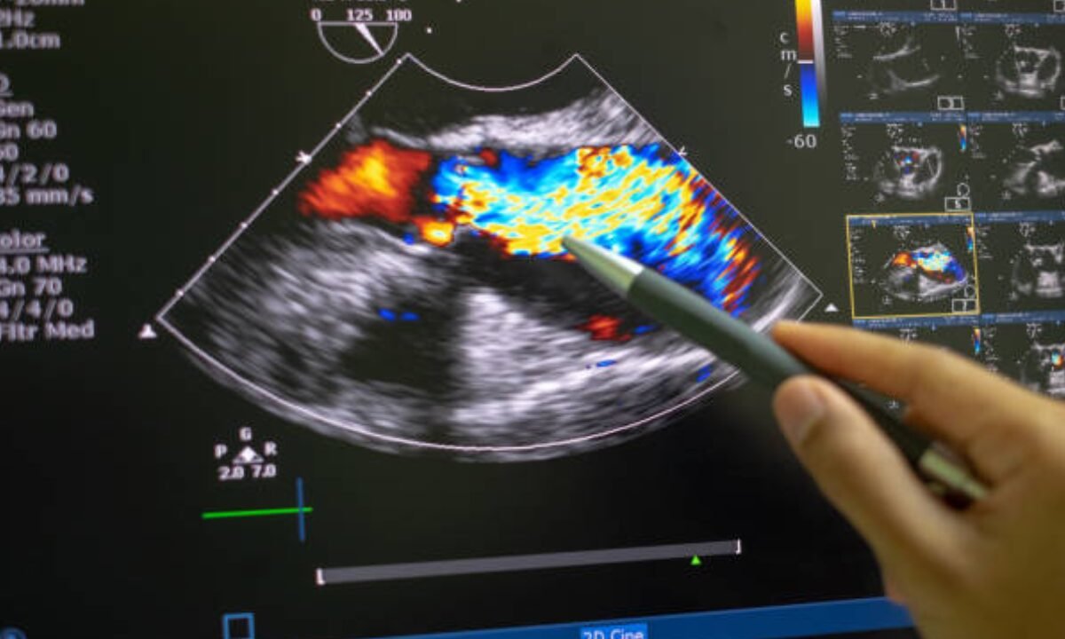
Ultrasound scans involve the use of sound waves, typically above the audible range, to produce images of the internal organs. A handheld device called a transducer emits these waves, which then bounce off tissues in the body, creating echoes. These echoes are then converted into real-time images, allowing medical professionals to examine various organs. The absence of radiation makes ultrasound safer for frequent use, and this non-invasive imaging method has become indispensable in detecting structural changes indicative of cancer.
Advantages of Ultrasound in Cancer Detection
Ultrasound scans offer numerous advantages, particularly in early cancer detection:
- Non-invasive and Painless: Unlike biopsies or other invasive diagnostic methods, ultrasounds are pain-free and do not require recovery time.
- Radiation-Free: Ultrasounds do not use radiation, which is especially beneficial for patients who require multiple scans over time.
- Cost-Effective: Ultrasounds are generally more affordable compared to MRI and CT scans, making them accessible for routine screenings.
- Real-Time Imaging: The ability to see live images allows for immediate assessment, which can be crucial for diagnosing rapidly progressing cancers.
- In cities like Bangalore, the affordability and ease of use make ultrasound scans a preferred choice for both patients and healthcare providers looking for early signs of cancer.
Types of Cancer Detectable by Ultrasound
Ultrasound scans are commonly used for detecting various types of cancer. Here are some notable applications:
- Breast Cancer: Breast ultrasounds help in identifying lumps and differentiating between cysts and solid masses. This scan is often used alongside mammography for more accurate diagnosis.
- Liver Cancer: Liver ultrasounds can detect tumors or other abnormalities, making it easier to diagnose liver cancer in its early stages.
- Thyroid Cancer: Thyroid ultrasounds assess nodules within the thyroid gland and help in differentiating between benign and malignant nodules.
- Pancreatic Cancer: Though less common, ultrasound imaging can sometimes identify pancreatic abnormalities.
- Gynecological Cancers: Pelvic ultrasounds can assist in detecting ovarian and endometrial cancers by visualizing abnormal growths in reproductive organs.
Regular ultrasound screening in Bangalore can play a significant role in the early detection of these cancers, especially for those at higher risk.
The Importance of Early Detection in Cancer Treatment
Early detection through ultrasound imaging is vital for successful cancer treatment. Identifying cancer in its initial stages allows for less aggressive treatments, potentially improving survival rates and quality of life. By catching the disease early, ultrasound scans make it possible for doctors to provide personalized treatment plans, which may include surgical intervention, chemotherapy, or radiation therapy as appropriate. Timely detection can also reduce the emotional and financial burden on patients and their families.
Choosing the Right Center in Bangalore
Selecting the right center is essential for accurate ultrasound results. When choosing a facility for cancer screening in Bangalore, consider the following factors:
- Accreditation and Credentials: Ensure the center is certified and employs experienced radiologists.
- Technology and Equipment: Look for centers with the latest ultrasound technology for optimal imaging quality.
- Reviews and Patient Feedback: Research reviews and patient testimonials to gauge service quality and diagnostic accuracy.
- Range of Services: A good diagnostic center should offer comprehensive cancer screening packages, including ultrasound.
With numerous options available, Bangalore residents have the advantage of selecting a center that meets their specific healthcare needs.
Future of Ultrasound Technology in Cancer Detection
Ultrasound technology continues to evolve, with advancements like 3D and 4D imaging offering more precise visuals. In addition, research is underway to enhance the diagnostic power of ultrasound with artificial intelligence (AI) integration. AI-assisted ultrasound can potentially increase the accuracy of cancer detection, making it easier for radiologists to identify early signs of malignancy.
In Bangalore and other tech-forward cities, ultrasound technology is set to benefit from ongoing advancements, improving cancer diagnostic capabilities in the coming years.
Conclusion
Ultrasound scans are a valuable tool in the fight against cancer, offering a non-invasive, accessible, and reliable means of early detection. In cities like Bangalore, where quality healthcare facilities are abundant, the role of ultrasound in cancer screening is more important than ever. From identifying potential issues in high-risk patients to providing real-time imaging that aids in personalized treatment, ultrasound scans have revolutionized early cancer detection. Embracing these diagnostic advancements and making routine ultrasound screenings a part of regular healthcare can empower individuals to manage their health proactively.
Koshikaa Screening Centre offers a wide range of comprehensive cancer screening services, ensuring accurate results and personalized health care. Contact us today at +91 7996666106