
Early detection of breast cancer is important and mammography helps to do so. It’s an essential part of most women’s routine healthcare as many women pass through their 40s and beyond. This guide aims to shine a light on the importance of mammography in Bangalore, what to expect during the procedure and its clear understanding.
What is Mammography?
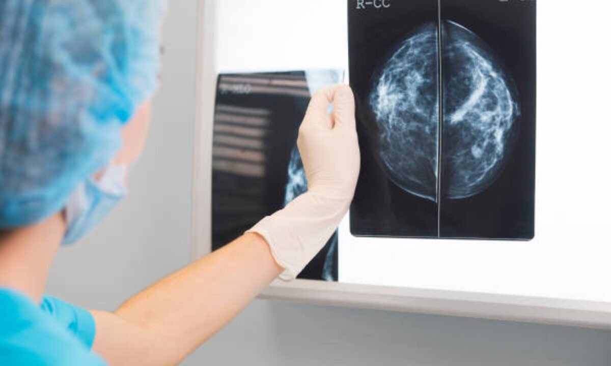
Mammography is a special method of medical imaging that uses low-dose X-rays to examine breast tissue. It’s a procedure that can help detect abnormalities such as tumors or it can show changes in tissue which can be breast cancer or other health problems.
In Bangalore, many healthcare facilities provide state-of-the-art mammography facilities for those who are aware and need early breast cancer detection. The availability of PET CT scans in Bangalore provides full-range images for patients, which is helpful in accomplishing the perfect diagnosis and correct treatment planning.
Why is Mammography Important?
Early detection is important because early detection greatly increases a patient’s chance of successful treatment. Here are a few reasons why mammography is an important procedure:
- Early Cancer Detection: Early treatment is possible because mammograms can detect tumours before they are felt.
- Lower Mortality Rates: It has been shown that regular screenings will reduce rates of breast cancer mortality.
- Peace of Mind: That you know your breast health status relieves anxiety and promotes proactive healthcare.
With the focus being on the city’s commitment to women’s health and early cancer detection, Bangalore has made mammography available. Most patients would want to get the best option available at a credible screening centre in Bangalore in terms of early detection of health issues. A full range of mammography services in Bangalore helps facilitate timely interventions with better outcomes in health care.
Who Should Get Mammograms?
Guidelines vary but typically, women begin having regular mammograms around age 40. However, people with a family history of breast cancer or other risk factors should speak with their healthcare provider about earlier screenings.
Guidelines for Mammography
1. Ages 40-44: Mammograms should be annual for women.
2. Ages 45-54: You are encouraged to undergo annual mammograms.
3. Ages 55+: Women can elect to switch their screening to biennial or stay on an annual basis at personal preference.
Knowing about mammography in Bangalore will help you know where you can get screened.
A Mammogram: What to Expect?
If it’s your first mammogram, here’s a step-by-step breakdown of what to expect:
Preparation
- Schedule Your Appointment: Schedule your mammogram as late as possible in the menstrual cycle to avoid scheduling it when breasts are most sensitive.
- Wear Comfortable Clothing: Wear a two-piece outfit as you will need convenience during the exam.
During the Procedure
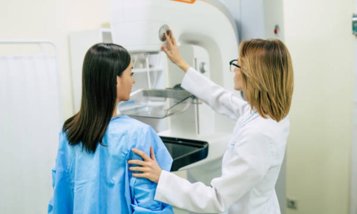
1. Positioning: The mammography machine will make you stand. Your breast will be placed on the machine’s plate so a technician will be able to guide you on how to position it.
2. Compression: The breast is then compressed to get clear images via the machine. That’s the only bad thing about it: it’s uncomfortable in a way, but just for a few seconds.
3. Imaging: Almost always it takes 20 to 30 minutes at once when several images from different angles are taken.
Bangalore’s mammography facilities have staff trained to make the experience as bearing as possible.
Understanding Results and Follow-Up
After the mammogram is finished, a radiologist goes through the images. Results are usually communicated within a few days.
Possible Outcomes
- Normal: You’ll be told to continue with regular screenings unless issues are detected.
- Abnormal: There may be further testing involved. Of course, cancer isn’t always the issue here; it could be benign conditions that require follow-up.
They can discuss what suggested follow-up may be, for example, ultrasound or biopsy, if any.
Benefits of Digital Mammography
Digital mammography in Bangalore is a step up from traditional film methods. It offers several advantages:
- Improved Image Clarity: Better detection rates could be achieved through enhancement.
- Speed: Instant processing and review of digital images is possible.
- Compression Comfort: However, some modern machines provide a more comfortable compression technique.
Digital technology is available at facilities that provide mammography in Bangalore and the patients are guaranteed to receive the best care.
Addressing Common Myths About Mammography
Myth: Mammograms are painful.
Fact: It is a brief procedure that comes with some discomfort during compression.
Myth: Mammograms are only necessary for women with a family history.
Fact: It can affect anyone so routine screening is important.
Myth: Mammograms can cause cancer.
Fact: The radiation is minimal and the risk is tiny when it comes to early detection.
Myths such as ‘mammography highlights normal breast tissue’ and ‘mammography is not useful for young women’ are dispelled by awareness campaigns on mammography in Bangalore enabling women to emphasize their health.
The Role of Self-Exams
Complementing self-exam in professional screenings is important. Checking your breasts regularly can help you spot changes, or abnormalities, early. Here’s how to perform a self-exam:
1. Look: Look at yourself in the mirror and if there is no change in breast shape, size, or colour, not just during bra fitting, but also stand in front of a mirror.
2. Lift Arms: See if there are any differences between raising your arms or between looking at and touching them.
3. Feel: Lie down and use the pads of your fingers to feel your whole breast area.
Mammograms are not meant to replace self-exams, as a supplement to this, it has become a good checkup for women as the regions like Bangalore are becoming more conscious about women’s health and are willing to look into this more.
Conclusion: Putting Your Breast Health First
Mammography: a key tool in the fight against breast cancer. Risks can be understood, you can determine what to expect with the procedure and what to do if the test comes back positive and you can make screening a regular part of your life, which will dramatically improve the odds of detection.
Improved accessibility to mammography in Bangalore gives more women greater opportunities to take charge of their health. No matter what your mammogram is – your first one or just a regular check-up, it’s best treated like an invaluable health investment. Regular screenings and breast health proactivity lead many women to receive early intervention in their breast care and healthier breast outcomes.
Koshikaa is renowned for its advanced diagnostics. One of its state-of-the-art PET CT scans in Bangalore is administered by using high-quality technology along with a pool of experienced professionals who ensure proper delivery.