
When it comes to protecting your long-term heart health, waiting for warning signs like chest pain or shortness of breath is a risky strategy.
Modern preventative medicine relies on proactive screening tools, and scheduling a non-invasive cardiac CT scan provides a clear, detailed window into your cardiovascular system before complications arise.
If you are looking to assess your personal heart risk, booking an advanced CT scan in Bangalore connects you with state of the art multi slice technology capable of identifying early plaque buildup. This localized approach allows healthcare teams to build customized lifestyle interventions that protect your vitality.
Traditional resting heart evaluations, such as standard electrocardiograms (ECGs) or baseline blood lipid checks, serve as useful surface-level markers but have clear diagnostic limitations. They measure your heart’s current electrical rhythm or circulating fat levels, but they cannot look directly through your chest wall to inspect the physical condition of your coronary arteries.
A person can have perfectly normal electrical patterns at rest while silently developing hardened mineral blockages within the arterial vessels that feed the heart muscle itself.
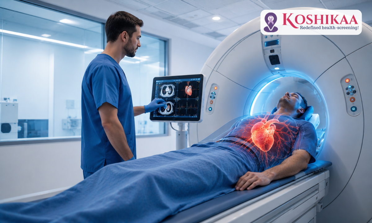
This is where advanced volumetric imaging transforms standard preventative medicine, as highlighted in the diagnostic cross sections shown above. By capturing hundreds of distinct, razor-thin visual slices of the moving heart and stitching them into an interactive 3D map, specialized scanning systems expose microscopic calcified or soft plaques years before they narrow an artery enough to spark an emergency.
Intercepting these cardiovascular changes at a structural stage empowers you to shift from passive monitoring to a highly targeted, data-backed approach to longevity.
Medical Disclaimer
The radiological guidelines, preparation pathways, and comparative diagnostic parameters detailed throughout this article are intended strictly for public health education and general awareness purposes. This material does not function as a replacement for expert medical advice, formal clinical evaluations, direct hospital diagnoses, or custom-tailored treatment prescriptions. Cardiovascular conditions are exceptionally dynamic and demand personalized risk staging under the direct supervision of a board-certified cardiologist or licensed medical specialist. Always consult a qualified physician to thoroughly evaluate your personal health history or metabolic baseline. Never ignore, delay, or bypass professional clinical consultations, as per the general educational frameworks discussed in this post.
Getting Ready: Pre-Scan Clinical Protocols
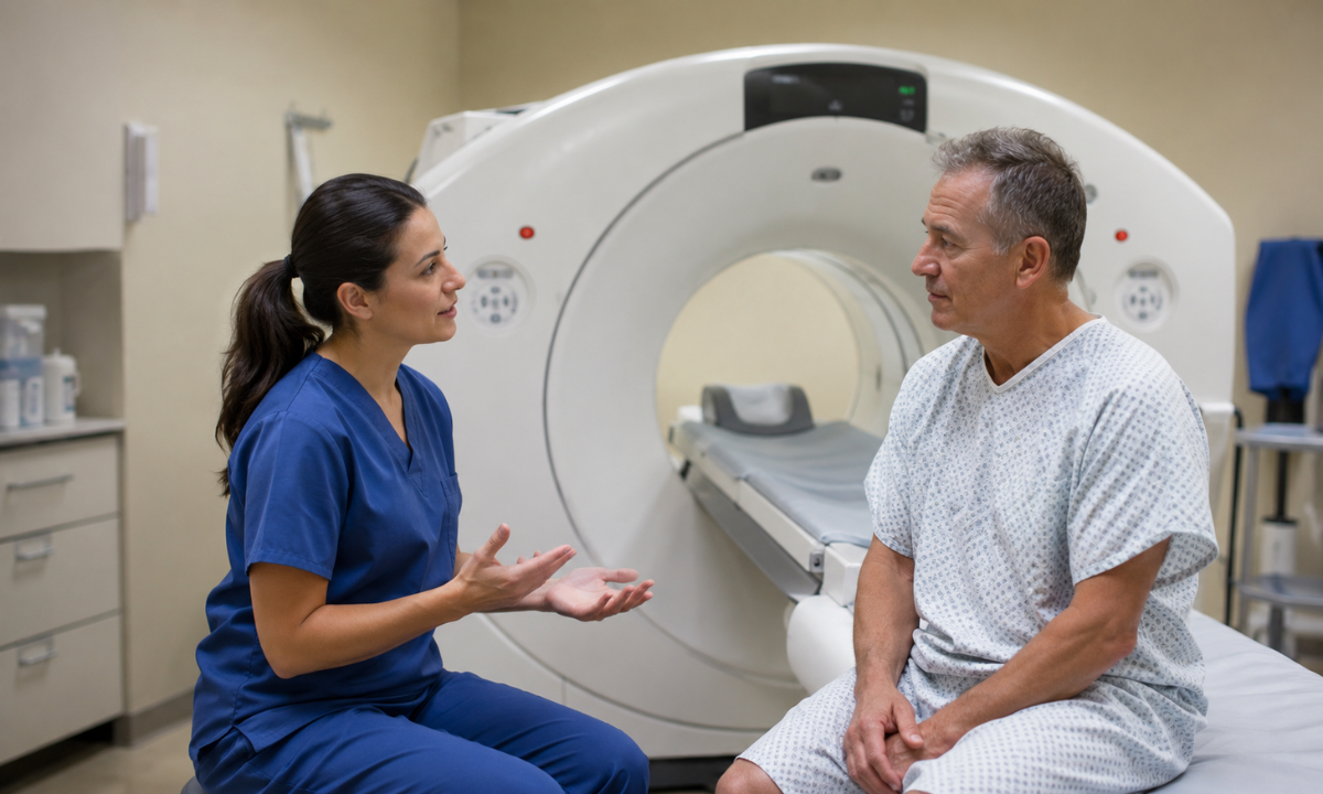
Achieving a pristine, motion-blur-free visual map of a beating heart requires strict adherence to biochemical and physiological guidelines before you even step inside the imaging suite. As the coronary arteries are constantly moving, proper cardiac CT scan preparation is entirely focused on creating the ideal internal environment, specifically stabilizing and lowering your resting heart rate so the high speed scanner can capture sharp snapshots between contractions.
The 24-Hour Stimulant Elimination Protocol
To prevent sudden spikes in heart rate or blood pressure that could compromise the clarity of the diagnostic images, you must eliminate all cardiovascular stimulants from your system for a full 24 hours before your appointment:
- Total Caffeine Restriction: Avoid coffee, energy drinks, green tea, black tea, and caffeinated sodas. Even decaffeinated beverages contain trace amounts of stimulants that can elevate your baseline pulse.
- Nicotine and Alcohol Cessation: Refrain from smoking, vaping, using nicotine patches, or consuming alcohol. Nicotine constricts blood vessels and artificially raises heart rate, which can lead to blurry imaging artifacts.
Avoid intense workouts, heavy cardiovascular training, or lifting heavy weights. Keeping your muscles and nervous system in a relaxed, steady state is key to a successful scan.
Dietary Boundaries and Fasting Windows
Managing your digestive activity is another crucial step in preparing your body for the safe administration of contrast materials:
- The 4-Hour Solid Food Fast: Do not consume solid food for at least four hours before your scan. Keeping your stomach empty minimizes the risk of mild nausea that can sometimes occur when the contrast dye enters the bloodstream.
While solid foods are restricted, you are highly encouraged to drink plenty of plain water up until the scan. Being well hydrated dilutes the contrast material and helps your kidneys flush it out efficiently after the imaging is complete.
Pharmacological Adjustments & Heart Rate Optimization
As high-resolution multi-slice systems capture the best images when your heart rate is consistently below 60 to 65 beats per minute, specific medical guidelines are put in place:
- Beta-Blocker Administration: Depending on your natural resting pulse, your healthcare team may provide temporary oral or intravenous medications (such as metoprolol) to slow down your heart rate and prolong the resting phase between heartbeats.
- Nitroglycerin Coordination: If you take medications for erectile dysfunction (such as sildenafil or tadalafil), you must discontinue them for 48 to 72 hours before the scan. This is as a mild spray of nitroglycerin is often given under the tongue right before the scan to widen the coronary arteries, and mixing these medications can cause a dangerous drop in blood pressure.
If you have diabetes and take metformin, you will be instructed to pause this medication on the day of the test and for 48 hours afterwards to protect your kidney function from contrast interaction.
Inside the Scanner: The Imaging Experience
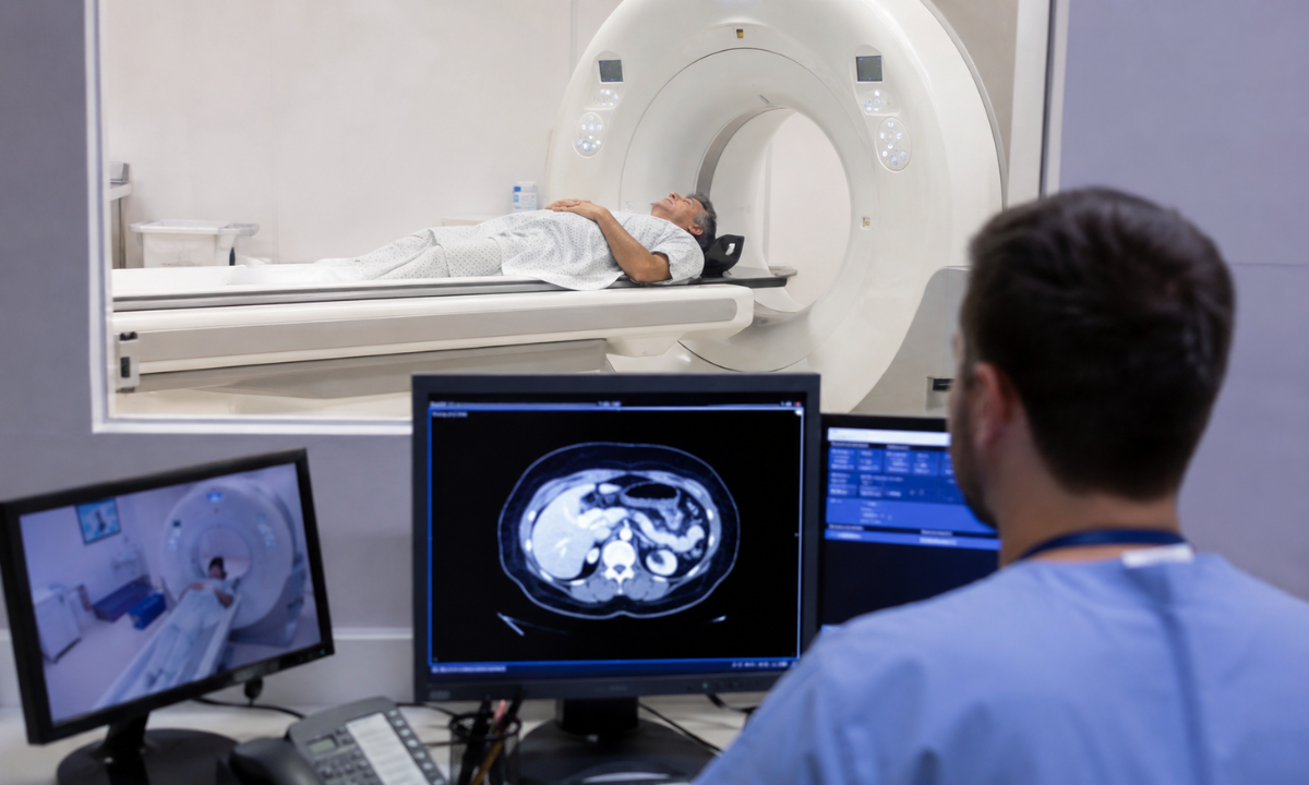
Stepping into the imaging suite can feel intimidating, but knowing what happens stage by stage transforms the process into a predictable, stress-free routine. The clinical cardiac CT scan procedure itself is remarkably fast, non-invasive, and completely painless, relying on a highly calibrated tracking sequence to capture perfect views of your coronary layout.
The Step-by-Step Scan Sequence
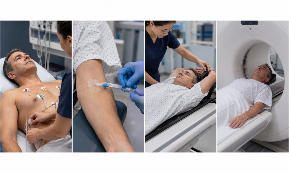
Once you enter the specialized imaging room, the medical team follows a precise roadmap to ensure absolute diagnostic accuracy:
- Electrode Synchronization: A technologist places small, circular adhesive patches called ECG leads across your chest. These patches continuously track your heart’s electrical activity, allowing the scanning system to coordinate its image capture perfectly with the brief pauses between your heartbeats.
- Intravenous Placement: A thin, flexible IV line is gently set into a vein in your hand or arm. This path will be used to introduce an iodine-based contrast solution that makes the internal channels of your arteries light up clearly on the final digital maps.
- Comfort Positioning: You will lie flat on your back on a motorized exam table. The technician will help position your arms safely above your head to keep them clear of the scanner’s line of sight, ensuring a completely unobstructed look at your chest area.
The Perfusion Phase and Breath-Holding Dynamics
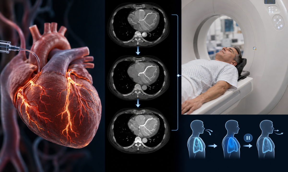
When the imaging sequence begins, the motorized table slowly moves your chest inside the open, doughnut-shaped scanner. The machine stays entirely open on both ends, preventing any feeling of heavy confinement:
- The Contrast Rush: As the iodine dye flows through the IV line, it is perfectly normal to experience a sudden, full-body wave of warmth or a brief metallic taste in your mouth. This sensation is harmless and fades completely within 15 to 30 seconds.
- Controlled Breath-Holds: The technologist will guide you through the intercom system, asking you to hold your breath for roughly 10 to 15 seconds at a time. Pausing your breathing keeps your diaphragm completely still, preventing the minor physical shifts that cause motion blur on high-resolution maps.
- Rapid Acquisition: While the inner ring of the machine rotates around you with a soft whirring or clicking sound, the actual high-speed image capture takes less than 15 seconds to complete.
The entire process inside the suite takes between 15 and 20 minutes from setup to completion. Once the technologist confirms the structural details are clear, the IV line is removed, allowing you to change clothes and return to your daily schedule immediately.
For a helpful visual walkthrough of the patient journey inside the imaging department, watch this video on what to expect during a hospital cardiac CT scan, which details the room layout and patient preparation steps.
Diagnostic Deep-Dive: Imaging Modalities Compared
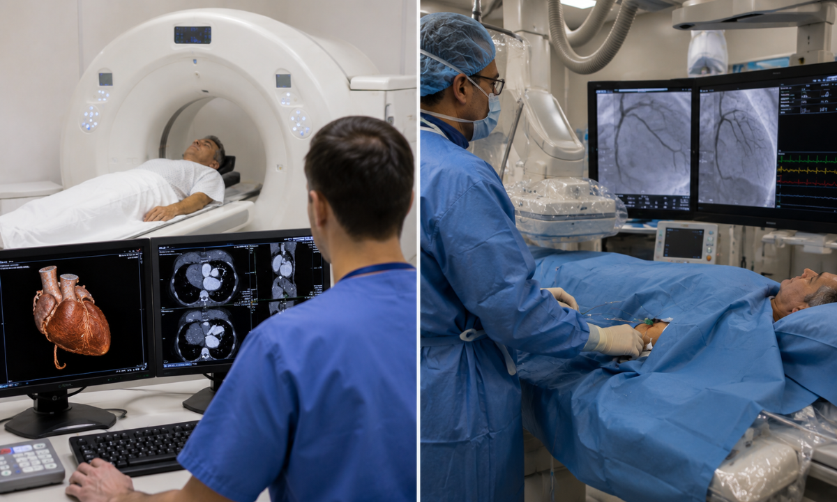
When mapping out your cardiovascular health strategy, it is essential to understand the structural differences between diagnostic paths.
Choosing between a non-invasive cardiac CT scan vs Angiogram performed in a hospital catheterization lab depends heavily on your current risk profile, baseline symptoms, and the overall clinical goal whether you are proactively screening for hidden plaque or reacting to an acute cardiac emergency.
Procedural Architecture and Clinical Focus
The two modalities operate on completely different mechanical and clinical principles. While one functions as a powerful, non invasive preventative mapping sweep, the other is an active surgical intervention designed for immediate tissue repair:
| Clinical Metric | Volumetric Cardiac CT Scan (CCTA) | Conventional Catheter Angiogram (ICA) |
|---|---|---|
| Invasiveness Level | Entirely non-invasive; requires only a standard arm IV line. | Minimally invasive surgery; requires a flexible catheter inserted through the groin or wrist. |
| Primary Screening Role | Early preventative interception and structural rule-out for low-to-intermediate risk profiles. | Emergency management, definitive diagnostics for high-risk patients, and immediate surgical planning. |
| Anatomical Visualization | Provides full 3D volumetric cross-sections of the artery walls, including soft and calcified plaque. | Provides 2D real-time contrast video showing the exact internal diameter of the vessel lumen. |
| Recovery Requirements | Zero downtime; patients can resume normal daily tasks immediately after the scan. | Requires several hours of absolute flat bedrest to ensure the arterial puncture site seals safely. |
| Procedural Risk Profile | Extremely low risk; avoids any mechanical danger to the heart walls or major arteries. | Small but distinct procedural risks, including arterial wall tearing, bleeding, or localized bruising. |
Diagnostic Performance and Preventive Utility
Understanding the data metrics of each approach helps patients see why multi-slice imaging has become a cornerstone of modern preventative screening frameworks:
| Diagnostic Marker | Volumetric Cardiac CT Performance | Conventional Angiogram Performance |
|---|---|---|
| Negative Predictive Value | Exceptionally high (reaching up to 99%); an ideal tool to completely rule out obstructive blockages. | Historically considered the absolute gold standard for confirming severe, complex blockages. |
| Plaque Type Characterization | Unique ability to identify soft, unstable non-calcified plaques before they rupture and cause an issue. | Focuses primarily on checking the visible reduction in blood flow caused by a physical blockage. |
| Therapeutic Capabilities | Strictly diagnostic; provides the structural data needed to guide lifestyle or medical therapy adjustments. | Dual-purpose; allows the interventional cardiologist to place a stent or perform angioplasty immediately. |
By utilizing these detailed comparative metrics, preventative care teams can match patients with the exact diagnostic tier they need. For individuals focused on proactive screening, the high negative predictive value of volumetric imaging provides absolute clarity without the physical stress or downtime of a catheter-based exploratory procedure.
Evaluating Financial and Value Factors and Investing in Proactive Health
When integrating advanced imaging into a preventative checkup framework, understanding the financial investment is a practical step in mapping out your healthcare journey. The total cardiac CT scan cost is not a fixed number; rather, it reflects a combination of high-precision hardware engineering, diagnostic software processing, and clinical expertise. Evaluating these components helps patients look past basic pricing to see the true long-term value of a preventative screening investment.
The Variables Driving Pricing Tiers
The final cost of your imaging session depends on several distinct medical and operational factors:
- The Specific Imaging Modality: A non-contrast Calcium Scoring session requires less processing and fewer materials than a comprehensive CT Coronary Angiography (CCTA), which builds an interactive 3D model of your blood vessels. In Bangalore, basic calcium tracking generally scales between ₹2,500 and ₹5,000, while a full CCTA sweep typically ranges from ₹9,000 to ₹15,000.
- Scanner Slice Configuration: The technical capacity of the machine plays a major role. Advanced facilities using 128-slice, 256-slice, or AI-powered 384-slice scanners charge a premium As their systems capture incredibly clear details at lightning speeds, keeping radiation exposure exceptionally low.
- Contrast Material Requirements: Using premium, non-ionic contrast dyes to make the internal pathways of your arteries stand out adds to the base pricing, but it gives radiologists the sharp detail needed to spot soft plaque buildup.
Having your detailed 3D heart maps analyzed by a senior, board-certified cardiothoracic radiologist ensures maximum accuracy, supporting a higher tier of professional fees.
Upfront Screening vs. Reactive Emergency Care
Viewing this diagnostic evaluation through the lens of preventative medicine changes how we look at the expense. Spending a predictable amount on advanced screening provides you with clear, actionable metrics to update your daily habits or treatment plans.
This proactive choice effectively shields your family from the sudden, massive financial and emotional strain of managing an unpredicted cardiovascular crisis.
| Financial and Health Dimension | Proactive Volumetric Screening | Reactive Emergency Intervention |
|---|---|---|
| Average Cost Footprint | Moderate, predictable outpatient diagnostic fee. | Exceptionally high; includes ICU stays, surgical suites, and specialized hardware. |
| Clinical Window | Performed early when soft plaque can be reversed through simple adjustments. | Performed in a crisis after a physical blockage has restricted blood flow. |
| Impact on Quality of Life | Zero recovery time or career disruption. | Requires significant hospital recovery, lifestyle disruption, and ongoing observation. |
Why Choose Koshikaa for Comprehensive Wellness?
Preventative healthcare requires a major shift from reactive treatment to proactive cellular and structural tracking. Selecting a premier Health screening centre in Bangalore that seamlessly integrates clinical blood panels with advanced multi-slice imaging ensures your metabolic and cardiovascular metrics are evaluated with absolute diagnostic precision.
Koshikaa’s state-of-the-art diagnostic ecosystem is purpose-built to eliminate guesswork, empowering individuals to understand their internal health profile long before physical symptoms ever develop.
Our advanced screening framework provides distinct patient advantages:
- Pioneering Age-Wise Profiling: We reject the ineffective “one-size-fits-all” approach to wellness. Our diagnostic panels are carefully customized based on your specific decade of life, gender parameters, and hereditary risk histories to ensure optimal clinical relevance.
- Integrated Multi-Slice Technology: By pairing high-sensitivity pathology panels with cutting-edge, low-radiation volumetric scanners, we ensure that deep cardiovascular risks are identified down to the millimeter.
- Rapid Digital Report Infrastructure: We prioritize your time and peace of mind by delivering clear, easy-to-read electronic and physical health summaries within 24 to 48 hours, complete with comprehensive reference tracking.
A preventative screening is only as good as its execution. Our medical network provides comprehensive diagnostic support, clear pre-scan preparation blueprints, and seamless laboratory coordination.
Conclusion
Taking charge of your cardiovascular health means making the transition from passive waiting to proactive health management. A volumetric heart sweep delivers the definitive internal roadmap required to expose calcified mineral tracking and silent arterial narrowing long before they manifest as an unpredicted crisis. By securing exact physical data early, you gain the precise clinical leverage needed to optimize your lifestyle, protect your natural vitality, and move forward with absolute long-term confidence.