When it comes to cardiovascular longevity, waiting for obvious warning signs like chest tightness or shortness of breath is a high-risk strategy.
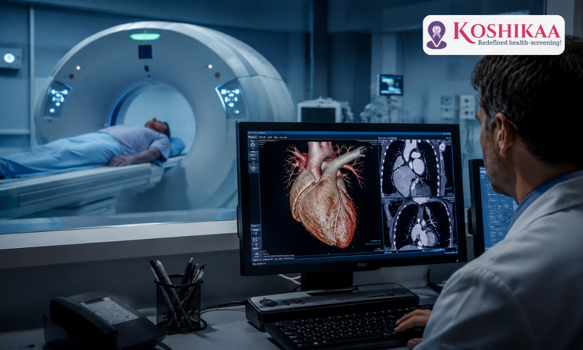
Your heart works tirelessly every second of the day, and understanding the condition of the intricate blood vessels that feed it is the ultimate foundation of proactive wellness. If you have ever wondered what CT angiography can reveal about your internal vascular health, it is best understood as a non-invasive, high-precision window inside your body.
Opting for an advanced, state-of-the-art CT scan in Bangalore, you eliminate the clinical guesswork, allowing specialized 3D imaging to map out your coronary arteries and safeguard your heart long before any physical symptoms ever emerge.
Traditionally, evaluating the deep pathways of the cardiovascular system required invasive catheterizations through the groin or wrist. Today, medical technology allows us to capture crystal-clear, cross-sectional views of your blood vessels in a matter of minutes. Whether you are tracking a family history of heart disease, investigating borderline stress test results, or precisely assessing your metabolic risk factors, this sophisticated imaging technique changes the entire narrative of heart care.
We at Koshikaa believe in empowering you with definitive data, transforming cardiovascular screening from a source of medical anxiety into a clear, actionable blueprint for lifelong structural vitality.
Medical Disclaimer
The clinical insights, procedural explanations, and vascular metrics presented in this guide are intended strictly for educational and informational purposes. This content does not constitute professional medical advice, a formal physician consultation, or a personalized clinical diagnosis. Individual cardiovascular risks, plaque stability levels, and aortic dimensions are deeply influenced by a patient’s specific metabolic profile, genetic markers, and underlying health conditions (such as chronic hypertension or diabetes). Always consult a qualified cardiologist or a board-certified physician to interpret your specific imaging results and to design a safe, appropriate preventative or therapeutic regimen. Never alter prescribed medications or delay seeking professional medical guidance based on the material read in this article.
How Does a CT Angiography Work?
To understand the sheer brilliance of a cardiac CT angiogram, it helps to realize that your heart is a moving target. It beats roughly 100,000 times a day, meaning that standard imaging tools often capture nothing more than a blur.
A CT angiography solves this problem by merging the rapid speed of advanced multi-slice computed tomography with a powerful mathematical trick called “ECG gating”, which syncs the scanner’s shutter perfectly with the resting phases of your heartbeat, essentially freezing the motion of your heart on screen.
But how do we see inside hollow, flexible blood vessels that are normally invisible on a standard X-ray? The secret lies in a highly calculated combination of contrast media and three-dimensional reconstruction.
Here is a look behind the scenes at the precise mechanical and chemical choreography that takes place during the scan:
- The Contrast Highway: Before the scanner begins spinning, a specialized, iodine-based contrast solution is introduced into a vein in your arm via an automated injector. This dye traveling through your blood works like a biological highlighter.
As iodine heavily absorbs X-ray beams, it makes the interiors of your coronary arteries illuminate with brilliant clarity against the surrounding muscle tissues of the heart.
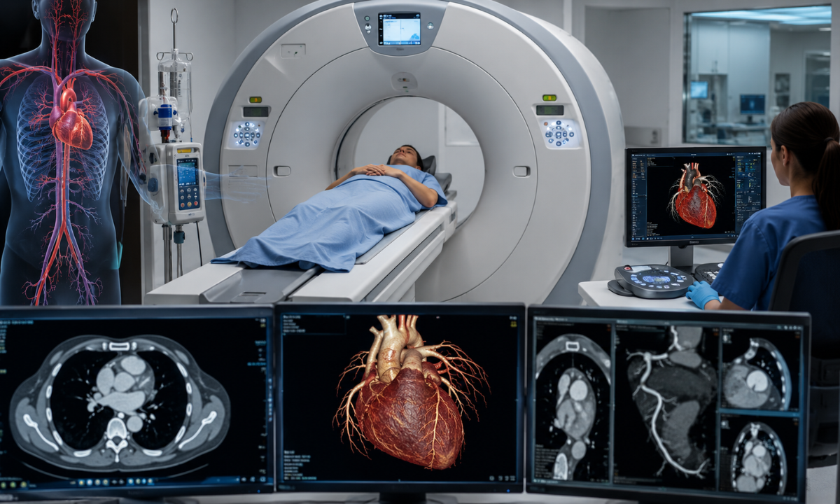
- The Symphony of Slices: As you slide gently into the open, doughnut-shaped gantry of the scanner, an X-ray source rotates around your chest at blinding speeds, frequently completing a full rotation in less than a third of a second. It shoots ultra-thin, fan-shaped beams through your body, capturing hundreds of individual cross-sectional “slices” or layers of your cardiac anatomy in a single breath-hold.
- Constructing the 3D Masterpiece: The raw data from these individual slices is instantly funneled into advanced imaging software. The computer stitches these layers back together, pixel by pixel, to build an interactive, high-definition 3D model of your heart and its branching arterial tree.
Looking at this digital model, radiologists don’t just see where blood is flowing; they can virtually “fly through” the inside of your arteries.
This allows them to examine the thickness of the vessel walls and detect the earliest formations of plaque with microscopic precision. It is the ultimate fusion of modern physics, computing power, and clinical biochemistry, all working together to map your internal health without a single surgical incision.
Proactive Prevention: Why is a Cardiac CT Angiogram Performed?
Historically, the medical system waited for a patient to suffer a cardiovascular event before looking directly at their coronary arteries. At Koshikaa, we view the heart through the lens of proactive longevity. A cardiac CT angiogram is not just a diagnostic tool for the symptomatic; it is the ultimate predictive screening mechanism for individuals who want to actively dictate their health span.
Physicians and proactive health strategists utilize this advanced imaging for three primary, high-impact reasons:
1. Unmasking the Silent Threat of Soft Plaque
When people think of arterial blockages, they often picture hard, calcified calcium deposits. However, the most dangerous form of cardiovascular disease begins as soft, lipid-rich (fatty) plaque. This soft plaque builds up quietly inside the lining of your arterial walls, completely hidden from traditional stress tests or standard electrocardiograms (ECGs), as it does not yet restrict resting blood flow.
A cardiac CT angiogram is one of the only non-invasive modalities capable of identifying this unstable soft plaque. Catching it in its infancy allows for aggressive, early lifestyle and nutritional interventions to stabilize the plaque wall, entirely preventing it from rupturing and causing a sudden cardiac event.
2. Resolving Inconclusive Diagnostic Voids
Standard cardiovascular screenings, such as a Treadmill Test (TMT), are functional assessments that measure how your heart behaves under physical stress.
Unfortunately, these tests frequently return borderline, ambiguous, or false-positive results, leaving patients stuck in a state of high anxiety. A CT angiogram bypasses functional guesswork by providing absolute structural clarity. It shows exactly what the physical pathways look like, instantly confirming whether an anomalous ECG reading is a structural emergency or a benign anatomical variation.
3. Evaluating High-Risk Genetic Profiles
For individuals with a strong familial history of early-onset cardiovascular disease, lifestyle optimization alone can sometimes mask underlying genetic predispositions.
If you have close relatives who suffered heart complications at an early age, or if you manage stubborn, complex lipid profiles, a cardiac CT angiogram serves as an essential baseline audit. It provides a definitive, clear look at your vascular reality, ensuring your preventative protocols are precisely tailored to your exact internal anatomy.
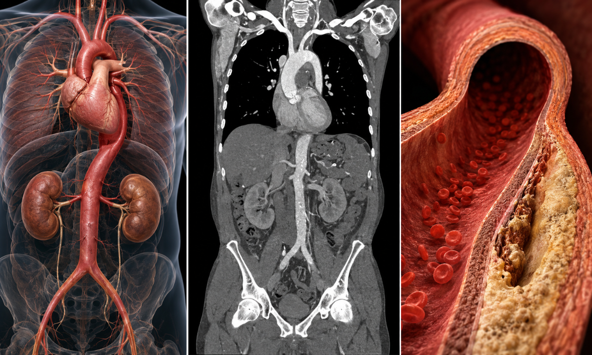
Expanding the View and Understanding the CT Aortogram
While a standard cardiac angiogram focuses narrowly on the microscopic arteries feeding the heart muscle itself, cardiovascular health requires us to look at the entire vascular architecture.
This is where a CT aortogram becomes vital. If the coronary arteries are the local streets, the aorta is the massive, multi-lane highway of your body.
The aorta is the largest blood vessel in the human body, originating directly from the left ventricle of the heart and arching downward through the chest and abdomen to deliver oxygen-rich blood to every single organ and limb. As it bears the direct force of blood being pumped straight out of the heart, its walls are under constant, immense pressure.
Through advanced vascular screening, a medical team can evaluate this critical pathway by looking at three distinct parameters:
1. The Mapping of CT Aortogram Anatomy
A complete aortic scan tracks this massive vessel across its entire geographical layout. Radiologists divide the CT aortogram anatomy into distinct structural zones to pinpoint vulnerabilities:
- The Ascending Aorta: The initial segment that travels upward from the heart is highly susceptible to high-pressure stress.
- The Aortic Arch: The curved top section that looks like an inverted “U,” where critical arteries branch off to supply blood to your brain, neck, and arms.
- The Descending Thoracic Aorta: The section that runs downward through the chest cavity.
The final stretch runs through the belly, which splits into the iliac arteries to feed your lower body and legs.
2. Clinical Insights via CT Aortogram Radiology
When evaluating a scan, specialized physicians look closely at the structural integrity of the vessel walls. Through CT aortogram radiology, doctors can identify dangerous conditions that standard blood tests or stethoscopes could never detect:
- Aneurysms: A dangerous ballooning or widening of an unreinforced section of the aortic wall, which risks a fatal rupture if left unchecked.
- Dissection: A life-threatening emergency where a small tear occurs in the inner layer of the aortic wall, causing high-pressure blood to split the layers of the vessel apart.
- Aortitis: Microscopic inflammatory changes or thickening of the arterial walls caused by systemic autoimmune conditions or deep-seated infections.
3. Defining a CT Aortogram Normal Result
| Aortic Structural Parameter | Normal Healthy Baseline | Signs of Structural Stress / Abnormalities |
|---|---|---|
| Aortic Diameter (Thoracic) | Typically stays between 3.5 cm to 4.0 cm (varies slightly by age, height, and gender). | Widening beyond 4.0 cm signals ectasia (stretching) or the formation of an aneurysm. |
| Aortic Diameter (Abdominal) | Typically measures less than 2.0 cm to 2.5 cm. | Any segment expanding beyond 3.0 cm is classified as an abdominal aortic aneurysm (AAA). |
| Vessel Wall Thickness | Uniformly thin, smooth, and highly flexible with zero structural disruptions. | Irregular thickening, ragged tears, or a “double lumen” look signaling a dissection. |
| Calcification / Plaque Burden | Clean, unobstructed interior pathways allow completely smooth blood flow. | Heavy, calcified plaque buildup or irregular atheromas along the inner lining, indicating advanced atherosclerosis. |
The Koshikaa Longevity Perspective: The primary reason a doctor will expand a cardiac scan to include an aortic evaluation is that conditions like thoracic or abdominal aneurysms are completely silent; they cause zero pain or symptoms until an actual rupture occurs.
Proactively identifying an enlarging aorta via advanced imaging allows for safe, long-term monitoring, blood pressure optimization, or timely elective interventions, entirely eliminating the risk of a sudden vascular emergency.
Walkthrough of The Procedure and Setup
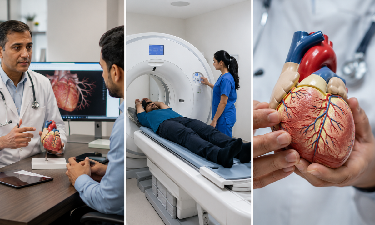
A common misconception among patients is that an advanced heart scan will be a lengthy, exhausting ordeal. In reality, the CT coronary angiography procedure is remarkably fast, highly coordinated, and entirely non-invasive, whether it is performed to evaluate the coronary arteries or the aorta.
If you are wondering how long an aortogram takes or how much time you will spend in the scanning room, the answer is often surprising: while the physical prep and screening window takes about 1 to 2 hours of clinic time, the actual image capture by the CT scanner takes less than 15 seconds.
Here is what you will experience from the moment you walk into the preparation suite:
1. The Pre-Scan Preparation (30–60 Minutes)
Before you approach the scanner, the medical team focuses on one core goal: stabilizing your heart rate. To capture crystal-clear 3D images without motion blur, your heart rate should ideally be smooth and steady, hovering under 60 to 65 beats per minute.
- Heart Rate Optimization: If your resting heart rate is a bit high due to natural pre-test anxiety, a nurse will administer a mild, short-acting medication (like a beta-blocker) to gently steady your pulse.
- The IV Line Setup: A small intravenous (IV) line will be placed in a vein in your arm. This connects to an automated injector that delivers the iodine contrast dye when the scan begins.
- Electrode Placement: Small, sticky ECG patches are placed on your chest to continuously track your heart rhythm, ensuring the scanner fires its beams at the exact millisecond your heart rests between beats.
2. Inside the Scanner Room (10–15 Minutes)
You will lie down comfortably on your back on a padded motorized table that moves smoothly through the wide, completely open, doughnut-shaped CT scanner. As the machine is fully open at both ends, it does not trigger the claustrophobia often associated with deep-tunnel MRI machines.
- The Warm Flash: As the contrast dye is injected, you will feel a distinct, warm, flushing sensation travel through your chest and body for about 30 seconds. This is a completely normal pharmacological reaction to the iodine medium and fades almost instantly.
- The Breath-Hold Command: The technician will communicate with you through an intercom system. You will be asked to take a deep breath and hold it perfectly still for roughly 10 to 12 seconds. While you hold your breath, the scanner rapidly rotates around you, collecting thousands of data points.
3. Post-Scan Recovery (15–30 Minutes)
Once the breath-hold is complete, the imaging process is officially over. The line in your arm is removed, and you will rest in a comfortable lounge for a brief observation period.
Our team will encourage you to drink a bottle or two of water right away. As the contrast dye is highly water-soluble, staying well-hydrated allows your kidneys to naturally flush the solution out of your body through your urine over the next few hours. There is no recovery downtime, no lingering drowsiness, and you can comfortably drive yourself home or head straight back to your normal daily routine.
Evaluating the Investment: Transparent Cost and True Value
Understanding the financial aspect of high-end diagnostic care is essential to feeling fully confident in your healthcare decisions. When individuals begin researching advanced cardiac screenings, one of the most prominent questions they face is: how much does a CT coronary angiogram cost?
We at Koshikaa believe in providing complete transparency so you can view this test not as an ambiguous medical expense, but as a high-return investment in your long-term health span.
The Pricing Landscape in Bangalore
As a CT coronary angiogram utilizes cutting-edge multi-slice imaging technology, specialized computer processing, and highly controlled contrast media, it carries a distinct price structure compared to basic screening methods.
In private diagnostic ecosystems across Bangalore, the cost matrix generally breaks down as follows:
- The Baseline Entry Range: Depending on the configuration of the medical facility and the resolution of the scanning hardware, a standalone CT coronary angiogram typically ranges from ₹8,000 to ₹15,000.
- Advanced Multi-Slice Scans: Premium diagnostic centers utilizing specialized 128-slice or 256-slice systems, which drastically minimize scan times and offer the absolute highest clarity, generally price the test toward the upper limit, between ₹12,000 and ₹15,000.
- Bundled Diagnostic Master Packages: Many proactive individuals choose to bundle the angiography with a comprehensive metabolic panel, calcium scoring, and lipid analysis. These complete vascular health checkups typically range between ₹15,000 and ₹25,000, offering a much higher value per individual test.
What Factors Influence the Price?
The total financial commitment for your cardiac imaging is rarely a single static number, as it is determined by several specific clinical variables:
- The Technology Tier: Centers using top-tier, low-radiation multi-slice scanners ensure maximum safety and sharper image resolution, which is factored into the test price.
- The Volume of Contrast Used: The exact amount and specific formulation of the non-ionic iodine dye required vary based on an individual’s body mass and physical requirements.
- The Scope of the Scan: Expanding a standard heart study to map the entire thoracic or abdominal architecture adds a layer of clinical reporting complexity, altering the ultimate baseline price.
Shifting from Expense to Long-Term ROI
When weighing the upfront cost of an advanced vascular scan, it is incredibly powerful to compare it directly to the alternative. The conventional, reactive approach to heart health involves waiting until a severe blockage leads to a hospital admission.
Looking at the true financial and emotional return on investment (ROI), the case for proactive screening becomes undeniable:
- The Proactive Alternative: Investing roughly ₹12,000 today provides an immediate, 3-dimensional blueprint of your arteries.
This allows you to catch unstable soft plaque early, making it entirely manageable through affordable adjustments in your nutrition, daily routine, and targeted supplement protocols.
- The Reactive Reality: Ignoring underlying cardiovascular risk factors, as a temporary diagnostic expense, exposes you to unexpected emergencies.
Emergency interventions like a conventional hospital angiogram, balloon angioplasty, or coronary artery stent placements can instantly surge to costs ranging from ₹2,500,000 to ₹5,000,000+, alongside months of forced recovery and compromised independence.
Note: Prices are subject to inflation, time and space. The ones mentioned here are mere estimates and may change depending on various variables and conditions. We at Koshikaa are always there to help; please contact us before making any financial decision.
Why Choose Koshikaa for Your Ultimate Cardiac Screening?
When it comes to mapping the intricate blood vessels of your heart and aorta, the technology you use and the team interpreting your results are paramount. At Koshikaa, we have entirely redefined what it means to step into a diagnostic space.
We believe that your health checkup shouldn’t be a cold, anxiety-inducing medical chore, but rather an empowering, positive milestone in your journey toward a long and active life.
As the best Health Screening Centre in Bangalore, we offer a patient-first ecosystem built on precision, warmth, and complete clinical clarity:
- State-of-the-Art Multi-Slice Technology: Our facility is equipped with advanced, ultra-fast multi-slice CT scanning technology. This allows us to capture high-definition, 3-dimensional views of your beating heart in a matter of seconds, while utilizing protocols designed to minimize radiation exposure.
- Deep Individual Context: We look far beyond the raw pixels on an imaging screen. Through our bespoke lifestyle and medical history questionnaires, we integrate your genetic predispositions, nutritional habits, and stress levels into your final clinical report, providing your physician with an incredibly rich, holistic health profile.
- Expert Radiographic Analysis: A high-resolution scan is only as powerful as the eyes interpreting it. Our board-certified radiologists specialize in cardiovascular and aortic imaging, ensuring that subtle anomalies like early-stage, non-calcified soft plaque are accurately identified and flagged.
From our comfortable, modern lounge spaces and zero-wait-time appointments to clear, transparent pricing and easy-to-read reports, we ensure that managing your cardiovascular wellness is a completely seamless and stress-free experience.
Conclusion
Your heart is the engine of your life, working quietly and continuously to sustain everything you do. Waiting for a severe, unexpected cardiac event to reveal that your internal plumbing is struggling is a reactive approach that can permanently disrupt your freedom and independence.
By prioritizing a quick, non-invasive, and highly advanced CT angiogram, you take back the reins of your health narrative.
You stop guessing about your arterial wellness and arm yourself with definitive, 3-dimensional facts. Whether your scan confirms pristine cardiovascular health or flags a minor warning sign that requires early, manageable lifestyle adjustments, having that data brings true peace of mind. Partner with Koshikaa today, map your baseline, and secure a strong, vibrant, and active future for decades to come.