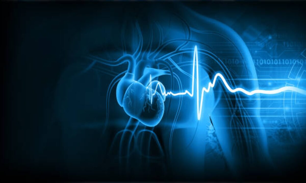
Heart diseases are major causes of death all around the world. Early detection is important and key to avoiding serious health problems. Several different heart health tests can be done that can help assess and indicate whether heart health is compromised or not before symptoms become too severe.
In this article, we explore essential tests for the early detection of heart diseases and what to expect from each test in comparison to an MRI scan.
Understanding Heart Diseases
Heart diseases are conditions of the heart’s structure and function. Coronary artery disease, heart failure, arrhythmias, and valvular diseases comprise these. Factors that increase the risk for heart disease, including high blood pressure, high cholesterol, obesity, smoking, and diabetes, can play an important role in developing heart disease.
Early detection is so important because many heart conditions are more readily treatable with early detection. However, regular checkups and diagnostic tests are an essential part of this process.
Early detection of heart disease is possible – Key tests.
1. ECG (electrocardiogram) or EKG
The test is called an electrocardiogram (ECG), which is simple and painless, and records the electrical activity of the heart. It is able to identify irregular heartbeats (arrhythmias) and can show signs that may indicate a previous heart attack or other overt heart condition.
What to Expect?
Small electrodes are placed on the chest, arms, and legs during an ECG to measure heart electrical activity. It should take about 10 minutes to complete and you probably won’t feel anything, but the adhesive on the electrodes. Moreover, an MRI scan in Bangalore uses detailed images of the structure and function of the heart to help detect possible abnormalities, beyond the scope of traditional methods.
An MRI scan uses detailed images of the structure and function of the heart to help detect possible abnormalities, beyond the scope of traditional methods.
2. Echocardiogram
An echocardiogram shows how the heart beats and the images of that beating. This test can tell the doctor if the heart’s valves are working properly if the heart’s chambers are functioning properly, and how the blood is moving through the heart.
What to Expect?
An echocardiogram technician applies a gel to your chest, looks and listens with a device called a transducer, and moves it to capture images of your heart. The procedure normally runs anywhere from 30 minutes to an hour. During the exam patients’ hearts will be seen on a monitor in real-time.
Here, an MRI scan is essential because of this particular advanced imaging technique to assess the condition of the heart when such things are not visible with other tests.
3. Stress Testing
Stress testing examines how the heart works when the heart is stressed. This test can show problems that may not be seen when the heart is at rest.
What to Expect?
Patients are always asked to walk on a treadmill while their heart rate, blood pressure, and ECG are monitored. The test’s purpose is to increase the heart’s workload and see how it responds.
4. Blood Tests
Cholesterol levels, blood glucose levels, and markers of inflammation or damage to the heart muscle are just a few things that several blood tests can help evaluate to assess your risk of heart disease.
What to Expect?
A healthcare provider will then draw your blood; generally by a sample taken from a vein in your arm. The results can also help you figure out your risk of developing heart disease and maybe even help plan further testing or treatment. In Bangalore, state-of-the-art facilities providing MRI scans in Bangalore are available for patients to have comprehensive cardiovascular check-ups.
5. Coronary Angiography
Minimally invasive procedure adding coronary angiography helps to see the Coronary artery. If there’s a strong suspicion of many blockages or other problems it is often performed.
What to Expect?
In this test, a catheter is slipped down into one of the blood vessels and sent down to the coronary arteries. X-ray images are then created by a contrast dye that visualizes blood flow. Normally, this takes about 30 to 60 minutes.
6. Heart MRI Scan
They are all safe – an MRI scan in Bangalore, with no radiation, shows the heart and surrounding structures in detail. It can evaluate heart function, and heart anatomy and help identify heart disease in its earlier stages.
What to Expect?
An MRI scan in Bangalore is painless and involves lying still on a table that slides into a cylindrical machine. The heart gets detailed images created using magnetic fields and radio waves. The procedure takes 30 to 90 minutes, as needed to get the images. Thorough heart assessments are offered in Bangalore by many hospitals and imaging centers.
7. CT Scan of the Heart
Calcium deposits in the coronary arteries can be measured in a CT scan and have been used to help determine heart disease risk. Detailed images of the heart’s blood vessels can be seen with a coronary CT angiography.
What to Expect?
A heart CT scan involves having you lie on a table in a scanner that slides you in and out. This often takes about 10 to 30 minutes and you may get a contrast dye put into your body to make the images clearer.
Monitoring, Lifestyle factors
Lifestyle also plays an important role in heart health and in addition to diagnostic tests. Reducing the risk of heart disease means keeping the body healthy by maintaining a healthy diet, exercise, and stress. Some healthcare providers may suggest periodically monitoring your blood pressure and cholesterol levels to check on heart health.
Healthcare Providers
If you have risk factors such as diabetes or high blood pressure, or if you have a family history of heart disease, it’s important to have open discussions with healthcare providers about heart health. Early detection tests can tell you things that will help you manage your health better. If your risk factors and symptoms are clear, your doctor may recommend some available tests.
Conclusion
Requirements for heart diseases are essential tests that can early detect heart diseases and it will positively affect treatment outcomes and reduce the chances of compounding. ECG, echocardiograms, stress tests, blood tests, and advanced imaging methods like MRI scans in Bangalore are vital, all of which test your heart health. Keep in mind that mastering the significance of these exams combined with working in concert with health care experts is a vital step for heart disease management as well as prevention.
Consequently, to live a longer and healthier life, heart health can be refined through essential tests and a healthy lifestyle. Regular screening and learning about your heart health are important parts of preventive health because heart disease remains one of today’s leading health issues. However, consulting about an MRI scan in Bangalore is not a reason to not be informed and proactive about your health so your heart stays healthy for years to come.
These services from Koshikaa provide a full body check-up in Bangalore to help users keep track of their health as a whole. Advanced facilities make sure that patients will get accurate assessments and personalized health insights.