
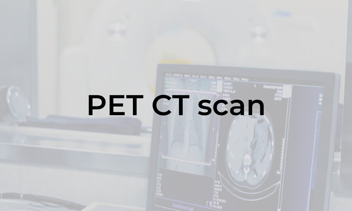
PET CT scans have transformed patient care by providing advanced diagnostic capabilities that aid in the early detection and precise monitoring of various medical conditions. These scans combine the strengths of Positron Emission Tomography (PET) and Computed Tomography (CT), offering a comprehensive view of metabolic and anatomical information. With a high demand for diagnostic imaging, PET CT scans are especially beneficial in assessing complex health issues, particularly for patients in cities with advanced medical services, like Bangalore. PET CT scan in Bangalore are becoming increasingly popular due to the city’s access to high-tech medical facilities and skilled professionals, bringing better diagnostic outcomes to a wide population. Let’s explore the impact of PET CT scans on patient care and the advantages they bring in specific areas of medicine.
What is a PET CT Scan?
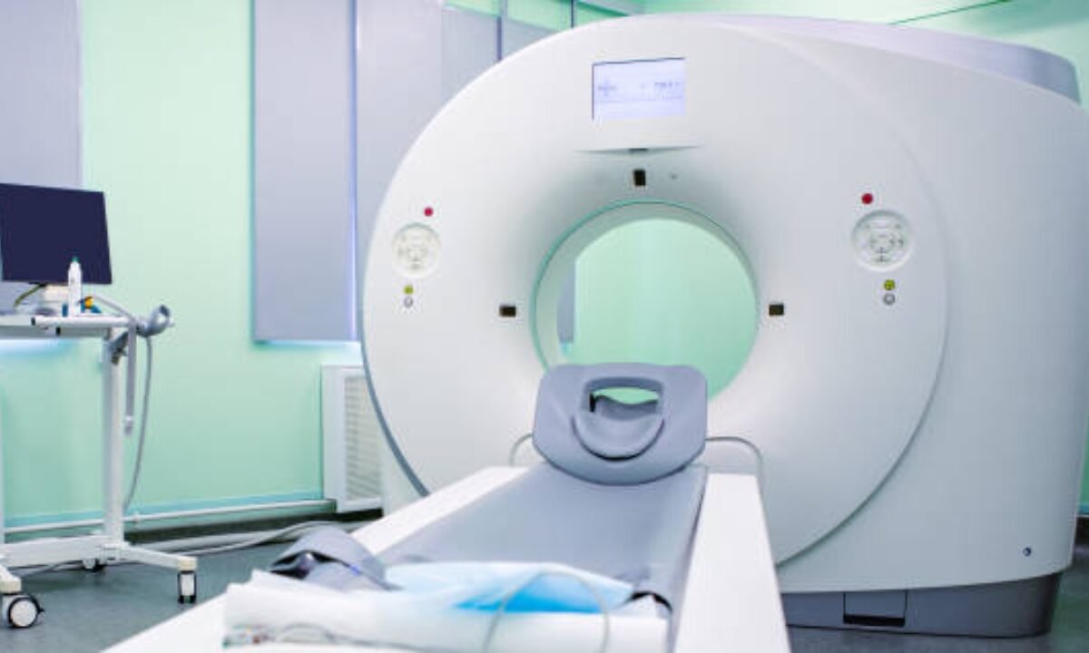
PET CT scans combine two imaging techniques—PET and CT—into a single, powerful tool. A PET scan detects changes in cellular metabolism by using a small amount of radioactive material that highlights areas of high chemical activity, often associated with diseases like cancer. A CT scan, on the other hand, provides detailed cross-sectional images of the body, allowing for precise localization of abnormalities. Together, PET CT scans offer a more accurate and detailed assessment than either scan alone.
The Role of PET CT Scans in Oncology
PET CT scans are invaluable in oncology, particularly for diagnosing and staging various cancers, as well as for tracking treatment progress. PET scans detect cancer cells due to their high metabolic rate, while CT scans help locate tumors within the body. When combined, PET CT scans give doctors in Bangalore and beyond a clearer understanding of a tumor’s behavior, aiding in customized treatment planning. This combination reduces the likelihood of exploratory surgeries, improving patient comfort and lowering healthcare costs.
Enhancing Neurological and Cardiovascular Diagnosis
PET CT scans are an invaluable diagnostic tool in both the fields of neurology and cardiology, providing critical insights that can significantly impact patient care.
In neurology, these scans are particularly beneficial for diagnosing neurodegenerative disorders. They enable healthcare professionals to detect conditions such as Alzheimer’s disease, epilepsy, and Parkinson’s disease. By examining brain metabolism, PET CT scans can identify abnormalities that might not be visible through standard CT imaging.
In cardiology, PET CT scans are equally important. They assist doctors in evaluating patients with coronary artery disease by identifying areas of the heart exhibiting reduced blood flow. It is essential to identify which parts of the heart muscle are still healthy and can be treated with the right medical or surgical options. By visualizing both blood flow and metabolic activity of heart tissues, PET CT scans help in developing comprehensive treatment plans that aim to restore heart function and prevent further cardiac damage. For instance, they could indicate whether a patient’s heart muscle is too damaged to benefit from procedures like angioplasty or bypass surgery, thus guiding the overall management strategy.
Overall, the integration of PET CT scans in both neurology and cardiology enhances the ability of healthcare providers to make accurate diagnoses, tailor treatment plans, and improve patient outcomes effectively.
PET CT Scans in Preventive Care and Early Diagnosis
In preventive medicine, PET CT scans are increasingly being used to detect diseases early, even before symptoms appear. Early detection is particularly important for diseases like cancer, where treatment success is closely tied to how early it is diagnosed. Bangalore, being a healthcare hub, offers PET CT scanning services that allow patients to take control of their health proactively. For people with a family history of certain diseases or risk factors, undergoing regular PET CT scans can make a significant difference in long-term health outcomes.
PET CT Scans and Patient Experience
PET CT scans not only enhance diagnostic accuracy but also improve the patient experience. In many cases, a single PET CT scan eliminates the need for multiple tests, which can be stressful, time-consuming, and expensive. Additionally, since PET CT scans are non-invasive, patients do not experience the same discomfort associated with biopsy or surgical procedures. Koshikaa offers high-quality PET CT scan services with advanced equipment that provides a streamlined, patient-centered experience.
Advancements in PET CT Technology and Its Impact on Healthcare
Advancements in PET CT technology are continually improving diagnostic capabilities. The latest machines offer higher resolution and faster scan times, enabling doctors to detect smaller tumors and subtle changes in the body. These advancements not only lead to more accurate diagnoses but also reduce radiation exposure, making the procedure safer for patients. In Bangalore, many diagnostic centers are equipped with state-of-the-art PET CT machines, ensuring that patients receive the most advanced care possible.
Preparing for a PET CT Scan: What Patients Should Know
Patients preparing for a PET CT scan should follow specific instructions to ensure the accuracy of the results. Generally, this includes fasting for a certain period before the scan and avoiding strenuous exercise. Patients should inform their doctors about any medications they are taking, as some can interfere with scan results. Bangalore’s screening centers provide comprehensive guidance to patients, helping them prepare for the procedure and addressing any concerns.
The Future of PET CT Scans in Medical Diagnostics
The future of PET CT scans looks promising, with ongoing research aimed at further enhancing their capabilities. New tracers and scanning protocols are being developed to improve specificity and reduce radiation exposure. These advancements are expected to make PET CT scans an even more integral part of patient care, particularly in cities like Bangalore, where technology adoption is high. As PET CT technology evolves, it will continue to revolutionize patient care, offering hope and improved outcomes for millions.
Conclusion
PET CT scans represent a significant advancement in diagnostic medicine, providing detailed and accurate insights that improve patient care. From oncology to preventive care, these scans have changed how doctors detect and treat diseases, offering a non-invasive option with minimal discomfort. In Bangalore, access to PET CT scans is making high-quality diagnostic services more available to a broad range of patients. By enhancing diagnostic accuracy, streamlining treatment plans, and allowing for early detection, PET CT scans are truly transforming healthcare, empowering patients and doctors alike to make well-informed decisions for better health outcomes.
Koshikaa Screening Centre offers a wide range of comprehensive cancer screening services, ensuring accurate results and personalized health care. Contact us today at +91 7996666106