Our brain is the most intricate organ. It is responsible for motion, memory, sensation, and numerous functions that define who we are. In case something happens in the brain, it is of paramount importance that the underlying issue is identified as fast and as properly as possible. Through technological developments, we have been offered new methods of viewing inside the brain. The Brain PET scan, a special form of imaging, is one of the most useful tools used by doctors to date, and also aids in the detection and diagnosis of a diverse range of Neurological Disorders.
What is a Brain PET Scan?

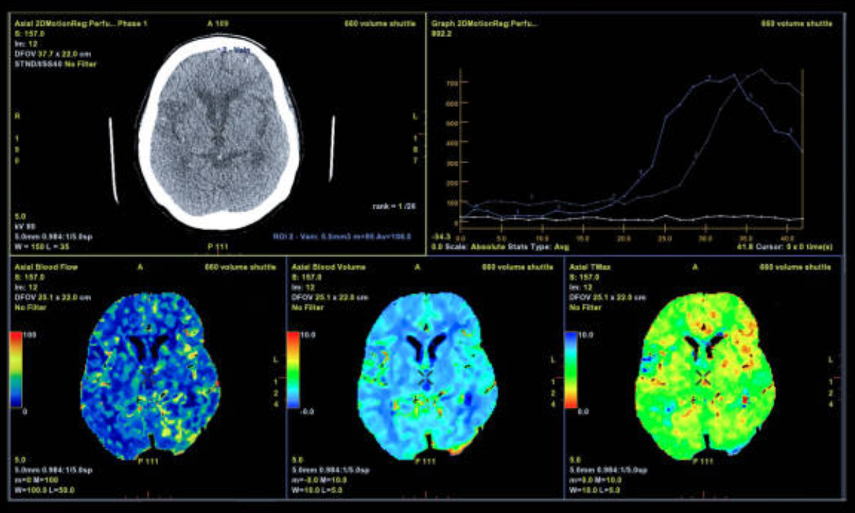
Brain PET scan, also known as Positron Emission Tomography scan, is a test that aids doctors in visualizing the way in which the brain is operating. In contrast with other scans, such as CT or MRI, which can depict what the brain looks like, PET scans can depict how various sections of the brain are operating in real-time. It is done by injecting the bloodstream with a small dose of some radioactive substance, in most cases glucose. As the brain consumes this glucose, the energy derived fills up the PET scanner to develop images in detail about brain functioning.
Diagnosing Neurological Disorders with Brain PET Scans
Brain PET scan is particularly useful in the diagnosis of different Neurological Disorders. Such disorders are Alzheimer’s Disease, Parkinson’s disease, epilepsy, and Brain Tumors. Physicians tend to select a PET scan in a situation where other scans cannot yield sufficient results.
1. Alzheimer’s Disease: Spotting Early Changes
Alzheimer’s is a form of dementia that involves memory, thinking, and behavior. It occurs gradually and may be insignificant at the initial stage. Brain shrinkage can be detected by the standard scans, e.g., MRI or CT, but usually in advanced cases. A Brain PET scan allows doctors to see the changes in the brain glucose consumption even before the symptoms become evident. This aids in the early detection of Alzheimer’s Disease, which will be treated more quickly and efficiently.
2. Identifying Brain Tumors
The other, and very significant use of the Brain PET scan is in identifying and tracking down the Brain Tumors. Brain tumors are benign or malignant. The two of them can bring about such interference in the brain functions through compression of the surrounding tissues. PET scan is beneficial in demonstrating the difference between the healthy and the bad tissue and enables doctors how big the tumors is, its position, and whether the disease is spreading or not. The piece of information is very essential, which can be used in the preparation of surgery, radiation or chemotherapy.
3. Diagnosis of Other Neurological Diseases
The PET scan of the brain is also useful in other conditions of Neurological Disorders besides the much-discussed Alzheimer’s Disease and Brain Tumors. In epilepsy, a PET scan can determine which parts of the brain are not functioning properly to cause the seizures, and then the surgeons will know which area of the brain to operate on. The scan also proves useful in the diagnosis and treatment of any movement disorder like Parkinson’s disease, where a positron emission tomography scan indicates the area of our body that has lost the activity of dopamine.
The Benefits of a PET Scan in Clinical Practice
The benefits of a PET scan are not limited to the detection of disease. PET scans are safe, in general, and non-invasive. The usage of radioactive material is largely negligible, and it does not remain in the body long enough. This renders the PET scans appropriate in the aspect of time-sequential follow-ups to be used to monitor or assess the response of a disease. The other crucial benefit of a PET scan is that it enables doctors to see the structure and functionality of the same scan, which gives them the full picture.
PET Scan in Bangalore: Getting to Advanced Care
More hospitals and imaging centers have recently established the PET scan in Bangalore. This is a welcome announcement to the patients because they will not have to go out of their way in search of quality brain images. The availability of a PET scan has enhanced the treatment and diagnosis of Neurological Disorders for many individuals. Patients will get the early diagnosis, specific treatment, and continuous monitoring sooner with improved access to this technology.
The Preparations for the Brain PET Scan
A Brain PET scan is usually a simple procedure to prepare for. The patients can be instructed to fast for a number of hours before the examination. It is necessary to inform the doctor about any medication consumed or if there is a chance of getting pregnant. The real test can last approximately between 30 minutes and one hour. Patients are resting on a moving bed in the scanner. It is relatively harmless, but there can be scary feelings in this closed room.
The Post PET Scan: What will happen?
Patients are normally able to resume their normal activities immediately after the Brain PET scan. A specialist analyses the results, examining which parts of the brain have more or less consumption of energy. This, as well as the symptoms of the patient and other history of the patient, aids doctors in coming up with a correct diagnosis. Depending on the hospital or clinic, it can take a couple of days to receive the final results.
Brain PET Scans Limitations
Although Brain PET scans are excellent in use, they possess certain limitations. In other instances, PET scans could fail to draw a clear picture concerning the different types of brain disorders. In addition, Brain PET scans can be costly, and not every hospital or clinic may offer such services, as this is slowly changing with the growth of services that include a PET scan in Bangalore.
Advancement of the Brain PET Scan Technology
Brain PET scan technology has a bright prospect. Scans are becoming increasingly accurate due to the improvement in imaging and safer tracers. The new tracers are also being created to fit specific Neurological Disorders, such as Alzheimer’s Disease or varying forms of Brain Tumors. It implies that more personalized treatment and even earlier diagnosis will happen soon.
Conclusion: Brain PET Scans and Their Current Place in Modern Medicine
The fastest and accurate diagnosis of Neurological Disorders is one of the greatest challenges in the field of medicine. Brain PET scan is a useful tool that enables doctors to visualize the functioning of the brain; hence, Alzheimer’s Disease and Brain Tumor, and numerous others can be easily identified. The advantages of PET scans, like early diagnosis, fine treatment planning, and the capacity to follow up on the diseases, are redefining patient care. As access increases, including advanced PET scans in Bangalore, greater numbers of people can enjoy the use of such an impressive technology.
Koshikaa provides superior services for PET scans in Bangalore, whose technology is of a high standard and can be used to improve the diagnosis and treatment of conditions like various neurological disorders and other patients’ ailments.