
Millions of people deal with headaches every single day. Most of the time, these headaches come from simple stress, lack of sleep, or dehydration. However, a headache that refuses to go away or changes how it feels can be a sign of a hidden problem inside the skull.
When a headache becomes a daily struggle, doctors need to look beyond standard painkillers. This is exactly when a CT scan for chronic headache becomes a necessary tool.
A CT scan is a fast and painless imaging test. It takes detailed pictures of your brain, blood vessels, and the bones inside your head. If you are searching for a reliable CT Scan in Bangalore, this imaging technology helps doctors find the exact root cause of your pain. By taking the guesswork out of your treatment, a brain scan serves as the Best Health Screening Centre in Bangalore for anyone suffering from severe, unexplained head pain.
The goal is to give you clear answers so you can finally get the right treatment and stop living in fear of your next headache episode.
Medical Disclaimer
The information provided in this guide is strictly for educational purposes and does not replace professional medical advice, diagnosis, or treatment. Always talk directly to a qualified doctor or neurologist about your specific headache symptoms and imaging needs. Never delay seeking professional clinical evaluation based on the contents of this article.
Specific Red Flags That Require a Brain Scan
While most daily headaches are harmless and treatable with rest, certain warning signs tell a doctor they need to look inside the skull immediately. Many health websites just give you a confusing list of symptoms without any context. We prefer to break down the exact physical signs that require a scan so you understand exactly what your body is trying to tell you.
The Major Warning Signs
Doctors look for specific patterns of pain that signal a structural issue rather than just stress or fatigue. The following table explains the most critical headache types that require immediate imaging.
| Headache Warning Sign | How It Actually Feels | Why Doctors Worry |
|---|---|---|
| The Thunderclap Headache | A severe explosive pain that reaches its absolute worst level within sixty seconds. Patients often describe it as feeling like they were struck by lightning. | This sudden pain can be a physical warning sign of a bleeding blood vessel in the brain. Finding this tiny bleed right away allows medical teams to stop it before it causes a stroke. |
| Neurological Body Changes | Head pain that happens alongside strange physical changes. This includes sudden blurry vision, weakness in one arm, difficulty speaking clearly, or a sudden loss of physical balance. | These body changes point directly to a physical mass or a blockage inside the skull pressing against the nerves that control your vision or your physical movement. |
| New Headaches After Age Fifty | An older adult starts getting severe and frequent headaches for the first time in their life. Standard headaches usually start when a person is in their twenties. | Older adults have a naturally higher risk of inflamed blood vessels or slow-growing masses. A clear picture helps doctors rule out these dangerous conditions immediately. |
What You Will Experience During the Scan
If you present with any of the warning signs mentioned above, your doctor will likely order a structural evaluation. The process is completely painless and designed for speed and comfort. Here is exactly what happens during your appointment.
- The Preparation Phase: You will have a quick consultation to discuss your complete medical history with our team. You do not need to fast or change your daily routine for a standard head scan.
- Contrast Dye Application: For some specific scans, a technician might give you a special contrast dye through a small IV in your arm. You might feel a brief warm sensation when the dye enters your body. This dye makes your blood vessels glow brightly on the monitor so the radiologist can see the detailed pathways.
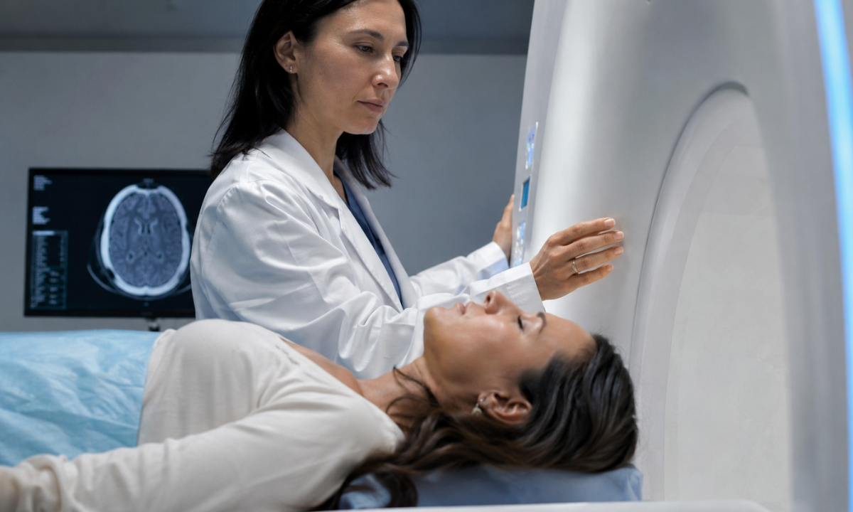
- The Imaging Process: You will lie flat on a sliding table that moves into a large circular scanner. The machine will circle your head to take fast cross-sectional pictures of your brain.
- Completion: You just need to remain completely still for about five to ten minutes. Once the scan is done, you can immediately return to your normal daily activities.
Koshikaa Safety Tip: Always tell your Koshikaa technician if you have any allergies or kidney issues before getting a scan with contrast dye. We use a highly detailed personalized health questionnaire to ensure your absolute safety before you even enter the scanning room. We guarantee your verified diagnostic report will be ready within 24 hours, so you do not have to wait in fear.
The Difference Between Migraines and Structural Headaches

One of the most common questions doctors hear is why they will not order a brain scan for a severe migraine. When your head is pounding, and you feel sick to your stomach, it is completely normal to fear that something is physically wrong inside your brain. Many patients specifically request a CT scan for migraine attacks to find relief and get answers.
However, doctors separate head pain into two completely different categories. Understanding this difference will save you time, money, and unnecessary worry.
Why Most Migraines Do Not Require Imaging
A standard migraine is what doctors call a primary headache. This means the headache itself is the main medical problem. It is caused by temporary chemical changes and active nerve signals in the brain. Because a standard migraine does not involve a physical object like a growing tumor or a bleeding blood vessel, a CT scan will simply show a completely normal and healthy brain.
A structural headache is a secondary headache. This means the headache is just a symptom of a hidden physical problem. This is exactly what a CT scan is built to find.
Comparing Standard and Atypical Migraines
To help you understand when a normal migraine crosses the line into a medical warning sign, we have created a simple breakdown of the symptoms.
| Headache Type | What You Actually Feel | Do You Need A Scan | The Medical Reason |
|---|---|---|---|
| The Standard Migraine | Throbbing pain usually on one side of your head. You might feel sick to your stomach and become very sensitive to bright lights or loud noises. | Usually No | The pain comes from nerve activity and chemical shifts. There is no physical mass or bleeding for the imaging machine to capture. |
| The Atypical Migraine | Your usual migraine suddenly changes. The pain is much worse than usual; it lasts for days instead of hours, or it stops responding to the medication that always worked before. | Yes | A sudden shift in your normal headache pattern strongly suggests that a new physical problem inside the skull might be triggering the pain. |
| Migraines With Motor Weakness | A migraine that happens alongside physical weakness in your arms or legs. This is very different from the common visual spots or flashing lights many migraine sufferers see. | Yes | Physical weakness can be a sign of a small stroke or a blood flow blockage. Doctors need a clear picture to rule out a physical brain injury. |
What Doctors Look For During Atypical Migraine Scans
When your migraine pattern changes and a doctor finally orders a scan, they are checking for physical blockages.
They use the imaging technology to ensure your spinal fluid is draining properly and that your blood vessels are not swollen or pressing against sensitive nerve endings. If the scan is clear, you and your doctor can confidently return to testing new migraine medications without the fear of a hidden disease.
Koshikaa Screening Advice: If you have suffered from migraines for years, keep a simple headache diary on your phone. Write down how often they happen and what they feel like.
If your pain pattern suddenly changes, bring this diary to your Koshikaa appointment. Our diagnostic team will use your exact health history to determine if a structural scan is the right step for your long-term brain health.
Understanding CT Scan Frequency and Safety
Many patients naturally worry about radiation when a doctor suggests a brain scan. The truth is that modern medical technology is incredibly safe and highly regulated. However, a very common and valid question we receive is how frequently a CT scan can be done or the indications for a CT Scan without causing long-term harm.
Doctors do not order these tests casually. They follow strict safety protocols to ensure that the benefits of finding a hidden problem far outweigh the minimal exposure from the machine.
How Doctors Decide When to Scan
Medical professionals divide brain imaging into two distinct categories to minimize your radiation exposure. The table below explains the difference between getting your first scan and needing additional tests.
| Scanning Phase | The Medical Purpose | Typical Frequency |
|---|---|---|
| Initial Baseline Scan | To capture the very first structural map of your brain when severe warning signs appear. | Usually, a one-time event to find or completely rule out an immediate physical danger. |
| Follow-Up Monitoring | To check if a previously discovered issue, like a small cyst or a treated blood vessel, is growing or changing shape. | Only ordered if your daily symptoms suddenly worsen or as part of a strict yearly review by a neurologist. |
How Modern Clinics Protect Your Health
If you need a scan, you should know that the technology has improved drastically. Hospitals and diagnostic centers use specific strategies to keep you completely safe during the procedure.
- Low Dose Technology: Brand new imaging machines use highly advanced computer software. This allows them to create perfect, high-resolution pictures using a tiny fraction of the radiation that older machines required just ten years ago.
- Targeted Imaging Areas: The technician will never scan your whole body to find a headache trigger. They program the machine to only scan the exact area of your skull that is necessary, keeping the rest of your body completely protected.
- Symptom-Based Decisions: A good doctor will never order a repeat scan just to check in. They will only recommend another imaging test if you experience brand new physical symptoms or if your current medication completely stops working.
Koshikaa Safety Guarantee: Your long-term health is our absolute priority. We utilize state of the art low radiation imaging machinery for every single patient. Our clinical team will thoroughly review your personal health questionnaire to ensure that any recommended scan is medically necessary and completely safe for your specific body.
Why Choose Koshikaa for Your Brain Health
When you are dealing with chronic head pain, waiting for medical answers can be the most stressful part of the entire process. Koshikaa is built specifically to remove that stress. We provide a completely different kind of diagnostic experience focused entirely on your physical comfort and your long-term safety.
- Personalized Safety Checks: Before you step into our facility, you will complete our unique personalized health screening questionnaire. This ensures our medical team knows your exact health history and specific risk factors so we can tailor the imaging process directly to your body.
- Advanced Low Radiation Machines: We utilize the newest imaging technology available. This allows our technicians to capture incredibly detailed pictures of your brain using the lowest possible radiation dose to keep you completely safe.
You should never have to sit at home for weeks, wondering what is causing your severe pain. We guarantee that your verified diagnostic report will be ready within 24 hours of your scan, so your doctor can start your treatment immediately.
Take Control of Your Headaches Today
Living with constant head pain takes a massive toll on your daily routine, your career, and your overall mental health. You do not have to keep guessing about what is happening inside your skull. If your headaches are changing, getting steadily worse, or arriving with strange new physical symptoms, it is time to take immediate action and find the true root cause.
Stop relying on basic painkillers that only mask the problem. Book a comprehensive diagnostic scan at Koshikaa today. Our clinical team is ready to give you the clear, accurate answers you need to build the right treatment plan and finally get back to enjoying your life without the constant fear of your next headache.