
Yes, cancer can absolutely be detected in a CT scan, and it remains one of the most powerful diagnostic tools available to modern medicine.
When doctors suspect the presence of an abnormal growth, they rely heavily on this technology to capture detailed, cross-sectional images of your internal organs, bones, and blood vessels. If you are trying to understand how can cancer be detected in CT scan technology, the answer lies in its ability to spot structural changes, abnormal tissue densities, and unusual blood flow patterns deep inside your body.
Finding a tumor early is the single most important factor in successful treatment, which is why scheduling a high-resolution CT Scan in Bangalore is often the first critical step your medical team will take.
Utilizing advanced imaging, you can transform what would otherwise be a complex internal mystery into a clear and actionable medical map, making it an essential component of the Best Health Screening Test in Bangalore for preventative wellness.
Let us explore exactly how this technology works and how it identifies hidden abnormalities throughout the body.
Medical Disclaimer
The information provided in this article is strictly for educational purposes and does not replace professional medical advice, formal diagnosis, or specialized treatment. Always consult directly with a qualified doctor or oncologist regarding your specific physical symptoms, health concerns, and screening eligibility. Never ignore medical warnings or delay seeking professional clinical care based on the contents of this guide.
Key Points at a Glance
- Immediate Detection: A CT scan is one of the most powerful modern tools for identifying tumors early, mapping their exact size, shape, and location before physical symptoms appear.
- How It Works: By taking hundreds of thin, cross-sectional 3D images, the scanner allows radiologists to look deep inside soft tissue and catch abnormal cellular growths that standard flat X-rays miss.
- Types of Tumors Found: This technology excels at spotting a wide range of conditions, from lung nodules and internal abdominal masses to early signs of Colon cancer via virtual colonoscopy.
- Enhanced Patient Safety: Modern facilities utilize Low-dose CT protocols, reducing radiation exposure by up to 75% while using advanced software to maintain crystal-clear image resolution.
How the Technology Spots Abnormal Tissue
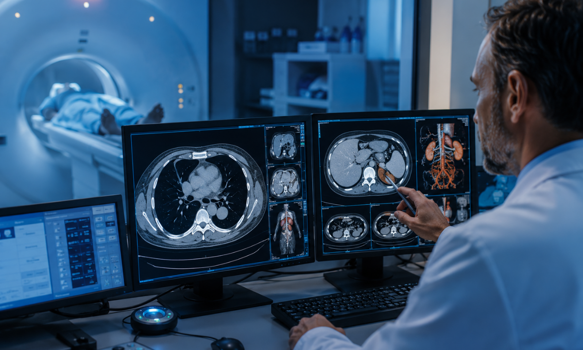
To understand how a scanner identifies potential tumors, it helps to look at how the machine takes pictures. A standard X-ray only takes a single, flat image from one angle. This causes your internal organs, muscles, and bones to overlap on the film. A modern scanner circles completely around your body, taking hundreds of thin, cross-sectional images. The computer then stacks these detailed slices together to create a complete 3D model of your internal anatomy.
When a radiologist reviews your images during a routine Cancer screening, they are looking for specific structural anomalies that indicate a mass is present. Healthy organs have predictable shapes, clean borders, and smooth textures. A cancerous tumor typically grows in an uncontrolled, chaotic pattern that disrupts these normal boundaries.
The imaging technology relies on a few key indicators to spot these dangerous growths.
- Irregular Physical Shapes: Healthy tissue grows in organized layers. Malignant tumors often form irregular, jagged lumps with rough edges that push aggressively into nearby muscles or healthy organs.
- Abnormal Tissue Density: Cancerous tumors are packed tightly with fast-multiplying cells. This makes them significantly denser than the soft, healthy tissue surrounding them, causing them to appear as distinct, dark or bright areas on the monitor.
- The Contrast Dye Reaction: Before your scan begins, a technician will often inject a safe contrast fluid into your bloodstream. Because tumors grow rapidly, they require a massive blood supply and quickly build a dense network of new blood vessels. The contrast dye rushes into this dense network, causing the hidden tumor to light up brightly on the screen.
- Organ Displacement: As a tumor increases in size, it takes up physical space inside your body cavity. The scan will show if a hidden mass is actively pressing against your stomach, crowding your lungs, or pushing your healthy blood vessels out of their natural alignment.
Tracking these physical changes, radiologists can spot small abnormalities early. When you book a specialized CT Scan in Bangalore, this advanced imaging process ensures that any unusual tissue activity is identified with perfect clarity.
Common Types of Tumors Found
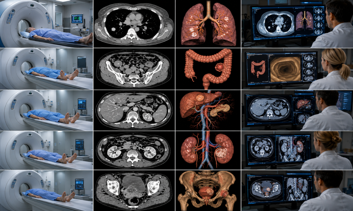
When exploring how effective this imaging technology is, it helps to look at the specific areas of the body where it performs best. Standard X-rays often miss small growths because internal organs overlap on the film. A modern scanner eliminates this problem by taking incredibly detailed 3D slices of the body. This makes it an indispensable tool for identifying tumors hidden deep inside your chest, abdomen, and pelvis.
Different types of scans are designed to look for specific abnormalities. We have outlined exactly what these specialized scans reveal and why they outperform older testing methods.
| Type of Body Scan | Target Area and Cancer Type | What the Scan Reveals |
|---|---|---|
| Specialized Chest Scan | Lung cancer and respiratory nodules | It easily detects tiny lung nodules that are only a few millimeters wide. Because it provides a clear cross-section of the lung tissue, it is far more accurate than a traditional chest X-ray for early detection among high-risk patients. |
| Virtual Colonoscopy | Colon cancer and intestinal polyps | This specialized abdominal scan pumps a small amount of air into the colon to inflate it. The computer then creates a highly detailed virtual fly-through of the intestine to spot precancerous polyps before they turn into dangerous tumors. |
| Targeted Abdominal Scan | Pancreatic and liver cancer | It provides a crystal clear view of the complex organs hidden deep in the abdomen. The scan highlights abnormal tissue density and maps the exact blood supply feeding a potential mass in the pancreas or liver. |
| Renal Imaging | Kidney and adrenal gland cancer | It creates a sharp outline of the kidneys and surrounding structures. This allows radiologists to confidently differentiate between a harmless fluid-filled cyst and a solid, potentially dangerous tumor. |
| Comprehensive Pelvic Scan | Ovarian and bladder cancer | It maps the entire pelvic region to evaluate unexplained masses. It is incredibly useful for seeing if an abnormal growth in the ovaries or bladder has begun to press against nearby healthy tissue or local lymph nodes. |
While a scanner cannot officially confirm if a mass is malignant without a physical biopsy, it provides the precise anatomical blueprint your doctor needs. By accurately identifying the size and exact location of a suspicious growth, this technology ensures that your medical team knows exactly where to focus their attention.
Making the Process Safer for Patients
One of the most common reasons patients hesitate to schedule a recommended imaging test is the fear of radiation exposure. While it is true that traditional scanners use X-ray energy to look inside the body, medical technology has advanced significantly to prioritize patient safety without sacrificing diagnostic accuracy.
Today, leading facilities use advanced imaging methods known as a low-dose CT scan to drastically reduce radiation exposure. This technological breakthrough ensures that you can get the vital answers you need while keeping your body perfectly safe.
Here is exactly how this modern technology makes your preventative health screenings safer.
- Smart Exposure Adjustments: Modern scanning machines do not blast a uniform amount of radiation through your body.
Instead, the computer uses smart sensors to measure your body thickness in real time. It automatically adjusts the X-ray beam to use the absolute minimum amount of energy required to capture a clear picture.
- Targeted Screening Protocols: When a doctor uses this technology for a routine Cancer screening, they can select specific low-exposure settings.
For example, screening your lungs with these modern protocols reduces the radiation dose by up to 75% compared to a standard chest scan, making it incredibly safe for annual checkups.
- Ultra Fast Imaging Speed: Older hospital machines required patients to lie perfectly still for several minutes while the scanner slowly gathered data.
New machines can scan your entire chest or abdomen in just a few seconds. As the process is so fast, your body is exposed to radiation for a tiny fraction of the time.
- Crystal Clear Software Resolution: In the past, reducing the radiation dose meant getting a blurry, grainy image. Modern systems solve this problem by using powerful reconstruction software. The computer takes a lower exposure scan and automatically cleans up the digital noise, delivering a crystal clear 3D image to your radiologist.
Eliminating the risks associated with older machines, this technology allows patients to safely undergo routine preventative testing. When you look for a modern CT Scan in Bangalore, choosing a facility that utilizes these low-exposure protocols ensures that you are protecting your long term health while keeping your current physical safety as the highest priority.
Why Choose Koshikaa? The Koshikaa Advantage for Early Detection
When it comes to identifying microscopic health changes, the quality of the imaging facility you choose makes all the difference. Koshikaa is recognized as a premier diagnostic center because we combine advanced technology with unmatched patient care. We do not just run routine tests. We provide the comprehensive and accurate answers your doctor needs to protect your overall health.
Here is exactly why patients and doctors trust Koshikaa for advanced diagnostic imaging.
- Advanced Low Radiation Technology: We utilize the latest generation of scanning machines that automatically minimize your radiation exposure. This guarantees that your preventative screening is completely safe while still delivering high-definition 3D images of your internal organs.
- Zero Wait Time Processing: We value your time and understand the deep anxiety of waiting for medical tests. Our streamlined process ensures you experience zero wait time when you arrive for your scheduled appointment, making your entire visit smooth and stress-free.
Your final images are never evaluated by basic technicians. Our dedicated team of specialized radiologists is an absolute expert at spotting the earliest microscopic signs of abnormal tissue. They provide your doctor with a perfectly accurate and detailed report so you can make fast and informed decisions about your treatment.
Conclusion
Early detection is the single most powerful tool you have for maintaining your long-term health and longevity. Finding a hidden abnormality early often means the difference between a simple and effective treatment and a major medical crisis. You do not have to wait for painful physical symptoms to appear before taking action to protect your body.
Take complete control of your preventative health today. Book an advanced scan at Koshikaa and get the clear and accurate answers you deserve. Our expert team is ready to provide a perfectly safe and comfortable screening experience that puts your peace of mind first.
Prioritize Your Preventative Health. Do NOT Compromise.