
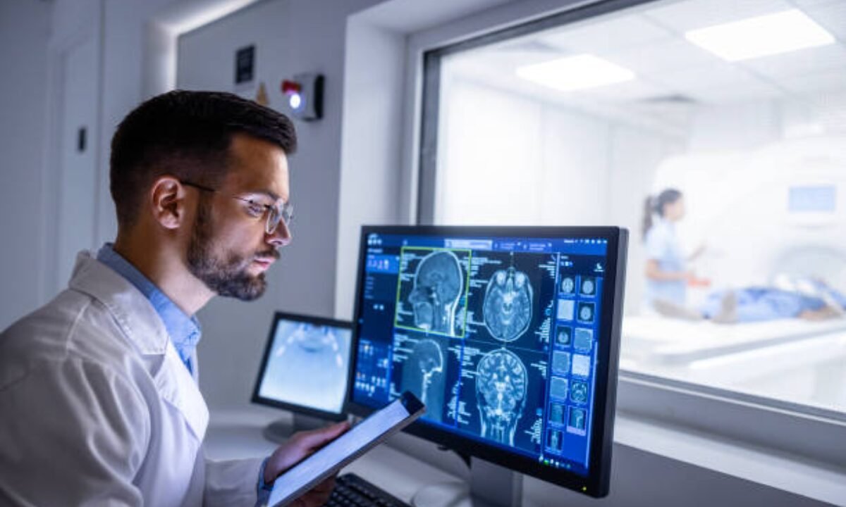
The faster the diagnosis is made in head injury cases, the better for the patient. One of the key tools doctors use is a CT scan for head injury. CT scans offer a view of the brain that is very clear to doctors and surgeons, and is a good way of finding out what is wrong with a patient quickly and then having him treated. This article covers the basics of CT scans and brain injuries – the procedures, cost of operation, what the scans reveal, and how they can be helpful for the subsequent recovery.
What is CT Scan of the Brain?
A CT scan brain is a special type of X-ray that takes detailed images of the brain and skull. CT stands for computed tomography, and this scan uses computer technology to turn X-ray images into cross-sectional pictures. This enables the doctors to get more details of your head than if you were to have a normal X-ray.
Why is it important?
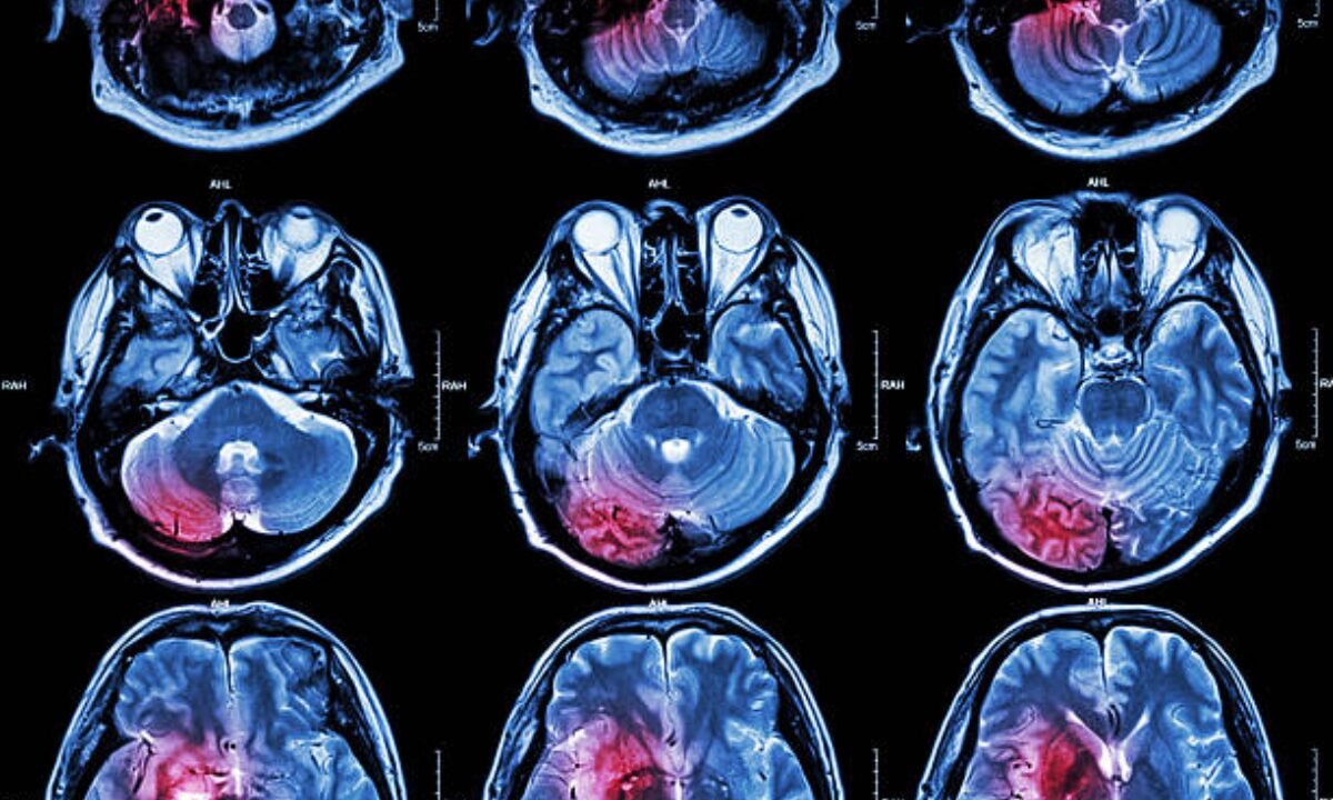
If a patient has a head trauma, among all diagnostics, a computed tomography for head injury is one of the quickest and accurate methods in evaluating:
- Bleeding in the brain
- Swelling
- Fractures
- Blood clots
- Brain tissue damage
All of the above problems are serious and life-threatening, thus requiring an immediate yet effective diagnosis.
When Do Doctors Recommend a CT Scan for Head Injury?
Doctors don’t use a CT scan brain for every bump or bruise, but they do recommend it when:
- The injury was severe (fall, accident, blow to the head)
- When an injured person has lost consciousness.
- They have symptoms like vomiting, memory loss, confusion, or severe headache
- Anyone with a known risk (elderly, blood thinner medication) gets hit in the head
Brain CT Scan Procedure
The brain CT scan procedure is very straightforward, painless, and fast. Here’s what typically happens:
1. Preparation: You might also be instructed to take off any items such as metals, rings, or glasses ready to make your treatments. They noted that sometimes contrast dye may be applied in order to highlight certain structures better when taking images.
2. Positioning: You will need to lie on a table that moves into the CT scan machine.
3. Scanning: The machine, referred to as an X-ray machine, circles around your head capturing several pictures from various angles.
4. Completion: The whole brain CT scan poses no threat and the entire process only takes 10 to 15 minutes.
Normal Brain CT Scan vs Abnormal Brain CT Scan
Normal Brain CT Scan:
A normal brain CT scan shows the brain’s structures in their usual shapes, sizes, and positions. There’s no excess fluid, bleeding, swelling, or evidence of injury.
Abnormal Brain CT Scan:
An abnormal brain CT scan might show bleeding, fractures, blood clots, swelling, or brain tissue damage. Doctors can immediately determine if there is something urgent, like a haemorrhage or a mass compressing brain tissue.
Example Comparison Table:
| Finding | Normal Brain CT | Abnormal Brain CT Scan |
|---|---|---|
| Brain tissue | Even, symmetrical | Swelling nd asymmetry |
| Bleeding or Clot | None | There are clots |
| Skull | Intact | Fractures |
| Fluid accumulations | Normal | Excessive (hydrocephalus) |
| Tumor | – | Mass palpable |
How Much Does a CT Scan Brain Cost?
CT scan brain cost can vary depending on several factors, such as the hospital, whether a contrast dye is used, the city, and if any emergency care is involved. On average places like Bangalore, prices may be a bit higher because of more facilities.
If you are searching specifically for a CT scan in Bangalore, it’s important to check with hospitals or diagnostic centers in advance about their prices.
CT Scan in Bangalore: What to Expect
Bangalore, being a major medical hub, has numerous hospitals and diagnostic centers where you can get a CT scan in Bangalore. Most centers offer:
- 24/7 emergency CT scan facilities
- Technically qualified personnel and radiologists
- Reports in a few hours
It is also common for these centers to give you electronic copies of your scan, which you can send to doctors all over the country.
Remember, when you need a CT scan for head injury, choosing a reputable center in Bangalore ensures accurate results and timely care.
Risks and Safety of Brain CT Scans
A CT scan for head injury is more exposed to radiation than common X-rays, and although the risk is small, it is significantly lower compared to the benefits in case of emergencies.
Precautions:
- Pregnant women should inform their doctor before a scan
- Allergic reactions to the dye (if used) can be rare, but can occur
- Modern scanners use the least amount of radiation to get good images
The main takeaway: the advantages of detecting and managing a major injury are drastically exceeding those of small dangers.
CT Scans and Traumatic Brain Injury Healing Stages
After a head injury, especially a major one, the traumatic brain injury recovery stages can vary from person to person. CT scans are an important part of this journey for doctors as they:
- Determine the kind of injury and how serious it is
- Monitor changes over time
- Guide treatments and interventions
Recovery usually goes through the following phases:
1. Acute Stage: Right after injury—focus on stabilization and immediate intervention guided by CT findings
2. Subacute Stage: Ongoing care, such as managing swelling or bleeding, often with repeat scans
3. Rehabilitation Stage: Prioritize long-term physical and brain healing with limited scans to see the progress.
In Summary
A brain CT scan is a life-saving diagnostic procedure to use for anyone subject to great head trauma. Its rapid, thorough images assist doctors in selecting the best treatment path, from the emergency room to the recovery process. The CT scan brain cost varies by location and facility, and big cities such as Bangalore provide credible, 24/7 care.
The ability to know the difference between a normal brain CT scan and an abnormal brain CT scan can help you ask the right questions and understand your care. The risks are minimal, but the advantages are huge in emergencies. As per usual, by the time you receive the results, you should consult with your doctor on your results and what comes next in the traumatic brain injury recovery stages.
Koshikaa provides high-quality CT lazy services of up-to-date techniques and qualified specialists, who provide accurate diagnostics for any medical condition.