Detailed Guide to Chest CT Scan: What You Should Know

CT scan in Bangalore

Table of Contents

A Chest CT scan may seem scary, but it is a quite common and necessary procedure for modern medicine. CT stands for computed tomography, which is a form of imaging that allows doctors to see the chest up close (far more detail than an X-ray). Many people have questions before their scan, and they want to know what’s involved in the test, what doctors are looking for, how to prepare themselves, and what comes after the test. In this post, everything is explained in easy terms to guide you into feeling knowledgeable and confident.

What is a Chest CT Scan?

PET CT scan in Bangalore

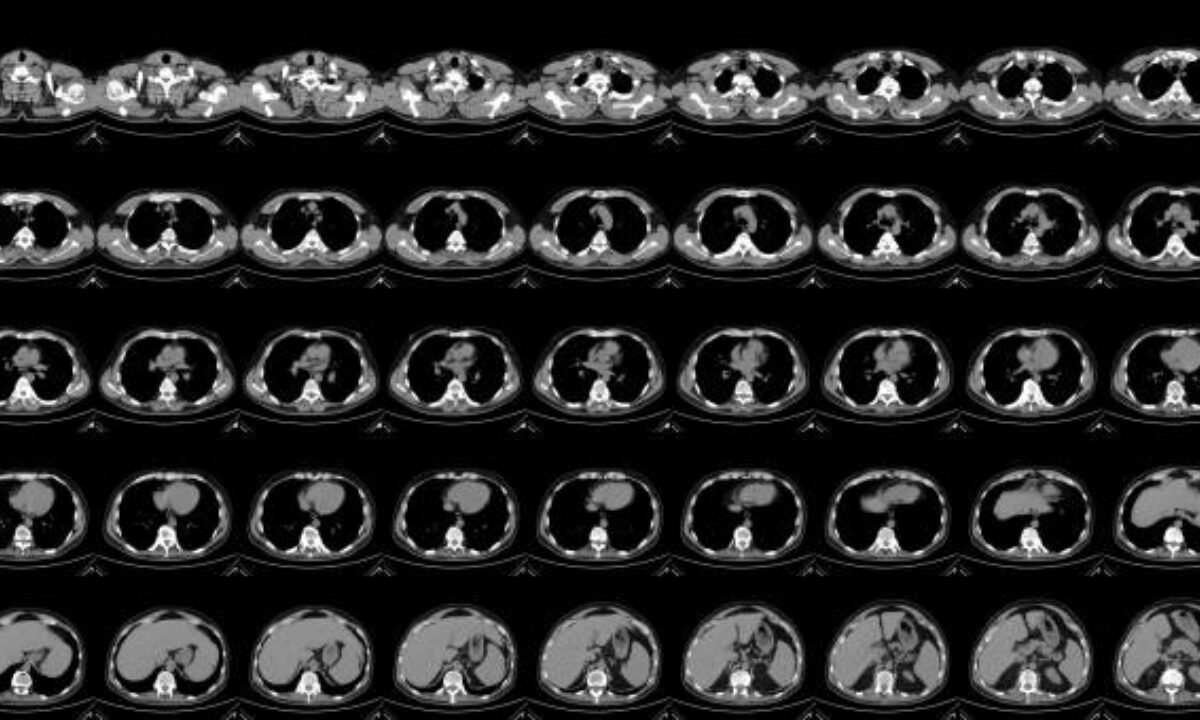
A Chest CT scan is an image test that takes painless X-rays and computer imaging to see the structures, bones, tissues, and blood vessels in the chest. In contrast with the standard X-rays, which only give one “shadowy” picture, the chest CT displays cross-sectional images – just imagine slices or layers – and a doctor would have better vision.

Simple Explanation

A Chest CT scan works like a loaf of bread cut into slices so you can see through each slice and see small details you wouldn’t notice from the outside.

More Detailed Explanation

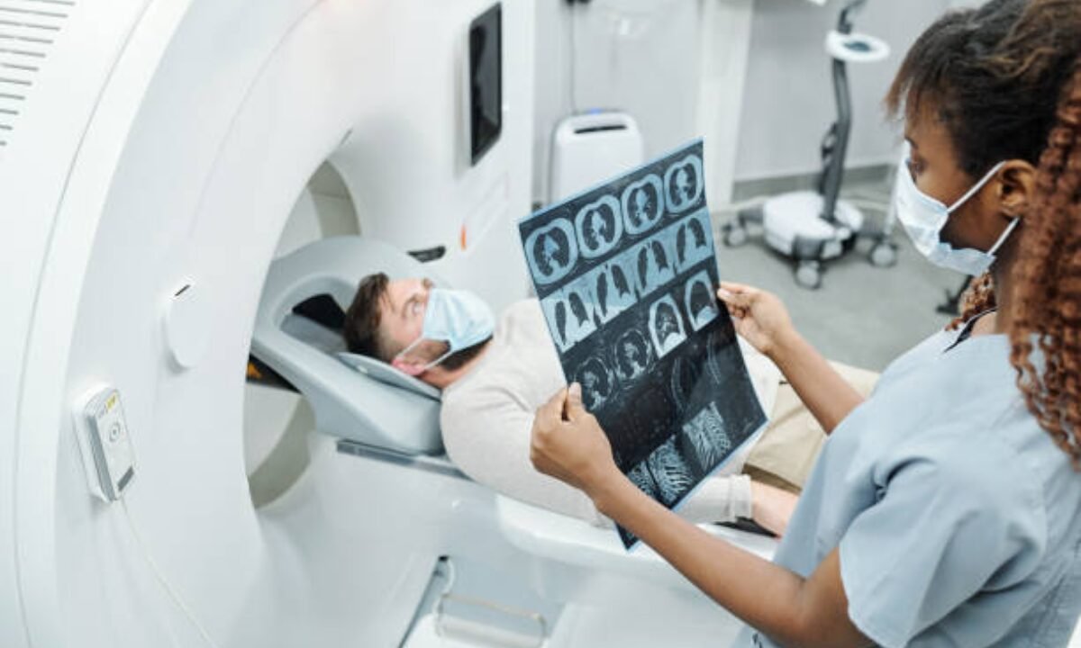
  • There are also improved scanning machines that you lie down on a table that slides in and out of a circular machine and you are scanned there.
  • The scanner makes several X-ray images from different angles.
  • A computer combines these images into an elaborate view of your chest from all directions.

For what reason do I need a Chest CT Scan?

There are many reasons why doctors request a Chest CT scan. They may prescribe it to you if you have symptoms such as shortness of breath, abnormal chest pain, or a (p) persistent cough, or if something abnormal was found on the X-ray of the whole chest.

Common Reasons Include:

  • Looking for infections such as TB or pneumonia.
  • detection of tumors, nodules, or lung cancer
  • Research on blood clots in the lungs
  • Diagnosing heart or blood vessel issues.
  •  Searching for injuries after an accident
  • Monitoring chronic lung conditions

What is a Chest CT Scan looking for?

A chest CT scan is primarily searching for anything abnormal in your chest. This includes:

  • Routine and hidden lung diseases.
  • Fluid collections
  • Swelling of lymphnodes
  • Abnormal blood vessels or enlarged heart chambers.
  • Cancerous or non-cancerous nodules

Types of Chest CT Scans

There are various chest CT scans. The most common are:

  • CT Chest Plain: This is a scan without the use of contrast dye. It provides fundamental but important information about lungs, chest wall, bones and heart.
  • CT Chest with Contrast: In this case, a dye is injected to make blood vessels and some tissues stand out. This makes it easier to locate blood clots or tumors.

When is CT Chest Plain Applied?

A CT chest plain is usually the first step. Doctors use it to:

  • Eliminate major problems such as big tumors or fractures as soon as possible.
  • Look for obvious infections or fluid.
  • Give a base scan to compare with future tests.

Preparing for the Scan

Correct preparation will help make your scan smoother and accurate:

  • Clothing: Wear comfortable, loose clothes. You might have to put on a hospital gown.
  • Jewelry and Metal Objects: Remove these before the scan because they may affect the pictures.
  • Eating and Drinking: If your scan requires the use of contrast dye, then you may be advised not to eat for a few hours.
  • Medical Conditions: Inform your doctor if you are allergic, have kidney problems, diabetes, or pregnant.

What to Expect during this Process? 

1. You will be asked to lie down on a moving table.

2. A technician will assist you in positioning for the best images.

3. Where a contrast dye is needed, this will be injected via a vein in your arm. This may trigger a warm feeling, but it is generally painless.

4. The table slides into the scanner (a big doughnut-shaped machine). You can hear humming or clicking sounds.

5. You will have to hold your breath for a few seconds.

6. The scan typically does not take more than 10 minutes.

After the scan, you can go home and continue with activities unless told otherwise. If you had contrast dye, drink lots of water to flush it out of your system.

Interpreting Your Chest CT Scan Report

After your scan is complete, a radiologist will look at the pictures and write a detailed chest CT scan report. This report is given to your doctor, who will explain the results to you.

Essential Aspects of a Chest CT Scan Report:

  • Findings: By noting anything strange, nodules, fluid, or an indication of infection.
  • Impression: A summary that will help your doctor to understand the main results quickly.
  • Recommendations: Tests if needed, after which.

What is a normal Chest CT Scan?

  • No unexpected growths or masses.
  • No signs of infection, bleeding, or fluid accumulation.
  • Lungs, blood vessels, heart, and chest wall are all healthy.

Common Results: Normal Chest CT Scan vs Abnormal Findings

Type of FindingWhat It Means       
Normal Chest CT ScanEverything looks healthy and typical
 Infection   There may be signs of pneumonia or TB
Tumor/Nodule  Possible need for further tests or biopsy
 Fluid   Could suggest infection or heart failure
Clots/Blood VesselIndicates possible pulmonary embolism

If your scan is normal, it’s a good sign—your doctor may look at other causes for your symptoms. If something strange is observed, do not panic; Generally, there is the need for further tests or follow-ups.

CT Scan in Bangalore

Good facilities are essential. When you are looking for a CT scan in Bangalore, research quality hospitals and imaging centers. There are numerous modern CT machines in advanced medical centers in Bangalore. Ask about:

  • Time taken to get your chest CT scan report
  • Whether they offer CT Chest plain and contrast scans, if necessary
  • Cost and whether your insurance covers the scan.

There are numerous credible places that provide CT scan in Bangalore and therefore you will probably receive good care and accurate results.

Risks and Side Effects

CT scans use more radiation than standard X-rays, but the risks are generally low, compared to the benefits. Never fail to inform your doctor if you are pregnant or may be pregnant. The contrast dye is generally safe but may cause:

  • Allergic reactions (rare)
  • Kidney problems in individuals who already have kidney problems.

If you feel sick after the scan, inform your doctor immediately.

Summary of Key Points

  • A Chest CT scan is a safe, fast, and very detailed method of seeing inside your chest.
  • It is used to diagnose infections, cancer, heart problems, and so on.
  • There are CT Chest plain scans and scans with contrast. Both have specific uses.
  •  A normal chest CT scan means all is well.
  • The chest CT scan report provides your doctor with the detailed answers required.
  • CT scan in Bangalore is a good medical center that offers quick and reliable results.

Understanding what is a Chest CT scan looking for and what to expect can make your experience much less stressful. If you have any concerns or questions about your scan, never hesitate to speak to your doctor.

Koshikaa offers the latest imaging services for CT scan in Bangalore with the help of the latest technology and radiologists.

MRI of the uterus is primarily used for diagnosing and evaluating conditions such as uterine fibroids, adenomyosis, and malignancies. It is particularly useful in the planning of surgical procedures and assessing the extent of gynecological diseases. This imaging test is valuable for its ability to deliver detailed cross-sectional images of the uterus and surrounding structures.
An MRI of the spleen can help diagnose various disorders affecting the spleen including splenomegaly (enlarged spleen), tumors, and other abnormalities. It provides clear, detailed images that are crucial for assessing the health of the spleen and planning potential surgical interventions.
Prostate MRI is a non-invasive imaging technique that provides detailed pictures of the prostate gland and surrounding tissues. It helps in the detection, staging, and monitoring of prostate cancer. Prostate MRIs are increasingly used to guide biopsy procedures and assist in the planning of treatment strategies for prostate health.
An MRI of the pancreas is used to identify issues such as pancreatitis, pancreatic cancer, and other pancreatic disorders. It offers high-resolution images that help in detecting small lesions and assessing the pancreatic ducts. This imaging technique is valuable for guiding biopsies and planning surgeries involving the pancreas.
MRI scans of the liver, kidney, and bile ducts provide detailed images that help diagnose various conditions, such as tumors, cysts, and blockages in the bile ducts. They are crucial for assessing organ damage, staging cancer, and planning liver or kidney-related surgeries. This type of MRI can also evaluate the health and function of these organs comprehensively.
Breast MRI is highly sensitive for detecting breast cancer, especially in individuals with dense breast tissue or a high risk of cancer. It is often used in conjunction with mammography to provide a more comprehensive evaluation. Breast MRIs can also monitor the response to breast cancer treatments and provide detailed pre-surgical mapping.
Brain and spine MRIs are critical tools for diagnosing and monitoring neurological conditions, including tumors, spinal cord injuries, and multiple sclerosis. These MRIs can reveal abnormalities in brain tissue and spinal cord structure that might not be visible with other imaging methods. They are essential for planning surgeries and assessing the effectiveness of treatments in neurology.
PET-CT scans of transplant organs like the liver, kidney, or pancreas are performed to assess metabolic activity, detect complications, and monitor post-transplant function. These scans play a vital role in the early detection of issues such as organ rejection, recurrence of disease, or infection, guiding timely interventions and optimizing transplant outcomes for patients.
PET-CT examinations of organs such as the liver, kidney, or prostate are crucial for assessing metabolic activity, detecting abnormalities, and monitoring disease progression. These scans help diagnose conditions such as liver metastases, renal cell carcinoma, or prostate cancer, guiding treatment decisions and monitoring response to therapy over time.
PET-CT scans of soft-tissue masses are utilized to assess metabolic activity and detect malignant tumors or other abnormalities. By combining PET imaging with CT imaging, these scans provide detailed information about tumor location, size, and metabolic characteristics, aiding in accurate diagnosis and treatment planning for soft-tissue cancers.
PET-CT scans are valuable for detecting cysts or fluid collections within the body, including cysts in the liver, kidneys, or other organs. These scans help characterize the nature and metabolic activity of cysts, guiding diagnosis and treatment planning for conditions such as cystic liver disease, renal cysts, or pancreatic cysts.
PET-CT scans of muscles are performed to assess metabolic activity and detect abnormalities such as inflammation, infection, or tumors. These scans provide valuable information about muscle function and pathology, aiding in the diagnosis and management of conditions like myositis, muscular dystrophy, or soft-tissue sarcomas.
PET-CT scans of the brain are utilized to evaluate brain metabolism and detect abnormalities such as tumors, epilepsy, or neurodegenerative diseases. By combining functional PET imaging with anatomical CT imaging, these scans offer comprehensive information about brain function and structure, guiding neurosurgical interventions and treatment decisions for neurological disorders
PET-CT scans of bones are commonly used to detect bone metastases in cancer patients, assess bone health in conditions like osteoporosis, and evaluate response to treatment. By combining PET and CT imaging, these scans provide detailed information about metabolic activity and anatomical changes in bone structures, aiding in accurate diagnosis and treatment planning for various bone-related conditions.
CT scans of the spine are conducted to assess the vertebrae, spinal cord, and surrounding soft tissues for conditions like herniated discs, spinal fractures, or spinal stenosis, aiding in the diagnosis and management of spinal disorders with detailed imaging essential for surgical planning and intervention.
CT scans of the pelvis are utilized to visualize the pelvic bones, reproductive organs, bladder, and surrounding structures for conditions such as fractures, tumors, or pelvic inflammatory disease, guiding treatment plans and surgical interventions with precise anatomical information.
CT scans of the neck are performed to evaluate structures such as the throat, thyroid gland, lymph nodes, and blood vessels for abnormalities like tumors, infections, or injuries, assisting in diagnosis and management of head and neck disorders with detailed imaging.
CT scans of the extremities are employed to assess bones, joints, and soft tissues in the arms or legs, aiding in the diagnosis of fractures, arthritis, or other musculoskeletal conditions, guiding orthopedic treatment and rehabilitation plans for optimal recovery.
CT scans of the chest are utilized to examine the lungs, heart, blood vessels, and surrounding structures for conditions such as lung nodules, pneumonia, or pulmonary embolism, facilitating accurate diagnosis and management of respiratory and cardiovascular diseases.
CT scans of the brain are conducted to assess brain structures, identify abnormalities like tumors, hemorrhages, or strokes, and aid in diagnosing neurological conditions, guiding neurosurgical interventions and treatment strategies with precise imaging of the brain’s anatomy.
CT scans of the abdomen are performed to visualize and evaluate the internal organs such as the liver, kidneys, pancreas, and intestines for conditions like tumors, infections, or injuries, providing detailed images essential for diagnosis and treatment planning.
Transplant organ ultrasound scans, including those of the liver, kidney, or pancreas, are performed to assess organ viability, detect complications, and monitor post-transplant function. These scans play a vital role in the early detection of issues such as organ rejection, vascular complications, or fluid collections, guiding timely interventions and optimizing transplant outcomes for patients.
Ultrasound examinations of organs such as the liver, kidney, or prostate are crucial for assessing organ structure, detecting abnormalities, and monitoring disease progression. These scans help diagnose conditions such as liver cirrhosis, kidney stones, or prostate enlargement, guiding treatment decisions and monitoring response to therapy over time.
Soft-tissue masses can be evaluated using ultrasound to determine their size, location, and characteristics. Ultrasound imaging helps differentiate between benign and malignant masses, guiding further diagnostic evaluation and treatment planning. By providing real-time imaging, ultrasound assists in the precise localization of soft-tissue masses for biopsy or surgical removal if needed.
Ultrasound scans are valuable for detecting cysts or fluid collections within the body, including cysts in the liver, kidneys, or other organs. These scans help characterize the nature and size of cysts, aiding in diagnosis and treatment planning for conditions such as cystic liver disease or renal cysts, and guiding interventions such as cyst drainage or sclerotherapy.
Musculoskeletal ultrasound scans are used to assess the health and integrity of muscles, identifying issues such as strains, tears, or muscle inflammation. These scans provide detailed images of muscle structures, allowing healthcare providers to diagnose muscle injuries accurately and develop personalized rehabilitation plans to promote recovery and restore function.
Ultrasound examinations of joints are essential for evaluating joint health, detecting abnormalities such as inflammation, fluid accumulation, or cartilage damage. By visualizing the joint structures in real-time, ultrasound helps diagnose conditions like arthritis, bursitis, or ligament injuries, guiding treatment decisions and monitoring disease progression over time.
Ultrasound scans of tendons are performed to assess for injuries, inflammation, or tears, aiding in the diagnosis and management of conditions such as tendonitis or tendon tears. These scans provide detailed images of tendon structures, helping healthcare providers determine the extent of damage and plan appropriate treatment strategies, including physical therapy or surgical intervention if necessary.
Pelvis X-rays play a crucial role in diagnosing injuries, diseases, and developmental conditions affecting the pelvic region. They provide detailed images of the hip bones, sacrum, and coccyx, aiding in the assessment of fractures, dislocations, or signs of arthritis. Pelvic X-rays are instrumental in guiding surgical interventions, monitoring healing progress, and evaluating conditions such as osteoporosis or tumors. By providing precise anatomical information, these imaging studies enable healthcare professionals to deliver personalized care and improve patient outcomes.
Teeth X-rays, known as dental radiographs, are indispensable tools for dentists to assess oral health. They aid in diagnosing dental decay, gum disease, and abnormalities in tooth structure or alignment. These images provide valuable insights into hidden dental issues, allowing for early intervention and prevention of complications. Additionally, dental X-rays are essential for planning treatments such as orthodontics, root canals, or dental implants, ensuring optimal outcomes and patient satisfaction.
Spine X-rays are pivotal in diagnosing spinal fractures, degenerative conditions, and spinal misalignments, providing critical information for treatment planning and monitoring spinal health. They offer detailed images of the vertebrae and surrounding structures, enabling healthcare professionals to identify issues such as scoliosis, disc herniation, or spinal stenosis
Skull X-rays are valuable for detecting fractures, assessing head trauma, and evaluating the alignment of facial bones, crucial in diagnosing conditions like fractures, tumors, or developmental abnormalities in the skull. They provide detailed views of the cranial structure, aiding clinicians in determining appropriate treatment strategies and ensuring patient safety.
For the heart, X-ray solutions can aid in the detection of various cardiac problems, ranging from mild to severe, including – Heart failure, Cardiac congestion and other heart-related conditions. In addition to cardiac assessments, X-ray solutions are also used for breast examinations to identify tumors or signs of breast cancer.

Bone X-rays are essential for diagnosing fractures and assessing bone alignment, aiding doctors in developing treatment plans and monitoring healing progress. They provide detailed images of the skeletal system, helping identify abnormalities or injuries that may not be visible through other imaging techniques

Abdominal X-rays are performed when patients experience symptoms such as significant nausea, ongoing vomiting, abdominal pain, flank or back pain, or abdominal swelling. These X-rays can help diagnose various conditions.

Frequently Booked Tests

Frequently Booked Checkups