How PET CT Helps in Diagnosing Inflammatory Heart Conditions

Full body checkup in Bangalore

Table of Contents

Heart diseases manifest differently because their symptoms can be subtle to the observer. Medics find it difficult to detect inflammatory heart diseases primarily because their symptoms do not announce themselves. Modern technology has given healthcare professionals access to better diagnostic tools for monitoring heart activity in patients. Doctors currently use the Cardiac PET CT scan as an imaging tool which provides accurate and early diagnosis of inflammatory heart diseases.

The Challenge of Diagnosing Inflammatory Heart Diseases

The heart inflammation disorders myocarditis or sarcoidosis display non-specific symptoms that include exhaustion together with shortness of breath, pain in the chest area and heart rhythm irregularities. ECG and echocardiogram together with blood tests often prove inadequate for appropriate heart diagnoses. The assessment process heavily depends on imaging technologies. MRI coupled with Cardiac PET CT scan provides medical professionals unique views of heart structure together with molecular activity.

Introducing PET CT: What Makes It Unique?

PET (Positron Emission Tomography) combined with CT (Computed Tomography) is an advanced imaging modality. Unlike regular CT or MRI, this scan tracks metabolic activity by using a mildly radioactive tracer. Inflammation in the heart causes certain cells to become more active and absorb more of the tracer, making inflamed areas stand out in the images. This ability to see active inflammation is why Cardiac PET CT scan is gaining attention in heart care.

How Is a PET CT Scan Done?

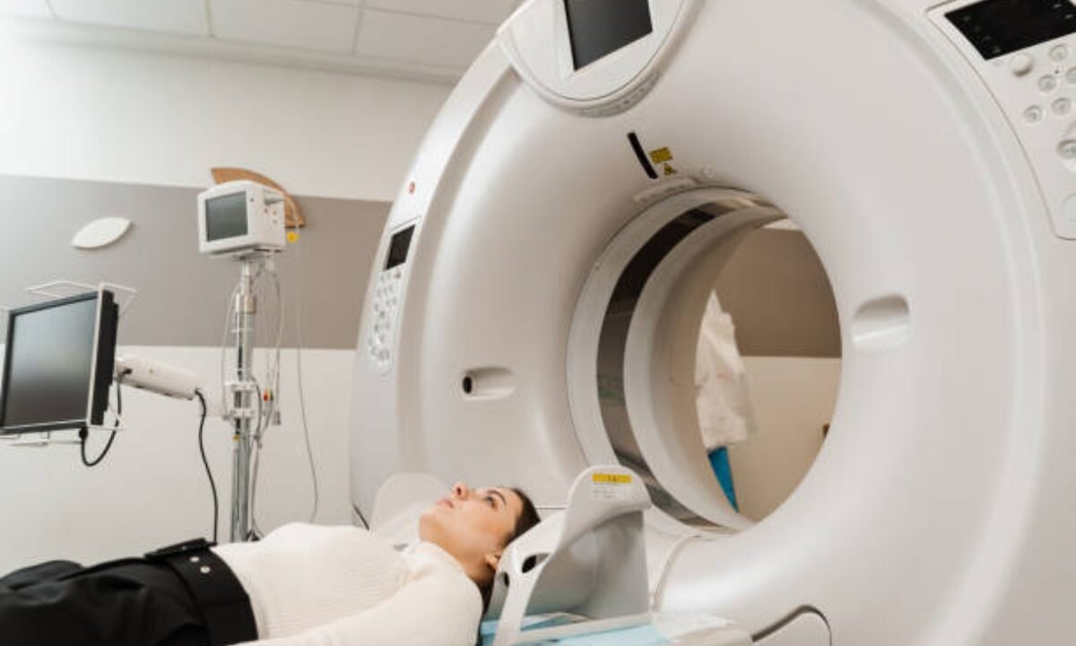
The process is straightforward and usually comfortable for the patient. To answer the common question: How is a PET CT scan done? The patient first fasts for a few hours. Then, a small amount of radioactive tracer is injected via a vein, often in the arm. As the tracer circulates, it collects in areas where the cells are most active—places where there’s inflammation.

During the scanning time, patients should lie on the designated table for 30 to 60 minutes. Through the PET CT scanner transportation, the table performs a gradual movement that generates detailed body pictures with specific attention to heart structures. The complete scan duration requires twenty to forty minutes to finish. A Cardiac PET scan poses no invasion to the body and creates no unpleasant experience. This Cardiac PET scan procedure is non-invasive and does not involve discomfort.

PET CT’s Role in Detecting Heart Inflammation

PET CT Scan in Bangalore

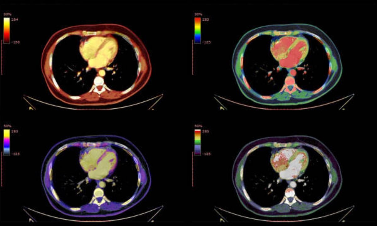
Inflammatory heart diseases can be complicated, but the cardiac pet scan procedure gives unique insights. For example, in sarcoidosis—a disease where clusters of immune cells form in organs, including the heart—classic imaging may miss early signs. PET CT shows exactly which areas in the heart are active with inflammation. Similarly, in myocarditis, it can detect inflamed heart muscles before they get permanently damaged.

Repeated scans also help track if the treatment is working by seeing the change in tracer uptake. This gives doctors and patients a clear idea about progress.

Why the PET CT Scan Stands Out

  • The detection capabilities of PET CT show active inflammation before standard results come back normal.
  • It indicates with precision the specific position of active heart inflammation.
  • Doctor assessments become better with PET CT because they show which patients will benefit from improved therapy or require medication modifications.
  • The success of heart treatment can be monitored through changes detected within tracer activities during the period.

The uncertain nature of inflammatory heart symptoms makes PET CT scans from Bangalore facilities especially helpful for medical accuracy.

What To Expect: The Cardiac PET CT Scan Procedure

Patients commonly wonder what will happen during the scan. The Cardiac PET CT scan procedure starts with careful preparation. You may be told to avoid caffeine and sugar for 12-24 hours before the test since these can affect how the tracer behaves. Blood sugar testing by medical staff requires interception before result measurement becomes necessary.

You will rest after the tracer injection until the substance completes its circulation. It is vital to remain motionless during scanning because your steady position creates better scanner images. The facility will monitor you briefly immediately after the examination period to verify your well-being.  But the radioactive tracer used is very safe and leaves your body quickly.

Local Relevance: PET CT Scan in Bangalore

Access to advanced cardiovascular imaging is growing in India. If you are searching for a PET CT scan in Bangalore, you will find that several reputable hospitals and diagnostic centers now offer this service. These centers combine advanced technology with skilled medical teams for the best outcomes.

When choosing where to get your scan, look for places with a good track record for heart care and modern equipment—especially for specialized scans like cardiac PET CT.

The Role of Full Body Checkup in Bangalore for Heart Health

The detection of heart inflammatory conditions should be at an early stage to stop further health complications from occurring. The standard medical checkup known as a Full body checkup in Bangalore includes vital heart examination services. Regular screening tests help identify both mental and physical heart abnormalities and risks for early medical intervention. Discussing the option of a Cardiac PET CT scan as part of a more comprehensive checkup is an excellent idea if there are symptoms or family history suggestive of heart inflammation.

Benefits and Limitations of Cardiac PET CT

Advantages

  • Non-invasive
  • Minimal discomfort
  • Sensitive for early inflammatory changes
  • Can guide and monitor treatment

Potential Limitations

  • Not available in every city (though PET CT scan in Bangalore is readily accessible)
  • Exposure to a small amount of radiation (generally considered safe)
  • Higher cost compared to standard tests

Conclusion: Simple, Accurate Answers to Complex Problems

In summary, inflammation in the heart is a complex, often silent problem. The rising availability and advancement of Cardiac PET CT scanners provides better opportunities to detect diseases earlier and enhance treatment outcomes. The understanding of PET CT scan processes and unique features helps patients select better options and enhances their readiness for such medical procedures. 

If you’re in a city with top medical facilities, like Bangalore, consider asking your doctor about the cardiac PET scan procedure if you have unexplained heart symptoms. Sometimes, a Full body checkup in Bangalore might uncover the need for more advanced imaging tests, and you’ll find high-quality PET CT scan in Bangalore centers to provide what’s needed for optimal care.

Modern diagnostic tools enable quick and clear and safe monitoring of your heart activity so you can obtain proper care right away. 

The Koshikaa Screening Centre stands out for its advanced imaging technologies and dedicated service to rapid accurate diagnostics and it thus becomes a prime selection for complete patient health evaluations.

MRI of the uterus is primarily used for diagnosing and evaluating conditions such as uterine fibroids, adenomyosis, and malignancies. It is particularly useful in the planning of surgical procedures and assessing the extent of gynecological diseases. This imaging test is valuable for its ability to deliver detailed cross-sectional images of the uterus and surrounding structures.
An MRI of the spleen can help diagnose various disorders affecting the spleen including splenomegaly (enlarged spleen), tumors, and other abnormalities. It provides clear, detailed images that are crucial for assessing the health of the spleen and planning potential surgical interventions.
Prostate MRI is a non-invasive imaging technique that provides detailed pictures of the prostate gland and surrounding tissues. It helps in the detection, staging, and monitoring of prostate cancer. Prostate MRIs are increasingly used to guide biopsy procedures and assist in the planning of treatment strategies for prostate health.
An MRI of the pancreas is used to identify issues such as pancreatitis, pancreatic cancer, and other pancreatic disorders. It offers high-resolution images that help in detecting small lesions and assessing the pancreatic ducts. This imaging technique is valuable for guiding biopsies and planning surgeries involving the pancreas.
MRI scans of the liver, kidney, and bile ducts provide detailed images that help diagnose various conditions, such as tumors, cysts, and blockages in the bile ducts. They are crucial for assessing organ damage, staging cancer, and planning liver or kidney-related surgeries. This type of MRI can also evaluate the health and function of these organs comprehensively.
Breast MRI is highly sensitive for detecting breast cancer, especially in individuals with dense breast tissue or a high risk of cancer. It is often used in conjunction with mammography to provide a more comprehensive evaluation. Breast MRIs can also monitor the response to breast cancer treatments and provide detailed pre-surgical mapping.
Brain and spine MRIs are critical tools for diagnosing and monitoring neurological conditions, including tumors, spinal cord injuries, and multiple sclerosis. These MRIs can reveal abnormalities in brain tissue and spinal cord structure that might not be visible with other imaging methods. They are essential for planning surgeries and assessing the effectiveness of treatments in neurology.
PET-CT scans of transplant organs like the liver, kidney, or pancreas are performed to assess metabolic activity, detect complications, and monitor post-transplant function. These scans play a vital role in the early detection of issues such as organ rejection, recurrence of disease, or infection, guiding timely interventions and optimizing transplant outcomes for patients.
PET-CT examinations of organs such as the liver, kidney, or prostate are crucial for assessing metabolic activity, detecting abnormalities, and monitoring disease progression. These scans help diagnose conditions such as liver metastases, renal cell carcinoma, or prostate cancer, guiding treatment decisions and monitoring response to therapy over time.
PET-CT scans of soft-tissue masses are utilized to assess metabolic activity and detect malignant tumors or other abnormalities. By combining PET imaging with CT imaging, these scans provide detailed information about tumor location, size, and metabolic characteristics, aiding in accurate diagnosis and treatment planning for soft-tissue cancers.
PET-CT scans are valuable for detecting cysts or fluid collections within the body, including cysts in the liver, kidneys, or other organs. These scans help characterize the nature and metabolic activity of cysts, guiding diagnosis and treatment planning for conditions such as cystic liver disease, renal cysts, or pancreatic cysts.
PET-CT scans of muscles are performed to assess metabolic activity and detect abnormalities such as inflammation, infection, or tumors. These scans provide valuable information about muscle function and pathology, aiding in the diagnosis and management of conditions like myositis, muscular dystrophy, or soft-tissue sarcomas.
PET-CT scans of the brain are utilized to evaluate brain metabolism and detect abnormalities such as tumors, epilepsy, or neurodegenerative diseases. By combining functional PET imaging with anatomical CT imaging, these scans offer comprehensive information about brain function and structure, guiding neurosurgical interventions and treatment decisions for neurological disorders
PET-CT scans of bones are commonly used to detect bone metastases in cancer patients, assess bone health in conditions like osteoporosis, and evaluate response to treatment. By combining PET and CT imaging, these scans provide detailed information about metabolic activity and anatomical changes in bone structures, aiding in accurate diagnosis and treatment planning for various bone-related conditions.
CT scans of the spine are conducted to assess the vertebrae, spinal cord, and surrounding soft tissues for conditions like herniated discs, spinal fractures, or spinal stenosis, aiding in the diagnosis and management of spinal disorders with detailed imaging essential for surgical planning and intervention.
CT scans of the pelvis are utilized to visualize the pelvic bones, reproductive organs, bladder, and surrounding structures for conditions such as fractures, tumors, or pelvic inflammatory disease, guiding treatment plans and surgical interventions with precise anatomical information.
CT scans of the neck are performed to evaluate structures such as the throat, thyroid gland, lymph nodes, and blood vessels for abnormalities like tumors, infections, or injuries, assisting in diagnosis and management of head and neck disorders with detailed imaging.
CT scans of the extremities are employed to assess bones, joints, and soft tissues in the arms or legs, aiding in the diagnosis of fractures, arthritis, or other musculoskeletal conditions, guiding orthopedic treatment and rehabilitation plans for optimal recovery.
CT scans of the chest are utilized to examine the lungs, heart, blood vessels, and surrounding structures for conditions such as lung nodules, pneumonia, or pulmonary embolism, facilitating accurate diagnosis and management of respiratory and cardiovascular diseases.
CT scans of the brain are conducted to assess brain structures, identify abnormalities like tumors, hemorrhages, or strokes, and aid in diagnosing neurological conditions, guiding neurosurgical interventions and treatment strategies with precise imaging of the brain’s anatomy.
CT scans of the abdomen are performed to visualize and evaluate the internal organs such as the liver, kidneys, pancreas, and intestines for conditions like tumors, infections, or injuries, providing detailed images essential for diagnosis and treatment planning.
Transplant organ ultrasound scans, including those of the liver, kidney, or pancreas, are performed to assess organ viability, detect complications, and monitor post-transplant function. These scans play a vital role in the early detection of issues such as organ rejection, vascular complications, or fluid collections, guiding timely interventions and optimizing transplant outcomes for patients.
Ultrasound examinations of organs such as the liver, kidney, or prostate are crucial for assessing organ structure, detecting abnormalities, and monitoring disease progression. These scans help diagnose conditions such as liver cirrhosis, kidney stones, or prostate enlargement, guiding treatment decisions and monitoring response to therapy over time.
Soft-tissue masses can be evaluated using ultrasound to determine their size, location, and characteristics. Ultrasound imaging helps differentiate between benign and malignant masses, guiding further diagnostic evaluation and treatment planning. By providing real-time imaging, ultrasound assists in the precise localization of soft-tissue masses for biopsy or surgical removal if needed.
Ultrasound scans are valuable for detecting cysts or fluid collections within the body, including cysts in the liver, kidneys, or other organs. These scans help characterize the nature and size of cysts, aiding in diagnosis and treatment planning for conditions such as cystic liver disease or renal cysts, and guiding interventions such as cyst drainage or sclerotherapy.
Musculoskeletal ultrasound scans are used to assess the health and integrity of muscles, identifying issues such as strains, tears, or muscle inflammation. These scans provide detailed images of muscle structures, allowing healthcare providers to diagnose muscle injuries accurately and develop personalized rehabilitation plans to promote recovery and restore function.
Ultrasound examinations of joints are essential for evaluating joint health, detecting abnormalities such as inflammation, fluid accumulation, or cartilage damage. By visualizing the joint structures in real-time, ultrasound helps diagnose conditions like arthritis, bursitis, or ligament injuries, guiding treatment decisions and monitoring disease progression over time.
Ultrasound scans of tendons are performed to assess for injuries, inflammation, or tears, aiding in the diagnosis and management of conditions such as tendonitis or tendon tears. These scans provide detailed images of tendon structures, helping healthcare providers determine the extent of damage and plan appropriate treatment strategies, including physical therapy or surgical intervention if necessary.
Pelvis X-rays play a crucial role in diagnosing injuries, diseases, and developmental conditions affecting the pelvic region. They provide detailed images of the hip bones, sacrum, and coccyx, aiding in the assessment of fractures, dislocations, or signs of arthritis. Pelvic X-rays are instrumental in guiding surgical interventions, monitoring healing progress, and evaluating conditions such as osteoporosis or tumors. By providing precise anatomical information, these imaging studies enable healthcare professionals to deliver personalized care and improve patient outcomes.
Teeth X-rays, known as dental radiographs, are indispensable tools for dentists to assess oral health. They aid in diagnosing dental decay, gum disease, and abnormalities in tooth structure or alignment. These images provide valuable insights into hidden dental issues, allowing for early intervention and prevention of complications. Additionally, dental X-rays are essential for planning treatments such as orthodontics, root canals, or dental implants, ensuring optimal outcomes and patient satisfaction.
Spine X-rays are pivotal in diagnosing spinal fractures, degenerative conditions, and spinal misalignments, providing critical information for treatment planning and monitoring spinal health. They offer detailed images of the vertebrae and surrounding structures, enabling healthcare professionals to identify issues such as scoliosis, disc herniation, or spinal stenosis
Skull X-rays are valuable for detecting fractures, assessing head trauma, and evaluating the alignment of facial bones, crucial in diagnosing conditions like fractures, tumors, or developmental abnormalities in the skull. They provide detailed views of the cranial structure, aiding clinicians in determining appropriate treatment strategies and ensuring patient safety.
For the heart, X-ray solutions can aid in the detection of various cardiac problems, ranging from mild to severe, including – Heart failure, Cardiac congestion and other heart-related conditions. In addition to cardiac assessments, X-ray solutions are also used for breast examinations to identify tumors or signs of breast cancer.

Bone X-rays are essential for diagnosing fractures and assessing bone alignment, aiding doctors in developing treatment plans and monitoring healing progress. They provide detailed images of the skeletal system, helping identify abnormalities or injuries that may not be visible through other imaging techniques

Abdominal X-rays are performed when patients experience symptoms such as significant nausea, ongoing vomiting, abdominal pain, flank or back pain, or abdominal swelling. These X-rays can help diagnose various conditions.

Frequently Booked Tests

Frequently Booked Checkups