
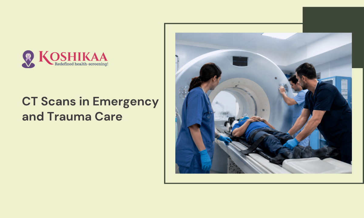
When a medical crisis strikes, doctors do not have the luxury of time. Whether a patient arrives with severe chest pain, a sudden loss of speech, or following a major car accident, the medical team must immediately understand what is happening inside the body.
Guessing is simply not an option when a life is on the line. This is exactly why a CT scan for emergencies is the most critical tool in a modern medical facility.
A CT scan provides a rapid and highly detailed map of the internal organs, bones, and blood vessels. For those seeking a reliable CT Scan in Bangalore, this imaging technology acts as the eyes of the emergency team. It allows doctors to spot hidden internal bleeding or blocked airways in a matter of seconds. As it delivers absolute clarity when every minute counts, this rapid imaging process is widely considered the Best Health Screening Centre in Bangalore for urgent trauma care.
The primary goal of this scan is to give surgeons the exact location of the problem so they can start saving a life immediately.
Medical Disclaimer
The information provided in this guide is strictly for educational purposes and does not replace professional medical advice, diagnosis, or emergency treatment. Always talk directly to a qualified doctor or emergency medical professional about specific physical symptoms or sudden health changes. Never delay seeking immediate clinical evaluation or calling an ambulance based on the contents of this article.
Rapid Action for Stroke Diagnosis
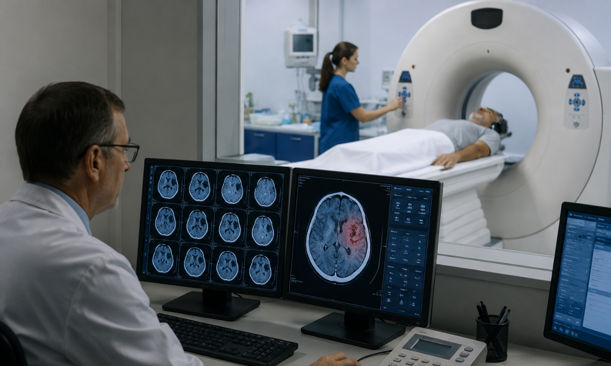
When a person suddenly loses the ability to speak, smile, or move one side of their body, every single second matters. A fast and accurate Stroke diagnosis is the absolute highest priority in an emergency room. When the ambulance arrives, the medical team will bypass almost every other test and rush the patient directly into a CT scanner.
The reason for this extreme speed is simple. There are two very different types of strokes. The life-saving medication for one type will cause massive harm to a patient suffering from the other type. The CT scan is the only way for a doctor to safely choose the correct medical path.
Understanding the Two Types of Strokes
Doctors use the scanning machine to instantly see which of the following two emergencies is happening inside the skull.
| Type of Stroke | What is Happening in the Brain | How the CT Scan Helps |
|---|---|---|
| The Bleeding Stroke | A weak blood vessel bursts open and starts leaking blood directly into the brain tissue. This creates dangerous physical pressure inside the skull. | Fresh blood shows up as a bright white spot on the scan monitor. If doctors see this white spot, they immediately know they must perform surgery to stop the bleeding. They absolutely cannot give the patient blood-thinning medications. |
| The Blocked Blood Vessel | A tiny blood clot gets stuck in a blood vessel and stops fresh oxygen from reaching a specific part of the brain. | The scan will show that there is no active bleeding. This gives the emergency doctor the exact physical proof they need to safely inject strong clot-busting medications to dissolve the blockage. |
The Emergency Patient Experience
If you or a loved one is rushed to the hospital with stroke symptoms, the imaging process is incredibly fast and highly coordinated.
- The Arrival: The ambulance crew will notify the hospital before you even arrive. The imaging team will have the scanning machine fully prepared and waiting for you.
- The Transfer: You will be moved directly from the ambulance stretcher onto the sliding scanner table. You do not need to change clothes or wait in a reception area.
- The Scan: The machine will circle your head rapidly. Because time is critical, the entire imaging process takes less than two minutes.
The emergency doctor reads the images on a computer monitor in real time while you are still on the table. They will make a final treatment decision before you are even moved to a regular hospital bed.
Finding the Source of a Vascular Emergency
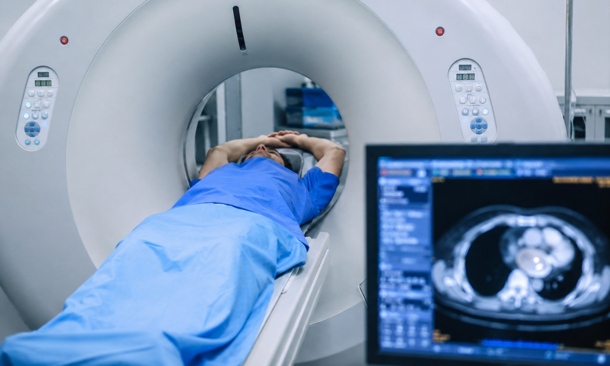
A Vascular emergency occurs when a major blood vessel tears, blocks, or bursts inside the body. Unlike a broken bone or a surface wound, you cannot see internal bleeding from the outside. If a patient is involved in a severe car accident or experiences a sudden and tearing pain in their chest, doctors must find the exact source of the internal damage instantly.
To solve this problem, the emergency medical team relies on a specific imaging technique that makes your bloodstream completely visible.
How Doctors See Inside Your Veins
When looking for a damaged blood vessel, standard X-rays are useless because blood and soft tissue blend on the image. A CT scan solves this by using a special medical contrast fluid. Here is exactly what the patient experiences and how the doctors use this technology to save lives.
- The Contrast Injection: A nurse or technician will inject a safe medical dye into a vein in your arm through a small IV. As the fluid enters your bloodstream, you will likely feel a sudden, harmless rush of warmth spreading through your body.
- The Glowing Map: As this dye travels through your circulatory system, the scanning machine takes rapid cross-sectional pictures. The contrast dye reacts with the imaging technology to make your blood vessels glow bright white on the doctor’s computer monitor.
- The Surgical Target: If there is a tear in an artery, the emergency doctor will actually see the glowing dye leaking out of the vessel on the screen. This provides the surgical team with a precise and brightly lit map. They know exactly where they need to cut and clamp before they even move you to the operating room.
Common Vascular Crises We Scan For
Emergency rooms use this glowing contrast method to identify several life-threatening conditions before they cause permanent damage.
| Type of Internal Injury | How It Typically Happens | What the Scan Actually Reveals |
|---|---|---|
| Abdominal Internal Bleeding | Severe physical trauma, such as a high-speed car crash or a bad fall. | The scan highlights pools of glowing fluid collecting around the liver or spleen. |
| Aortic Dissection | Chronic high blood pressure weakens the wall of the main artery leading from the heart, causing it to tear. | The images show a distinct rip in the thick wall of the blood vessel before it completely bursts open. |
| Deep Vein Thrombosis | A large blood clot forms deep inside the leg veins and threatens to travel upwards. | The contrast dye flows around the clot, showing a clear dark gap in an otherwise glowing white vein. |
Instantly highlighting these dangerous blockages and leaks, the CT scan removes all the guesswork from vascular surgery and drastically improves the patient’s chances of a full recovery.
Clearing the Air in a Respiratory Crisis
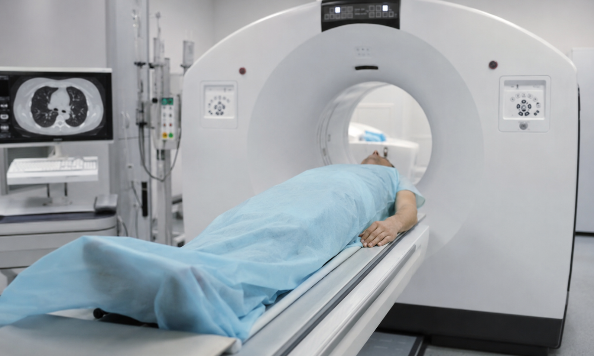
When a patient arrives at the emergency room gasping for air, the medical team faces a major challenge. A severe Respiratory crisis can be caused by many different things. It might be a sudden asthma attack, a massive infection, or physical damage inside the chest. While a standard chest X-ray is a helpful starting point, it only provides a flat picture where the heart, bones, and lungs all overlap. A CT scan takes hundreds of detailed slices of the chest, allowing doctors to look deep inside the lung tissue and airway passages with perfect clarity.
Finding the Hidden Causes of Breathing Problems
Emergency doctors rely on this advanced imaging because it immediately reveals life-threatening lung problems that regular X-rays often miss completely. Here is exactly how the scanning machine helps doctors clear the airway and restore normal breathing.
- Dangerous Lung Clots: Sometimes a blood clot travels from the leg directly into the lungs. This blocks fresh oxygen from entering the blood and puts massive strain on the heart.
The CT scanner uses a quick burst of contrast dye to highlight the exact location of the blockage. This allows doctors to safely administer powerful clot-busting drugs to clear the airway.
- Severe Lung Infections: When a patient has a massive infection like advanced pneumonia, the tiny air sacs in their lungs fill with thick fluid and pus.
The scan shows this fluid as dense white patches on the computer monitor. This detailed view helps the doctor see exactly how much healthy lung tissue is left and whether the patient needs to be placed on a mechanical ventilator to survive.
- Physical Chest Trauma: If a patient is injured in a bad fall or a high-speed car accident, a broken rib can easily puncture the lung wall. This causes air to leak out and crush the healthy lung tissue.
The scan instantly shows these trapped air pockets. This tells the emergency surgeon exactly where they need to insert a plastic chest tube to relieve the pressure and let the lung expand normally again.
A Fast and Painless Process
If you or a loved one is already struggling to breathe, the idea of lying flat inside a medical machine can feel very scary.
However, emergency chest scans are designed to be incredibly fast. The technician will guide you through the process quickly. You will usually only need to hold your breath for about three to five seconds while the machine circles your chest. The extreme speed of modern scanners ensures the medical team gets the answers they need without causing the patient any additional panic or discomfort.
The Koshikaa Advantage for Urgent Diagnostics
When an emergency happens, you need a facility that is built entirely for speed and accuracy. Koshikaa is designed to handle urgent diagnostic needs with absolute precision. We have optimized every single step of our imaging process to ensure that you and your doctors get clear answers without any unnecessary delays.
- Advanced Low Radiation Technology: We use the newest imaging machines available on the market. These machines capture crystal clear pictures of your internal organs while using a tiny fraction of the radiation required by older hospital scanners. This keeps you safe even if you need multiple scans during a complex recovery process.
- Zero Wait Time Processing: In a medical crisis, waiting for a report can be dangerous. Our dedicated team processes your emergency scans instantly. We ensure that your verified diagnostic images and reports are handed directly to your medical team immediately so they can begin life-saving treatments without hesitation.
- Expert Technical Staff: Our imaging technicians are specifically trained to handle high-stress emergencies. They know exactly how to safely move a patient who is in severe pain and how to capture perfect images on the very first try.
This means you spend less time on the scanning table and more time getting the direct medical help you need.
Conclusion
A medical emergency is the worst possible time to start researching where to get a reliable imaging test. Having a trusted diagnostic center in mind before a crisis hits is a crucial part of protecting your family. Whether you need a routine check of a stubborn headache or rapid imaging during a sudden physical trauma, knowing exactly where to go saves precious time and prevents panic.
Take control of your health today by choosing a facility that prioritizes both speed and absolute safety. Trust Koshikaa for all of your preventative and urgent diagnostic needs. Our advanced technology and expert clinical team are always ready to provide the clear answers your doctors need to keep you safe and healthy for the long term.