When Should You Get a Mammogram? A Simple Guide for Women

MRI scan in Bangalore

Table of Contents

Prevention Early breast health depends on early detection. A special type of X-ray scan called a mammogram searches for changes that occur in breast tissue before you can feel them. The primary component of breast cancer screening is regular mammograms, which contribute much to treatment. This simple guide informs individuals about who should be tested, how to conduct the tests, and where to seek treatment.

Recommended Age for Mammograms

Health authorities do not coincide completely, but they share some milestones:

  • The majority of recommendations suggest annual or alternate-year mammography of average-risk women beginning at age 40.
  • Women normally have an option of changing every two years after age 55, provided that prior results are normal.
  • Experts emphasize that every woman ought to have a personal plan – those who have dense tissue or family history can begin earlier.
  • Age rules are a beginning. Effective communication between you and your provider using the shared approach can lead you to the right starting point at the right time for you.

Individual screening and risk factors

Various variables may define the time of screening:

1. Family History: 30 or 35: screening may be necessary in a first-degree relative with breast cancer.

2. Genetic Mutations: Women who have BRCA1 or BRCA2 mutations undergo more vigilant monitoring.

3. History of chest treatment: Past radiation of the chest or benign breast disease should be treated with more imaging.

4. Breast Density: Breasts that are dense mask tiny tumors; there is a possibility of additional ultrasound or MRI.

When you discuss the risks with your provider, you will allow your provider to offer recommendations unique to your needs beyond age limits.

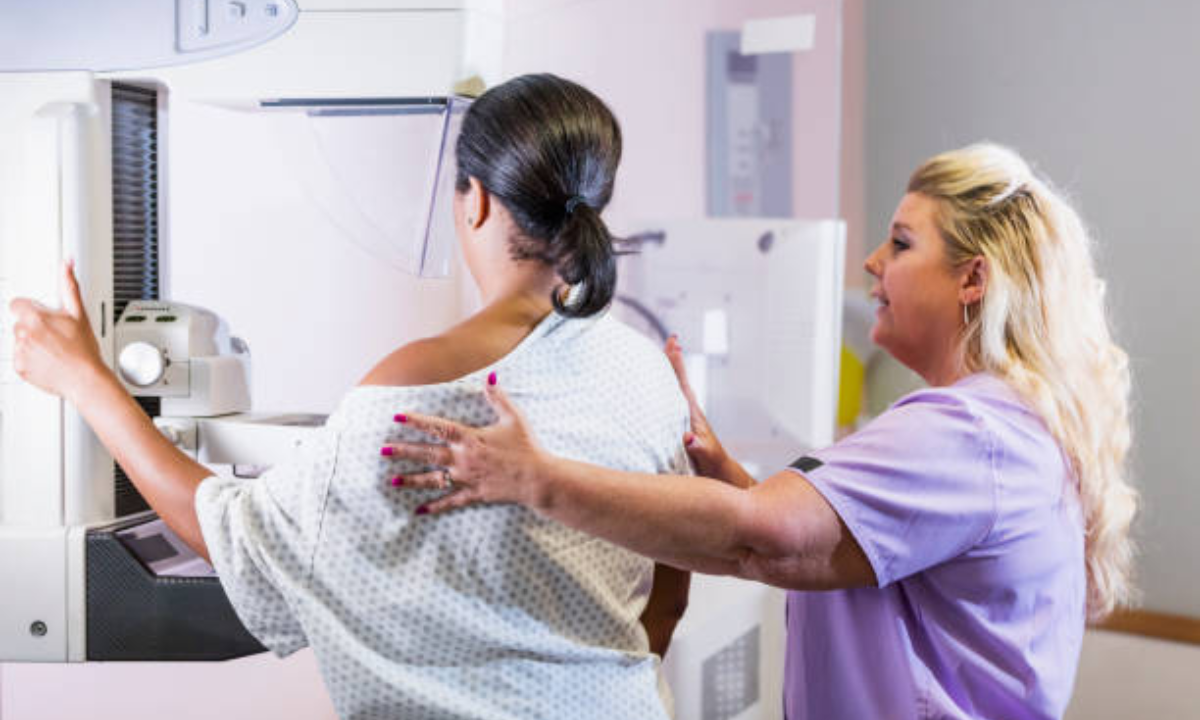
Knowledge of the mammogram test procedure

Mammography

The test itself takes not so much, approximately 20 minutes:

1. A technologist places each of the breasts, one at a time, on a flat support plate.

2. The tissue is evenly spread with a second plate that produces gentle compression.

3. Each breast is typically done in two positions, i.e., top-to-bottom and side-to-side.

4. The photos are assessed at the location; subsequent glimpses can be done, where necessary. 

The fact that people have some idea about the medical operation that is performed in a mammogram makes them prepared to endure mild pressure, and they understand why they might only experience little discomfort during the session.

Mammogram preparation tips

The readiness enhances comfort and facilitates the mammogram process. Here are some tips to follow: 

• Take the test a week after your period is over; the breasts are not as tender.  

• Do not wear deodorants, perfumes, powder, or lotions near your armpits or your breasts. Those can appear as artifacts on the images.  

• Put on a two-piece top since you can undress easily in the exam room.  

• If going to a new center, bring former imaging reports. A comparison of the scans provides an informative background.  

• Inform personnel of implants or surgeries within the past days since it impacts the technician dealing with the application of compression and positioning.  

The procedures assist in the generation of clear pictures and reduce the possibility of the need to retake the test.

Advancements in Imaging  

This early detection is very important since tumors that are detected at the earliest stage are usually easy to treat and have high survival rates. Women who do not show symptoms receive an early detection mammogram in search of microcalcifications or micro masses. Instructions come in line with screening recommendations and emphasize that you repeat the examination every year or two.  

Technology has been able to change film mammography to digital mammography, which transforms X-rays into high-resolution digital images. A digital mammography test is now being done in many radiology centers, which allows radiology experts to adjust the contrast and zoom in on points of interest. This increases the visibility of the lesion, particularly in dense breasts, and tends to reduce the requirement for a call back.

Accessing Services  

Being in India, and a place with plenty of options (the tech capital), you are all set. Browse to the search box, put the phrase mammography in Bangalore, and browse through the credentials of the mammography provider, like the accreditation done nationally or internationally. Most hospitals in urban settings have multi-disciplinary breast units with imaging, biopsy, and oncology services co-located. 

Affordable mammography in Bangalore is available in government health schemes, privately owned diagnostic chains, and community camps every now and then. Prices may differ significantly; thus, it is necessary to compare a package of charges that includes imaging, a visit to a doctor, and a follow-up report. This should prepare you so that you receive maximum care, to your surprise.

What comes after the Exam

Following the scan, the BI-RADS scale is used as a radiologist writes a report. The categories go as low as 0 (need more imaging) to as high as 6 (known cancer). The majority of screening would be BI-RADS 1 (normal) or BI-RADS 2 (benign findings), and regular follow-ups are sufficient. BI-RADS 3-6 categories frequently need additional examinations, including ultrasound, MRI, or precise biopsy. Such stages of follow-up might appear alarming, but they reduce confusion and aid in the selection of proper treatment.

Elevate Long-Term Success and Assistance

Breast health is a constant task. With your initial screening, you may set up reminders on your phone, calendar, or clinic to keep you on schedule. Conduct monthly self-checks, pay attention to changes as they happen, and combine screenings with healthy lifestyle choices to minimize the risk of developing this disease: maintain a balanced diet, lead an active lifestyle, limit the amount of alcohol consumed, and quit smoking.

Discuss risk factors, both familiar and genetic mutation, with your provider openly and bring up dense breast tissue. Request supplementary imaging consistency through ultrasound or MRI to diagnose cancer at an early age.

In addition, pay attention to the following advice:

  • No deodorants, lotion, or powder on the day of the exam.
  • Wear a two-piece outfit to have an easy time changing.
  • The quality of communication enhances the quality of the image.
  • Have routine follow-ups despite normal results. 

You can have the best of both worlds with regular mammograms when combined with a healthy lifestyle. It is also important to get in touch with a support group or attend a workshop to keep track.

Question on all the steps of mammograms to be aware of the procedure.

Search to find reputable services and programmes (low-cost or free), and local health organizations and advocacy services.

Conclusion

Being aware of when you should start screening, how to go about it, and what to expect takes the control of your health into your own hands. Request your physician to schedule a mammogram and devise a regimen that is right for you. The probability of early diagnosis and treatment of breast cancer increases when healthy habits are added to the checks.

Koshikaa Screening Centre is patient-centric and well-advanced in digital technology for mammography in Bangalore. Quality exams are offered by trained technologists and board-certified radiologists. Your test will result in an online review or a telemedicine follow-up that eliminates travelling and waiting. Screening is also less costly due to financial aid and easy payment schemes.

MRI of the uterus is primarily used for diagnosing and evaluating conditions such as uterine fibroids, adenomyosis, and malignancies. It is particularly useful in the planning of surgical procedures and assessing the extent of gynecological diseases. This imaging test is valuable for its ability to deliver detailed cross-sectional images of the uterus and surrounding structures.
An MRI of the spleen can help diagnose various disorders affecting the spleen including splenomegaly (enlarged spleen), tumors, and other abnormalities. It provides clear, detailed images that are crucial for assessing the health of the spleen and planning potential surgical interventions.
Prostate MRI is a non-invasive imaging technique that provides detailed pictures of the prostate gland and surrounding tissues. It helps in the detection, staging, and monitoring of prostate cancer. Prostate MRIs are increasingly used to guide biopsy procedures and assist in the planning of treatment strategies for prostate health.
An MRI of the pancreas is used to identify issues such as pancreatitis, pancreatic cancer, and other pancreatic disorders. It offers high-resolution images that help in detecting small lesions and assessing the pancreatic ducts. This imaging technique is valuable for guiding biopsies and planning surgeries involving the pancreas.
MRI scans of the liver, kidney, and bile ducts provide detailed images that help diagnose various conditions, such as tumors, cysts, and blockages in the bile ducts. They are crucial for assessing organ damage, staging cancer, and planning liver or kidney-related surgeries. This type of MRI can also evaluate the health and function of these organs comprehensively.
Breast MRI is highly sensitive for detecting breast cancer, especially in individuals with dense breast tissue or a high risk of cancer. It is often used in conjunction with mammography to provide a more comprehensive evaluation. Breast MRIs can also monitor the response to breast cancer treatments and provide detailed pre-surgical mapping.
Brain and spine MRIs are critical tools for diagnosing and monitoring neurological conditions, including tumors, spinal cord injuries, and multiple sclerosis. These MRIs can reveal abnormalities in brain tissue and spinal cord structure that might not be visible with other imaging methods. They are essential for planning surgeries and assessing the effectiveness of treatments in neurology.
PET-CT scans of transplant organs like the liver, kidney, or pancreas are performed to assess metabolic activity, detect complications, and monitor post-transplant function. These scans play a vital role in the early detection of issues such as organ rejection, recurrence of disease, or infection, guiding timely interventions and optimizing transplant outcomes for patients.
PET-CT examinations of organs such as the liver, kidney, or prostate are crucial for assessing metabolic activity, detecting abnormalities, and monitoring disease progression. These scans help diagnose conditions such as liver metastases, renal cell carcinoma, or prostate cancer, guiding treatment decisions and monitoring response to therapy over time.
PET-CT scans of soft-tissue masses are utilized to assess metabolic activity and detect malignant tumors or other abnormalities. By combining PET imaging with CT imaging, these scans provide detailed information about tumor location, size, and metabolic characteristics, aiding in accurate diagnosis and treatment planning for soft-tissue cancers.
PET-CT scans are valuable for detecting cysts or fluid collections within the body, including cysts in the liver, kidneys, or other organs. These scans help characterize the nature and metabolic activity of cysts, guiding diagnosis and treatment planning for conditions such as cystic liver disease, renal cysts, or pancreatic cysts.
PET-CT scans of muscles are performed to assess metabolic activity and detect abnormalities such as inflammation, infection, or tumors. These scans provide valuable information about muscle function and pathology, aiding in the diagnosis and management of conditions like myositis, muscular dystrophy, or soft-tissue sarcomas.
PET-CT scans of the brain are utilized to evaluate brain metabolism and detect abnormalities such as tumors, epilepsy, or neurodegenerative diseases. By combining functional PET imaging with anatomical CT imaging, these scans offer comprehensive information about brain function and structure, guiding neurosurgical interventions and treatment decisions for neurological disorders
PET-CT scans of bones are commonly used to detect bone metastases in cancer patients, assess bone health in conditions like osteoporosis, and evaluate response to treatment. By combining PET and CT imaging, these scans provide detailed information about metabolic activity and anatomical changes in bone structures, aiding in accurate diagnosis and treatment planning for various bone-related conditions.
CT scans of the spine are conducted to assess the vertebrae, spinal cord, and surrounding soft tissues for conditions like herniated discs, spinal fractures, or spinal stenosis, aiding in the diagnosis and management of spinal disorders with detailed imaging essential for surgical planning and intervention.
CT scans of the pelvis are utilized to visualize the pelvic bones, reproductive organs, bladder, and surrounding structures for conditions such as fractures, tumors, or pelvic inflammatory disease, guiding treatment plans and surgical interventions with precise anatomical information.
CT scans of the neck are performed to evaluate structures such as the throat, thyroid gland, lymph nodes, and blood vessels for abnormalities like tumors, infections, or injuries, assisting in diagnosis and management of head and neck disorders with detailed imaging.
CT scans of the extremities are employed to assess bones, joints, and soft tissues in the arms or legs, aiding in the diagnosis of fractures, arthritis, or other musculoskeletal conditions, guiding orthopedic treatment and rehabilitation plans for optimal recovery.
CT scans of the chest are utilized to examine the lungs, heart, blood vessels, and surrounding structures for conditions such as lung nodules, pneumonia, or pulmonary embolism, facilitating accurate diagnosis and management of respiratory and cardiovascular diseases.
CT scans of the brain are conducted to assess brain structures, identify abnormalities like tumors, hemorrhages, or strokes, and aid in diagnosing neurological conditions, guiding neurosurgical interventions and treatment strategies with precise imaging of the brain’s anatomy.
CT scans of the abdomen are performed to visualize and evaluate the internal organs such as the liver, kidneys, pancreas, and intestines for conditions like tumors, infections, or injuries, providing detailed images essential for diagnosis and treatment planning.
Transplant organ ultrasound scans, including those of the liver, kidney, or pancreas, are performed to assess organ viability, detect complications, and monitor post-transplant function. These scans play a vital role in the early detection of issues such as organ rejection, vascular complications, or fluid collections, guiding timely interventions and optimizing transplant outcomes for patients.
Ultrasound examinations of organs such as the liver, kidney, or prostate are crucial for assessing organ structure, detecting abnormalities, and monitoring disease progression. These scans help diagnose conditions such as liver cirrhosis, kidney stones, or prostate enlargement, guiding treatment decisions and monitoring response to therapy over time.
Soft-tissue masses can be evaluated using ultrasound to determine their size, location, and characteristics. Ultrasound imaging helps differentiate between benign and malignant masses, guiding further diagnostic evaluation and treatment planning. By providing real-time imaging, ultrasound assists in the precise localization of soft-tissue masses for biopsy or surgical removal if needed.
Ultrasound scans are valuable for detecting cysts or fluid collections within the body, including cysts in the liver, kidneys, or other organs. These scans help characterize the nature and size of cysts, aiding in diagnosis and treatment planning for conditions such as cystic liver disease or renal cysts, and guiding interventions such as cyst drainage or sclerotherapy.
Musculoskeletal ultrasound scans are used to assess the health and integrity of muscles, identifying issues such as strains, tears, or muscle inflammation. These scans provide detailed images of muscle structures, allowing healthcare providers to diagnose muscle injuries accurately and develop personalized rehabilitation plans to promote recovery and restore function.
Ultrasound examinations of joints are essential for evaluating joint health, detecting abnormalities such as inflammation, fluid accumulation, or cartilage damage. By visualizing the joint structures in real-time, ultrasound helps diagnose conditions like arthritis, bursitis, or ligament injuries, guiding treatment decisions and monitoring disease progression over time.
Ultrasound scans of tendons are performed to assess for injuries, inflammation, or tears, aiding in the diagnosis and management of conditions such as tendonitis or tendon tears. These scans provide detailed images of tendon structures, helping healthcare providers determine the extent of damage and plan appropriate treatment strategies, including physical therapy or surgical intervention if necessary.
Pelvis X-rays play a crucial role in diagnosing injuries, diseases, and developmental conditions affecting the pelvic region. They provide detailed images of the hip bones, sacrum, and coccyx, aiding in the assessment of fractures, dislocations, or signs of arthritis. Pelvic X-rays are instrumental in guiding surgical interventions, monitoring healing progress, and evaluating conditions such as osteoporosis or tumors. By providing precise anatomical information, these imaging studies enable healthcare professionals to deliver personalized care and improve patient outcomes.
Teeth X-rays, known as dental radiographs, are indispensable tools for dentists to assess oral health. They aid in diagnosing dental decay, gum disease, and abnormalities in tooth structure or alignment. These images provide valuable insights into hidden dental issues, allowing for early intervention and prevention of complications. Additionally, dental X-rays are essential for planning treatments such as orthodontics, root canals, or dental implants, ensuring optimal outcomes and patient satisfaction.
Spine X-rays are pivotal in diagnosing spinal fractures, degenerative conditions, and spinal misalignments, providing critical information for treatment planning and monitoring spinal health. They offer detailed images of the vertebrae and surrounding structures, enabling healthcare professionals to identify issues such as scoliosis, disc herniation, or spinal stenosis
Skull X-rays are valuable for detecting fractures, assessing head trauma, and evaluating the alignment of facial bones, crucial in diagnosing conditions like fractures, tumors, or developmental abnormalities in the skull. They provide detailed views of the cranial structure, aiding clinicians in determining appropriate treatment strategies and ensuring patient safety.
For the heart, X-ray solutions can aid in the detection of various cardiac problems, ranging from mild to severe, including – Heart failure, Cardiac congestion and other heart-related conditions. In addition to cardiac assessments, X-ray solutions are also used for breast examinations to identify tumors or signs of breast cancer.

Bone X-rays are essential for diagnosing fractures and assessing bone alignment, aiding doctors in developing treatment plans and monitoring healing progress. They provide detailed images of the skeletal system, helping identify abnormalities or injuries that may not be visible through other imaging techniques

Abdominal X-rays are performed when patients experience symptoms such as significant nausea, ongoing vomiting, abdominal pain, flank or back pain, or abdominal swelling. These X-rays can help diagnose various conditions.

Frequently Booked Tests

Frequently Booked Checkups