Looking for PET Scan in Bangalore with quick, accurate reports and caring staff at a clean, reliable place like Koshikaa PET CT Scan Centre.

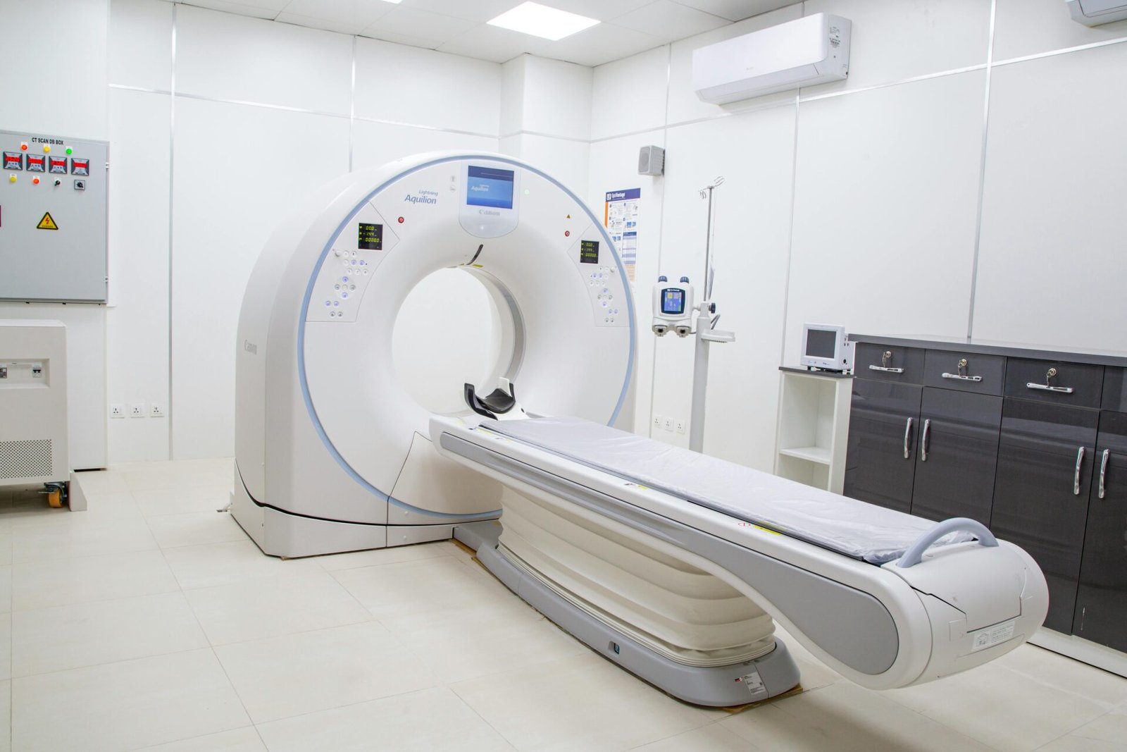
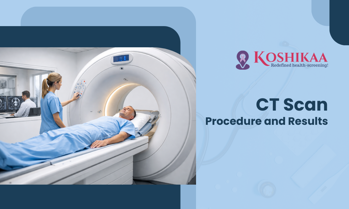
A PET-CT scan stands for positron emission tomography state of the art is a medical test that combines two scans:
Together, they give a complete view of both the structure and activity of your organs. It’s commonly used to detect cancer, check heart or brain problems, or find infections. You get a small tracer injection, lie in a scanner for 20–60 minutes, and the test is done. It’s safe with minimal radiation.
At Koshikaa, we provide different types of PET CT scans to suit your medical needs:
This scan checks your entire body for cancer spread or other diseases in one go.
It helps doctors evaluate brain function, tumours, epilepsy, or dementia effectively.
This scan assesses blood flow and detects any heart-related problems.
Used mainly for cancer detection, staging, and checking how well treatment is working.
It looks for cancer spread to bones or any bone disorders.
This common scan uses FDG tracer to find active cancer cells in the body.
It is done for detecting neuroendocrine tumours.
This scan is used to detect and stage prostate cancer accurately.
Doctors may advise a PET CT scan to:
✔️ Detect cancer and see if it has spread
✔️ Check how well cancer treatment is working
✔️ Diagnose heart problems like reduced blood flow
✔️ Identify brain conditions such as Alzheimer’s


The average cost of a PET CT scan in Bangalore ranges from ₹15,000 to ₹30,000, depending on:
Type of scan (whole body, brain, cardiac)
Radiotracer used
Hospital or diagnostic centre location
At Koshikaa Diagnostics, we provide affordable PET CT scans with clear pricing and no hidden charges.
Bone PET-CT (Positron Emission Tomography – Computed Tomography) using NaF-18 is a diagnostic imaging technique used to assess bone health and detect abnormalities such as fractures, infections, tumors, and metastases. NaF-18 is a radioactive tracer that binds to bone mineral, allowing PET-CT scans to highlight areas of increased bone activity or metabolism. This imaging modality is particularly useful in oncology for staging and monitoring skeletal metastases and in orthopedics for evaluating bone diseases and injuries.
Regional PET-CT is a scan that focuses on one part of the body instead of the whole body. It combines PET and CT to show both how that area looks and how it works.
PET cardiac viability imaging is a scan used to check if the heart muscle is still healthy and active in people with heart problems like blocked arteries.
FDG PET is a scan that shows how active your cells are. A small amount of radioactive sugar (FDG) is injected into your blood. Active cells, like cancer cells, absorb more FDG and show up brighter in the scan.
PSMA PET/CT scan is a special test used mainly for prostate cancer. A small amount of a radioactive tracer is injected, which sticks to prostate cancer cells. The PET scan shows where the cancer is active, and the CT scan shows the exact location inside the body.
DOTANOC PET/CT scan is a special test used to find and monitor neuroendocrine tumors (NETs). A small amount of a tracer called DOTANOC is injected, which sticks to NET cells. The PET scan shows where the tumor is active, and the CT scan shows its exact location in the body.
FAPI is a special tracer used in cancer scans. It targets a protein called FAP, which is found in support cells around tumors. These cells are often active in many types of cancer.
Utilizing PET CT Scan in Bangalore enables effective visualization of various structures and conditions within the body,


Booking an Affordable PET CT Scan at Koshikaa Diagnostic scan center bangalore is easy and takes less than a minute. Just follow these 4 simple steps:
Enter your name, mobile number, and email. It helps us contact you quickly and confirm your appointment.

Our team will call you to confirm your appointment and guide you on any instructions for the scan.

Once confirmed, you’ll receive the appointment details, center address, and reporting time directly to your phone or inbox.

Visit the center at your scheduled time. Our expert team will ensure a smooth, comfortable experience.
✔️ Fasting: Do not eat for at least 6 hours before the scan.
✔️ Medications: Inform your doctor about any ongoing medicines.
✔️ Clothing: Wear comfortable clothes without metal zips or buttons.
SCAN DURATION: The actual PET scan procedure typically takes about 30 minutes, but the entire process may last up to two – three hours due to the time required for the radiotracer to be absorbed by the body
TO-BE-MOMMYS and BREAST FEEDING MOMMYS!! Please Inform your provider about the possibility of pregnancy or breastfeeding and follow the instructions advised by them
| Feature | PET CT Scan | MRI Scan | CT Scan |
|---|---|---|---|
| Shows | Organ function + structure | Soft tissue structure | Body structure (bones, organs) |
| Best For | Cancer, heart, brain metabolism | Brain, spine, muscles | Bones, lungs, abdomen |
| Radiation? | Yes (minimal) | No | Yes |
| Scan Time | 30–90 mins | 30–60 mins | 5–15 mins |
| Contrast Use | Sometimes | Sometimes | Sometimes |
I was thoroughly impressed with my lung PET CT scan here. The staff was friendly, the technician skilled, and results were promptly provided with clear explanations
Koshikaa for thyroid PET CT scan surpassed my expectations. The staff was professional and caring, the technician thorough, and the facility clean and comfortable. Highly recommended.
My Head and Neck PET CT scan experience here was seamless. The knowledgeable technician answered all my questions, and results were promptly delivered with follow up to ensure understanding.


Get advanced PET CT scan designed for early disease detection, early cancer detection, staging, and treatment monitoring. Koshikaa delivers accurate results.
Visit our centre for professional PET-CT services in a calm, patient-friendly setting focused on comfort, precision, and quality care.
A PET-CT scan is used to combine two images a functional image from the PET (Positron Emission Tomography) and a structural image from the CT (Computed Tomography) to provide a comprehensive view of the body.
Doctors suggest a PET-CT Scan to:
Identify Infections/Inflammation: Locate areas of infection or unexplained inflammation.
A PET-CT scan works by performing two scans in one machine:
The scan detects abnormal metabolic activity at a cellular level, which can manifest as:
No. While widely known and primarily used in oncology for cancer detection, staging, and monitoring, PET-CT scans are also vital for diagnosing:
A PET-CT scan should only be taken when specifically ordered by a doctor, typically for patients who:
The cost of a PET-CT scan in Bangalore varies based on the type of scan, the tracer used (FDG, PSMA, etc.), and the facility.
General Price Range in Bangalore: The average cost typically ranges from ₹15,000 to ₹30,000.
The entire process, from injection to completion, can take 2 to 3 hours.
It is not a question of which is “better,” but rather which is more appropriate for the clinical need:
The two technologies are often complementary. For cancer, the PET-CT scan is usually preferred for staging and tracking treatment response because it can detect cellular changes earlier than structural-only scans.
This is the #1 concern, especially regarding fasting, diet, and exercise (all crucial for FDG tracer accuracy).
Explains the importance of low blood sugar to ensure the tracer (FDG) goes to the target cells, not muscle or fat.
Diabetes significantly affects the tracer’s accuracy. This needs a specific, clear answer.
Addresses common safety concerns about exposure to others (especially pregnant women/children).
Clarifies medication rules, which can be confusing (especially steroids or diabetic drugs).
A common diagnostic question is to clarify what the structural (CT) and functional (PET) parts detect.
PET CT scans are generally safe and low-risk, but like all medical tests, they have some potential side effects:
You may need to avoid or delay a PET CT scan in certain situations unless it’s medically necessary:
In all cases, consult the Koshikaa physician before proceeding.
At Koshikaa Screening in Bangalore, our PET CT Scan Pricing starts approximately:
These prices vary by scan type and radiotracer used. You can call and get PET Scan Costing with Koshikaa Diagnostics directly on the following number: +91 7996666104
No, the PET scan itself is not painful. The only minor discomfort you might feel is:
The actual scanning is painless and non-invasive.
It’s not that one is “better,” they serve different purposes:
PET-CT Scan: Shows metabolic activity and function of tissues, especially helpful for cancer detection, staging, or monitoring treatment response.
MRI Scan: Provides detailed anatomy and soft tissue contrast (e.g., brain, spine, joints) without radiation.
So, for cancer or metabolic diseases, PET CT is more useful for soft tissue anatomy, and MRI is preferred. Often, both are complementary.
You should wear comfortable, loose clothing without metal zips, buttons, belts, or accessories. Metal can interfere with imaging quality.
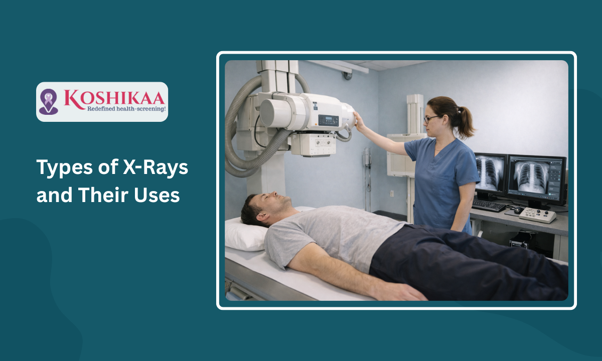
Bone X-rays are essential for diagnosing fractures and assessing bone alignment, aiding doctors in developing treatment plans and monitoring healing progress. They provide detailed images of the skeletal system, helping identify abnormalities or injuries that may not be visible through other imaging techniques