Essential Insights for Your Ultrasound: What You Must Know.

ultrasound scan in bangalore

Table of Contents

Today ultrasound scans serve as a fundamental foundation of medical practice by delivering safe yet effective means to diagnose and monitor different health conditions. The use of sound waves to produce body images through non-invasive technology grants doctors professional medical knowledge.  The article provides a complete overview about everything you need to learn regarding ultrasounds for both new and experienced readers.  

What is an Ultrasound Scan?

The diagnostic imaging process produces internal body visuals through high-frequency sound waves to reveal body structures. Ultrasound scanning avoids the use of radiation thus providing pregnancy-friendly and protected observation opportunities to sensitive patient groups. The medical field utilizes this procedure in multiple specialities including obstetrics and cardiology as well as other medical fields.  

Modern ultrasound technology improvements allow doctors to combine diagnostic instruments with ultrasound examinations for analyzing patient health extensively.  

How Does an Ultrasound Work?  

full body checkup in bangalore

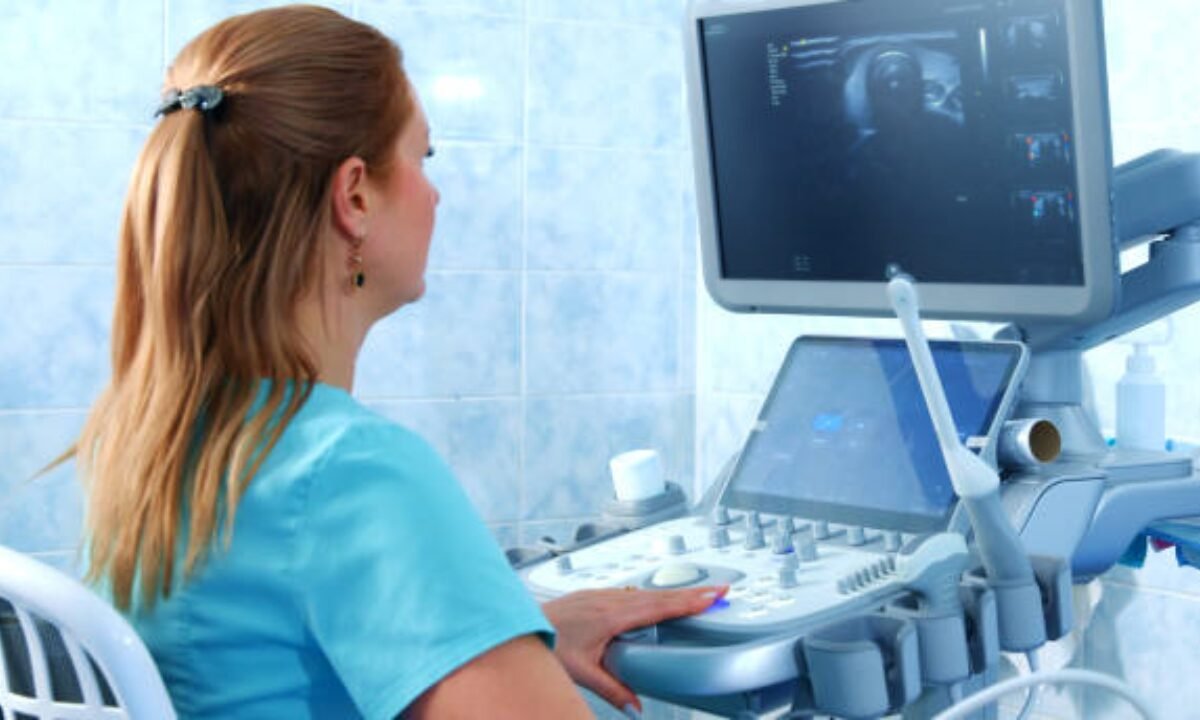
The operation of ultrasound scans depends on sound waves that interact based on tissue densities since tissues with different density levels reflect sound waves in different ways.  The sound waves generated from the transducer enable the creation of images that appear on a screen.  

Before starting the scan the healthcare professional applies gel as a conductive agent to the skin then employs a handheld transducer to scan the chosen area. The equipment allows evaluation of both soft tissues and visuals of organ function along with arterial and venous blood flow monitoring.  

When Do You Need an Ultrasound Scan?

Medical professionals use ultrasound scans as a diagnostic tool across different clinical situations particularly:  

Pregnancy scans serve as one of the main applications when using ultrasounds for pregnancy monitoring. The test provides continuous health assessments that protect the mother and her developing baby throughout the entire pregnancy period.  

Abdominal Issues Existence can be assessed through ultrasound testing as it detects gallstones and liver conditions and evaluates other organ health.  

Ultrasounds can serve to inspect both muscles together with ligaments and tendons. Medical practitioners in sports medicine utilize ultrasounds to detect soft tissue injuries in patients.  

Doctors use echocardiograms which function like ultrasound devices to measure heart conditions together with their mechanical functioning.  

Benefits of an Ultrasound Scan 

1. Non-invasive and Pain-free  

The diagnostic method of ultrasound scanning operates without any harm because it is completely non-invasive to patients and produces no physical discomfort. A transducer along with gel application provides everything that the procedure requires.  

2. No Radiation Exposure  

Because ultrasound technology uses sound waves instead of radiation it delivers complete safety for every part of the population including mothers with children and young individuals.  

 3. Cost-effective  

Medical professionals choose ultrasound as their primary diagnostic tool because it provides better affordability compared to MRI or CT scan imaging particularly during initial health evaluations.  

4. Real-time Imaging

The moving visual information generated by ultrasound scans proves exceptional for guiding procedures including biopsy procedures and blood flow visualization purposes.  

Limitations of Ultrasound Scans  

  • The usefulness of ultrasound scans cannot be overstated but patients should know that these tests have specific boundaries of functionality.  
  • Ultrasound methods cannot always identify essential small details which healthcare providers need MRI or CT scans to reveal.  
  • The travel of ultrasound waves is restricted in materials that have dense tissue structure and within air spaces. The brain and lungs cannot be adequately visualized through ultrasound procedures because of these constraints.  

Preparing for an Ultrasound Scan

The process of ultrasound scan preparation remains basic in the majority of scenarios. The needed procedures for testing may vary based on the specific part under examination. Here’s a quick guide:  

Abdominal Ultrasound

A full fast accompanied by fluid restriction is necessary when getting an abdominal ultrasound scan during the several hours before testing begins. The diagnostic results of some abdominal scans might need the patient to maintain a full bladder.  

Pelvic Ultrasound

Doctors request women to consume water before pelvic scanning because it helps their bladder reach a full state. The visualization benefits from a complete bladder.  

Other Types of Ultrasounds

Most diagnostic scanning procedures require no special preparation in advance. Choose outfits that are both comfortable and not restrictive so that scanner access to the scanning area remains uncomplicated.  

Visitors to any health screening centre in Bangalore or other facilities will receive pre-exam instructions from healthcare providers.  

Ultrasounds Beyond Diagnosis

Sonography serves two purposes: doctors perform this imaging technique during medical condition diagnosis and routine preventive examinations. The routine use of medical imaging tools allows healthcare providers to detect potential dangers at early stages so they become prescriptions for survival.  

In Cancer Detection 

Ultrasonography functions as a vital element for preliminary diagnostic imaging even though it does not identify cancer on its own. The majority of individuals need scans for initial diagnosis to determine appropriate advanced medical procedures. Consult your healthcare provider before getting a cancer detection test in Bangalore because they will determine if an ultrasound scan is correct as your starting point. 

Ultrasound in Comprehensive Full Body Checkup in Bangalore

A complete body examination serves as the optimal method for people to gain control over their health condition. Through early detection of potential health issues, symptoms may not progress into severe problems.  

During a full-body health checkup patients receive ultrasound evaluations that focus on liver, renal and cardiac organ assessment. These diagnostic imaging procedures act as effective tools to detect unspotted abnormalities within the body.  Your selection of a full body checkup in Bangalore should include evaluation of what ultrasound tests are included in the offering.  

The Selection of a Proper Health Screening Facility Requires Attention

The selection of medical establishments for ultrasound procedures proves essential. Health screening centres that are trusted both in Bangalore and other areas maintain advanced equipment and qualified technicians who produce accurate results.  

The selection of a diagnosis center should be based on their capability to provide trustworthy results along with established standards for medical care. Reading evaluations from patients and obtaining your doctor’s reference recommendations creates a great way to guarantee you will get a positive experience.  

Frequently Asked Questions About Ultrasounds  

1. Are Ultrasound Scans Painful?

All ultrasounds occur without any pain because they are completely painless tests.  

2. How Long Does the Procedure Take?  

Most medical ultrasound examinations need a duration between 15 to 30 minutes yet specific complicated testing may require additional time.  

3. Who Shouldn’t Get an Ultrasound?

All individuals possess suitable factors for ultrasound scanning except those without safety risks.  

Final Thoughts  

The medical diagnostic technique known as ultrasound scanning provides basic yet strong capabilities for healthcare assessment and detection of different medical conditions. The process behind ultrasounds assists both those getting complete body checkups and those specifically pursuing cancer detection tests in Bangalore.  

As a trusted health screening centre, Koshikaa provides broad diagnostic testing which includes ultrasound scan in Bangalore to assist in proactive health care management.

MRI of the uterus is primarily used for diagnosing and evaluating conditions such as uterine fibroids, adenomyosis, and malignancies. It is particularly useful in the planning of surgical procedures and assessing the extent of gynecological diseases. This imaging test is valuable for its ability to deliver detailed cross-sectional images of the uterus and surrounding structures.
An MRI of the spleen can help diagnose various disorders affecting the spleen including splenomegaly (enlarged spleen), tumors, and other abnormalities. It provides clear, detailed images that are crucial for assessing the health of the spleen and planning potential surgical interventions.
Prostate MRI is a non-invasive imaging technique that provides detailed pictures of the prostate gland and surrounding tissues. It helps in the detection, staging, and monitoring of prostate cancer. Prostate MRIs are increasingly used to guide biopsy procedures and assist in the planning of treatment strategies for prostate health.
An MRI of the pancreas is used to identify issues such as pancreatitis, pancreatic cancer, and other pancreatic disorders. It offers high-resolution images that help in detecting small lesions and assessing the pancreatic ducts. This imaging technique is valuable for guiding biopsies and planning surgeries involving the pancreas.
MRI scans of the liver, kidney, and bile ducts provide detailed images that help diagnose various conditions, such as tumors, cysts, and blockages in the bile ducts. They are crucial for assessing organ damage, staging cancer, and planning liver or kidney-related surgeries. This type of MRI can also evaluate the health and function of these organs comprehensively.
Breast MRI is highly sensitive for detecting breast cancer, especially in individuals with dense breast tissue or a high risk of cancer. It is often used in conjunction with mammography to provide a more comprehensive evaluation. Breast MRIs can also monitor the response to breast cancer treatments and provide detailed pre-surgical mapping.
Brain and spine MRIs are critical tools for diagnosing and monitoring neurological conditions, including tumors, spinal cord injuries, and multiple sclerosis. These MRIs can reveal abnormalities in brain tissue and spinal cord structure that might not be visible with other imaging methods. They are essential for planning surgeries and assessing the effectiveness of treatments in neurology.
PET-CT scans of transplant organs like the liver, kidney, or pancreas are performed to assess metabolic activity, detect complications, and monitor post-transplant function. These scans play a vital role in the early detection of issues such as organ rejection, recurrence of disease, or infection, guiding timely interventions and optimizing transplant outcomes for patients.
PET-CT examinations of organs such as the liver, kidney, or prostate are crucial for assessing metabolic activity, detecting abnormalities, and monitoring disease progression. These scans help diagnose conditions such as liver metastases, renal cell carcinoma, or prostate cancer, guiding treatment decisions and monitoring response to therapy over time.
PET-CT scans of soft-tissue masses are utilized to assess metabolic activity and detect malignant tumors or other abnormalities. By combining PET imaging with CT imaging, these scans provide detailed information about tumor location, size, and metabolic characteristics, aiding in accurate diagnosis and treatment planning for soft-tissue cancers.
PET-CT scans are valuable for detecting cysts or fluid collections within the body, including cysts in the liver, kidneys, or other organs. These scans help characterize the nature and metabolic activity of cysts, guiding diagnosis and treatment planning for conditions such as cystic liver disease, renal cysts, or pancreatic cysts.
PET-CT scans of muscles are performed to assess metabolic activity and detect abnormalities such as inflammation, infection, or tumors. These scans provide valuable information about muscle function and pathology, aiding in the diagnosis and management of conditions like myositis, muscular dystrophy, or soft-tissue sarcomas.
PET-CT scans of the brain are utilized to evaluate brain metabolism and detect abnormalities such as tumors, epilepsy, or neurodegenerative diseases. By combining functional PET imaging with anatomical CT imaging, these scans offer comprehensive information about brain function and structure, guiding neurosurgical interventions and treatment decisions for neurological disorders
PET-CT scans of bones are commonly used to detect bone metastases in cancer patients, assess bone health in conditions like osteoporosis, and evaluate response to treatment. By combining PET and CT imaging, these scans provide detailed information about metabolic activity and anatomical changes in bone structures, aiding in accurate diagnosis and treatment planning for various bone-related conditions.
CT scans of the spine are conducted to assess the vertebrae, spinal cord, and surrounding soft tissues for conditions like herniated discs, spinal fractures, or spinal stenosis, aiding in the diagnosis and management of spinal disorders with detailed imaging essential for surgical planning and intervention.
CT scans of the pelvis are utilized to visualize the pelvic bones, reproductive organs, bladder, and surrounding structures for conditions such as fractures, tumors, or pelvic inflammatory disease, guiding treatment plans and surgical interventions with precise anatomical information.
CT scans of the neck are performed to evaluate structures such as the throat, thyroid gland, lymph nodes, and blood vessels for abnormalities like tumors, infections, or injuries, assisting in diagnosis and management of head and neck disorders with detailed imaging.
CT scans of the extremities are employed to assess bones, joints, and soft tissues in the arms or legs, aiding in the diagnosis of fractures, arthritis, or other musculoskeletal conditions, guiding orthopedic treatment and rehabilitation plans for optimal recovery.
CT scans of the chest are utilized to examine the lungs, heart, blood vessels, and surrounding structures for conditions such as lung nodules, pneumonia, or pulmonary embolism, facilitating accurate diagnosis and management of respiratory and cardiovascular diseases.
CT scans of the brain are conducted to assess brain structures, identify abnormalities like tumors, hemorrhages, or strokes, and aid in diagnosing neurological conditions, guiding neurosurgical interventions and treatment strategies with precise imaging of the brain’s anatomy.
CT scans of the abdomen are performed to visualize and evaluate the internal organs such as the liver, kidneys, pancreas, and intestines for conditions like tumors, infections, or injuries, providing detailed images essential for diagnosis and treatment planning.
Transplant organ ultrasound scans, including those of the liver, kidney, or pancreas, are performed to assess organ viability, detect complications, and monitor post-transplant function. These scans play a vital role in the early detection of issues such as organ rejection, vascular complications, or fluid collections, guiding timely interventions and optimizing transplant outcomes for patients.
Ultrasound examinations of organs such as the liver, kidney, or prostate are crucial for assessing organ structure, detecting abnormalities, and monitoring disease progression. These scans help diagnose conditions such as liver cirrhosis, kidney stones, or prostate enlargement, guiding treatment decisions and monitoring response to therapy over time.
Soft-tissue masses can be evaluated using ultrasound to determine their size, location, and characteristics. Ultrasound imaging helps differentiate between benign and malignant masses, guiding further diagnostic evaluation and treatment planning. By providing real-time imaging, ultrasound assists in the precise localization of soft-tissue masses for biopsy or surgical removal if needed.
Ultrasound scans are valuable for detecting cysts or fluid collections within the body, including cysts in the liver, kidneys, or other organs. These scans help characterize the nature and size of cysts, aiding in diagnosis and treatment planning for conditions such as cystic liver disease or renal cysts, and guiding interventions such as cyst drainage or sclerotherapy.
Musculoskeletal ultrasound scans are used to assess the health and integrity of muscles, identifying issues such as strains, tears, or muscle inflammation. These scans provide detailed images of muscle structures, allowing healthcare providers to diagnose muscle injuries accurately and develop personalized rehabilitation plans to promote recovery and restore function.
Ultrasound examinations of joints are essential for evaluating joint health, detecting abnormalities such as inflammation, fluid accumulation, or cartilage damage. By visualizing the joint structures in real-time, ultrasound helps diagnose conditions like arthritis, bursitis, or ligament injuries, guiding treatment decisions and monitoring disease progression over time.
Ultrasound scans of tendons are performed to assess for injuries, inflammation, or tears, aiding in the diagnosis and management of conditions such as tendonitis or tendon tears. These scans provide detailed images of tendon structures, helping healthcare providers determine the extent of damage and plan appropriate treatment strategies, including physical therapy or surgical intervention if necessary.
Pelvis X-rays play a crucial role in diagnosing injuries, diseases, and developmental conditions affecting the pelvic region. They provide detailed images of the hip bones, sacrum, and coccyx, aiding in the assessment of fractures, dislocations, or signs of arthritis. Pelvic X-rays are instrumental in guiding surgical interventions, monitoring healing progress, and evaluating conditions such as osteoporosis or tumors. By providing precise anatomical information, these imaging studies enable healthcare professionals to deliver personalized care and improve patient outcomes.
Teeth X-rays, known as dental radiographs, are indispensable tools for dentists to assess oral health. They aid in diagnosing dental decay, gum disease, and abnormalities in tooth structure or alignment. These images provide valuable insights into hidden dental issues, allowing for early intervention and prevention of complications. Additionally, dental X-rays are essential for planning treatments such as orthodontics, root canals, or dental implants, ensuring optimal outcomes and patient satisfaction.
Spine X-rays are pivotal in diagnosing spinal fractures, degenerative conditions, and spinal misalignments, providing critical information for treatment planning and monitoring spinal health. They offer detailed images of the vertebrae and surrounding structures, enabling healthcare professionals to identify issues such as scoliosis, disc herniation, or spinal stenosis
Skull X-rays are valuable for detecting fractures, assessing head trauma, and evaluating the alignment of facial bones, crucial in diagnosing conditions like fractures, tumors, or developmental abnormalities in the skull. They provide detailed views of the cranial structure, aiding clinicians in determining appropriate treatment strategies and ensuring patient safety.
For the heart, X-ray solutions can aid in the detection of various cardiac problems, ranging from mild to severe, including – Heart failure, Cardiac congestion and other heart-related conditions. In addition to cardiac assessments, X-ray solutions are also used for breast examinations to identify tumors or signs of breast cancer.

Bone X-rays are essential for diagnosing fractures and assessing bone alignment, aiding doctors in developing treatment plans and monitoring healing progress. They provide detailed images of the skeletal system, helping identify abnormalities or injuries that may not be visible through other imaging techniques

Abdominal X-rays are performed when patients experience symptoms such as significant nausea, ongoing vomiting, abdominal pain, flank or back pain, or abdominal swelling. These X-rays can help diagnose various conditions.
Search Bar

Frequently Booked Tests

Frequently Booked Checkups