The PET CT scan procedure is one of the most advanced diagnostic tools available today, helping doctors detect diseases at an early stage. From cancer to neurological disorders, it combines accuracy with detail.
Here, we will cover the meaning of a PET CT scan, how the procedure works, the cost involved, and a detailed step-by-step process that might surprise you.
PET CT scan
A PET CT scan stands for Positron Emission Tomography – Computed Tomography. It combines two powerful technologies in one test.
- A PET scan shows how your cells are functioning, highlighting abnormal activity.
- A CT scan captures detailed images of the anatomy, including bones, tissues, and organs.
This dual approach enables doctors to view both structure and function simultaneously, providing a comprehensive picture.
It is widely used in detecting cancer, heart disease, neurological conditions, and infections. For patients who require a more extensive check-up, a PET CT scan of the whole body can reveal hidden issues that might otherwise go unnoticed.
PET CT Scan Procedure Explained

The PET CT scan procedure usually follows four main steps:
Step 1: Preparation
Patients are advised to fast for 4–6 hours, avoid strenuous exercise, and remove any metal objects, such as jewellery. Comfortable clothing is recommended.
Step 2: Injection of Radioactive Tracer
A small dose of a tracer, usually FDG (a type of glucose), is injected into a vein. This tracer highlights active cells, especially cancer cells, since they consume more glucose. After the injection, patients wait 30–60 minutes for the tracer to circulate.
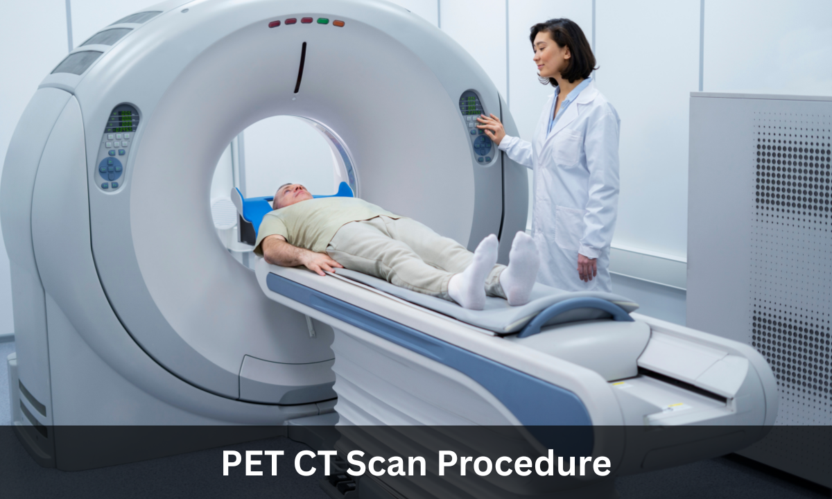
Step 3: The Scan
You will lie on a cushioned table that slides into the PET CT machine. The scanner then takes detailed PET CT scan images, combining both cell activity and anatomical structure. The process is painless and usually takes 20–30 minutes.
Step 4: After the Test
Once done, patients can drink water to flush out the tracer and return to their daily routine. Doctors analyze the PET CT scan report to guide treatment or further tests.
Difference between PET CT Scan Test, MRI, and CT Scans
| Feature | MRI | CT Scan | PET CT Scan Test |
|---|---|---|---|
| Focus | Soft tissues | Cross-sectional anatomy (bones, organs, tissues) | Both structure and function |
| What it Shows | Detailed images of soft tissues like the brain, muscles, and ligaments | Detailed structural anatomy | Cell activity and anatomical detail together |
| Best Use | Brain, spine, joints, soft tissue injuries | Trauma, fractures, chest, and abdominal imaging | Cancer staging, treatment planning, therapy monitoring |
| Unique Advantage | Excellent for soft tissue clarity | Quick and widely available | Detects disease at a cellular level while showing the body structure |
PET CT Scan Cost in India
The PET CT scan cost in India varies based on factors such as hospital type, city, and whether it’s for a specific body part or a whole-body scan.
Patients looking for a PET CT scan in Bangalore have access to advanced technology, offering high accuracy and faster results at trusted diagnostic centres.
Choosing the Best PET CT Scan Centre in Bangalore
Selecting the right centre ensures accurate results. The best PET CT scan centres in Bangalore use updated machines, follow strict safety standards, and employ skilled radiologists who can interpret complex reports with precision.
It is also essential to choose a health screening centre in Bangalore that offers comprehensive care, quick appointments, and reliable reporting.
At Koshikaa, patients receive trusted diagnostic services with a focus on accuracy and patient comfort, making it a preferred choice for many.
Final Thoughts
The PET CT scan procedure is safe, effective, and one of the best tools for early detection of diseases. It not only helps doctors diagnose conditions but also tracks treatment progress with precision.
If you are looking for a PET CT scan in Bangalore, consulting a trusted health screening centre in Bangalore like Koshikaa ensures timely and accurate results for better care and peace of mind.
FAQs on PET CT Scan
1. Is a PET-CT scan just for cancer?
No. While it is widely used in cancer detection and staging, PET CT scans also help diagnose heart problems, infections, and neurological disorders.
2. How do you prepare for a PET-CT scan?
You should fast for 4–6 hours, avoid physical activity, and wear loose-fitting clothes. Always inform your doctor if you have diabetes or are pregnant.
3. What is a PET CT scan of the whole body?
This scan evaluates the entire body in one test, useful for checking if cancer has spread or for identifying hidden conditions.
4. Are there any side effects of a PET CT scan?
Side effects are rare. The radioactive tracer is safe and usually leaves the body within 24 hours. Drinking water helps flush it out.
5. How soon will I get my PET CT scan report?
Reports are usually available within 24–48 hours, depending on the centre’s workload and the complexity of findings.