A growth scan is a reassuring pregnancy ultrasound. It shows your baby’s growth, weight, size, amniotic fluid, and placenta health. For many expectant moms, this scan gives peace of mind and a clear view of the baby’s development.
We often hear: “When is the best month for a growth scan?” or “How many growth scans do I need?” Every pregnancy is unique. This blog covers why the scan matters, what to expect, when to have it, and how it helps your safe pregnancy.
Key Points at a Glance
- What a growth scan measures and why it matters.
- The ideal month for scheduling a growth scan.
- How many growth scans may be needed in pregnancy?
- What happens during an abdominal and pelvic ultrasound scan?
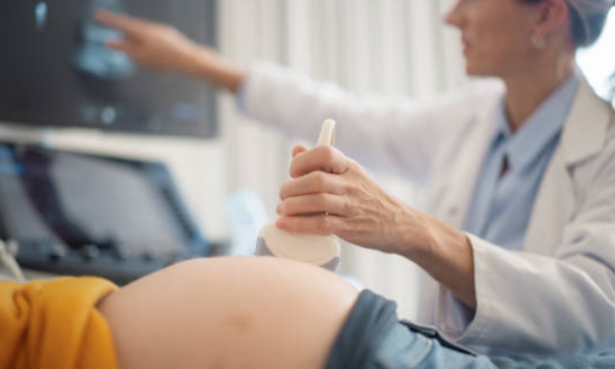
1. Why We Do a Growth Scan

We use a growth scan ultrasound to check your baby’s physical development, amniotic fluid levels, and the placenta’s condition. It helps us detect any concerns early, like growth restriction or macrosomia, so that we can manage your pregnancy proactively.
2. When Should You Have It?
We’re often asked, “Which month is best for a growth scan?” We usually do it in the late second or early third trimester—around 28 to 32 weeks. This window allows us to measure fetal growth accurately.
3. How Often Is It Recommended?
Wondering how many growth scans during pregnancy you will have? In uncomplicated pregnancies, usually just twice. However:
- If there are concerns (e.g., high blood pressure, low movement), we may repeat scans every 4–6 weeks.
- In conditions like gestational diabetes or previous growth issues, scans may be more frequent.
4. What Happens During the Scan?
During the ultrasound scan of the abdomen, gel is applied, and the transducer moves across your belly. A pelvic USG scan may be added if further detail is needed.
| Parameter | What It Tells Us |
|---|---|
| Biparietal Diameter (BPD), Head Circumference (HC) | Head size and development |
| Abdominal Circumference (AC) | Nutrient absorption, possible growth restriction |
| Femur Length (FL) | Length of long bones, body proportion |
| Amniotic Fluid Index (AFI) | Adequacy of fluid for the baby’s comfort and movement |
| Placental Position & Health | Ensures oxygen and nutrient supply |
5. Benefits of the Growth Scan

We want you to understand just how valuable this scan is:
- Peace of mind: Affirm your baby’s healthy progress.
- Early detection: Spot potential issues like inadequate growth or excess fluid.
- Informed planning: Helps us prepare in case extra care is needed at delivery.
- Reassurance: Each normal scan reassures both parents and care providers.
6. Example Scenario
Imagine you are at 30 weeks, and we notice your baby’s abdominal circumference is smaller than expected. We schedule a follow-up growth scan in 2 weeks. This closely monitored approach helps us ensure your baby’s steady growth and safety without unnecessary alarm.
7. Practical Tips
- Stay hydrated before your scan: good amniotic fluid enhances image clarity.
- Wear comfortable clothing: two-piece outfits are easiest for abdominal access.
- Bring your prior scan reports: helpful for comparison.
- Ask questions: We encourage you to understand each measurement and its implications.
- If you are seeking an ultrasound scan in Bangalore, choose a certified facility with experienced sonographers to ensure accuracy.
8. FAQs
Q1: Does the growth scan use radiation?
No, it doesn’t. A growth scan ultrasound uses safe, non-ionizing sound waves. These waves create images of your baby without exposing you to harmful radiation. That’s why it’s considered one of the safest ways to monitor your pregnancy, even if you need multiple scans.
Q2: Does a growth scan guarantee a complication-free birth?
Not exactly. A growth scan helps us detect problems early and provides reassurance about your baby’s progress. However, it doesn’t predict every possible complication. Instead, it works best when combined with regular check-ups, blood tests, and your overall clinical history.
Q3: What if fluid levels are high or low?
Fluid levels matter a lot. If they are too low, we recommend increasing hydration, monitoring closely, or further tests. If they are too high, it might signal gestational diabetes or other conditions. In both cases, regular follow-up scans help us keep your baby safe.
Q4: Can the scan predict the baby’s weight accurately?
Yes, but with some limitations. A growth scan gives us an estimated foetal weight by combining head, abdomen, and femur measurements. While these estimates can be off by about 10–15%, they are excellent for tracking trends in your baby’s growth. We use them along with other health indicators before making decisions.
9. Finding the Right Facility
Whether in Bangalore or elsewhere, ensure your ultrasound scan abdomen or pelvic ultrasound scan is done with high-resolution machines and certified professionals. In Bangalore, many centres of excellence offer advanced imaging, but always check for accreditation, equipment quality, and patient reviews.
Closing Thoughts
We have walked through what a growth scan during pregnancy entails, its purpose, timing, frequency, procedure, benefits, and practical pointers. Remember, this scan isn’t just a measurement; it’s peace of mind in real time.
If you are looking for an expert-guided ultrasound scan in Bangalore or simply curious about how Koshikaa supports expecting moms through each stage, know that your journey deserves personal care and professional guidance at every step.