MRI Scan Centre Near Me: 7 Smart Tips to Find the Best One Fast (Without Overpaying!)

Blood test

Table of Contents

Introduction:

When one seeks an “MRI scan centre near me”, it may be overwhelming, especially when trying to solve health concerns, and time is of the essence. With all these hundreds of diagnostic centers sprouting up in each city, how can you ever trust any of them? And more to the point, how do you find a good one that is near and affordable too?

In this guide, we will walk you step by step through how to get the best MRI scan centers close to your location: faster and smarter. We will answer your most burning questions, share tips you need to know and provide you with expert advice so that you can save your time, pocket, and nerves.

What Is an MRI Scan & Why Choosing the Right Centre Matters

MRI scan in Bangalore

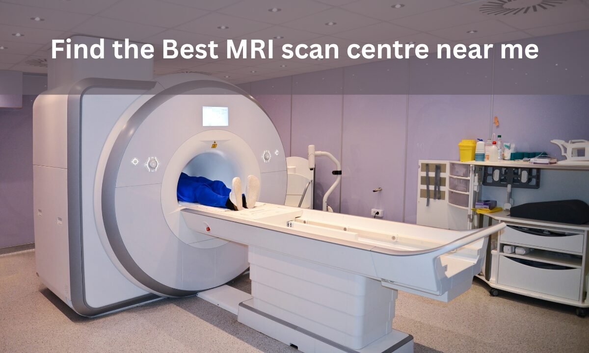
MRI scan, i.e., Magnetic Resonance Imaging scan, is a very vital diagnostic test that makes detailed images of your body through the use of strong magnets and radio waves. It’s non-invasive and radiation-free, but it works only if it is done by a reputed diagnostic centre using cutting-edge machines and skilled operators.

A quality scan can detect a condition early and improve treatment. A not-so-good scan, however, could result in a misdiagnosis or delay. That is why being able to locate a reputed “MRI scan centre near me” is more important than you can imagine.

Key Factors to Consider When Searching “MRI Scan Centre Near Me”

Here are 7 essential things you should check before choosing a nearby MRI scan centre:

Accreditation & Certifications

Look for centres accredited by NABL (National Accreditation Board for Testing and Calibration Laboratories) or NABH. These certifications ensure high standards of equipment, hygiene, and reporting.

Proximity & Accessibility

Choose a centre within 3-5 km from your location if possible. This reduces commute stress, especially for elderly or injured patients.

Machine Technology (1.5T vs 3T)

  • 1.5 Tesla: Ideal for most standard scans, affordable.
  • 3 Tesla: Offers higher clarity, used for detailed neurological, musculoskeletal, or vascular studies.

Availability of Radiologists

An efficient centre has in-house experienced radiologists who can interpret the scans quickly, meaning that you do not wait too long for the report.

Cost Transparency

The cost may vary from ₹3000 to ₹ 25000, depending on the scan type. Request a total price, including the additional price incurred, by contrast, reports, or urgent delivery.

Report Turnaround Time

Standard turnaround is 12–24 hours. Some give urgent reports in 4-6 hours for serious cases.

Patient Reviews & Ratings

Always ensure that you confirm Google reviews, Practo, Justdial, or a local listing platform to hear from genuine patients.

Quick Stats on MRI Usage in India:

  • Over 1.7 million MRI scans are done annually in India.
  • Bangalore, Mumbai, and Delhi have the highest number of MRI centres.
  • More than 60% of patients now book your scan online using aggregator platforms.

FAQs: Answers You Need Before Booking an MRI

How do I find the best MRI scan centre near me?

Compare centers on Google Maps, and online medical directories such as Practo or Koshikaa based on proximity, price and reviews.

Can I walk in, or do I need an appointment?

The majority of the MRI centers will need a doctor’s prescription and an appointment before any scanning, especially with the use of contrast MRI or a whole body scan.

How long does an MRI scan take?

According to the body part that needs to be scanned, an MRI scan can last between 20-60 minutes. Whole-body or contrast-enhanced scans are likely to be more time-consuming.

Is it painful or uncomfortable?

MRI scans are painless. One has to do this while lying still in an enclosed tunnel, which can be uncomfortable for claustrophobic individuals. Open MRI machines are available in some centers.

Are home pickup and drop-off services available?

Some of the advanced centers provide ambulance/cab tie-ups for the patients, especially the aged and immobile. Always ask.

Real-Life Scenario:

It’s Rajesh, a 52-year-old businessman in Bengaluru. He googled “MRI scan centre near me” and received a first quote of ₹9,000 from a hospital. After reviewing the options online and comparing centers using Koshikaa, he located a certified diagnostic lab that was only 2 km away from his home, providing the exam at Rs 4,800, including same-day report generation. He saved money, he saved time, and he saved stress all in the process of doing a 10-minute online comparison.

How to Quickly Find the Best MRI Scan Centre Near You (Action Plan)

  • Step 1: Use your GPS-enabled phone to search “MRI scan centre near me”
  • Step 2: Shortlist top 3 based on ratings (4.2+), distance (within 5 km), and availability
  • Step 3: Call to check machine type (1.5T or 3T), cost, and report time
  • Step 4: Ask about radiologist qualifications and whether they offer urgent reports
  • Step 5: Book an appointment online or via phone to avoid long waiting times

Pro Tip:

If you’re based in Bangalore and want an updated list of certified centers, you can check platforms like Koshikaa, where centers are verified, and you can filter by cost, distance, and reviews. For a trusted and budget-friendly MRI scan centre near me, visit this detailed guide on Koshikaa: MRI Scan Near Me.

Conclusion:

Leave it to chance when it comes to your health. Do you think it’s not possible to find a good and affordable MRI scan centre near you? Think again! With the help of online tools and platforms, this is not just possible, but easier than ever. Accreditation, technology, and patient reviews should be prioritized before the booking.

Take some time to research and compare. You may be grateful for your future health.

MRI of the uterus is primarily used for diagnosing and evaluating conditions such as uterine fibroids, adenomyosis, and malignancies. It is particularly useful in the planning of surgical procedures and assessing the extent of gynecological diseases. This imaging test is valuable for its ability to deliver detailed cross-sectional images of the uterus and surrounding structures.
An MRI of the spleen can help diagnose various disorders affecting the spleen including splenomegaly (enlarged spleen), tumors, and other abnormalities. It provides clear, detailed images that are crucial for assessing the health of the spleen and planning potential surgical interventions.
Prostate MRI is a non-invasive imaging technique that provides detailed pictures of the prostate gland and surrounding tissues. It helps in the detection, staging, and monitoring of prostate cancer. Prostate MRIs are increasingly used to guide biopsy procedures and assist in the planning of treatment strategies for prostate health.
An MRI of the pancreas is used to identify issues such as pancreatitis, pancreatic cancer, and other pancreatic disorders. It offers high-resolution images that help in detecting small lesions and assessing the pancreatic ducts. This imaging technique is valuable for guiding biopsies and planning surgeries involving the pancreas.
MRI scans of the liver, kidney, and bile ducts provide detailed images that help diagnose various conditions, such as tumors, cysts, and blockages in the bile ducts. They are crucial for assessing organ damage, staging cancer, and planning liver or kidney-related surgeries. This type of MRI can also evaluate the health and function of these organs comprehensively.
Breast MRI is highly sensitive for detecting breast cancer, especially in individuals with dense breast tissue or a high risk of cancer. It is often used in conjunction with mammography to provide a more comprehensive evaluation. Breast MRIs can also monitor the response to breast cancer treatments and provide detailed pre-surgical mapping.
Brain and spine MRIs are critical tools for diagnosing and monitoring neurological conditions, including tumors, spinal cord injuries, and multiple sclerosis. These MRIs can reveal abnormalities in brain tissue and spinal cord structure that might not be visible with other imaging methods. They are essential for planning surgeries and assessing the effectiveness of treatments in neurology.
PET-CT scans of transplant organs like the liver, kidney, or pancreas are performed to assess metabolic activity, detect complications, and monitor post-transplant function. These scans play a vital role in the early detection of issues such as organ rejection, recurrence of disease, or infection, guiding timely interventions and optimizing transplant outcomes for patients.
PET-CT examinations of organs such as the liver, kidney, or prostate are crucial for assessing metabolic activity, detecting abnormalities, and monitoring disease progression. These scans help diagnose conditions such as liver metastases, renal cell carcinoma, or prostate cancer, guiding treatment decisions and monitoring response to therapy over time.
PET-CT scans of soft-tissue masses are utilized to assess metabolic activity and detect malignant tumors or other abnormalities. By combining PET imaging with CT imaging, these scans provide detailed information about tumor location, size, and metabolic characteristics, aiding in accurate diagnosis and treatment planning for soft-tissue cancers.
PET-CT scans are valuable for detecting cysts or fluid collections within the body, including cysts in the liver, kidneys, or other organs. These scans help characterize the nature and metabolic activity of cysts, guiding diagnosis and treatment planning for conditions such as cystic liver disease, renal cysts, or pancreatic cysts.
PET-CT scans of muscles are performed to assess metabolic activity and detect abnormalities such as inflammation, infection, or tumors. These scans provide valuable information about muscle function and pathology, aiding in the diagnosis and management of conditions like myositis, muscular dystrophy, or soft-tissue sarcomas.
PET-CT scans of the brain are utilized to evaluate brain metabolism and detect abnormalities such as tumors, epilepsy, or neurodegenerative diseases. By combining functional PET imaging with anatomical CT imaging, these scans offer comprehensive information about brain function and structure, guiding neurosurgical interventions and treatment decisions for neurological disorders
PET-CT scans of bones are commonly used to detect bone metastases in cancer patients, assess bone health in conditions like osteoporosis, and evaluate response to treatment. By combining PET and CT imaging, these scans provide detailed information about metabolic activity and anatomical changes in bone structures, aiding in accurate diagnosis and treatment planning for various bone-related conditions.
CT scans of the spine are conducted to assess the vertebrae, spinal cord, and surrounding soft tissues for conditions like herniated discs, spinal fractures, or spinal stenosis, aiding in the diagnosis and management of spinal disorders with detailed imaging essential for surgical planning and intervention.
CT scans of the pelvis are utilized to visualize the pelvic bones, reproductive organs, bladder, and surrounding structures for conditions such as fractures, tumors, or pelvic inflammatory disease, guiding treatment plans and surgical interventions with precise anatomical information.
CT scans of the neck are performed to evaluate structures such as the throat, thyroid gland, lymph nodes, and blood vessels for abnormalities like tumors, infections, or injuries, assisting in diagnosis and management of head and neck disorders with detailed imaging.
CT scans of the extremities are employed to assess bones, joints, and soft tissues in the arms or legs, aiding in the diagnosis of fractures, arthritis, or other musculoskeletal conditions, guiding orthopedic treatment and rehabilitation plans for optimal recovery.
CT scans of the chest are utilized to examine the lungs, heart, blood vessels, and surrounding structures for conditions such as lung nodules, pneumonia, or pulmonary embolism, facilitating accurate diagnosis and management of respiratory and cardiovascular diseases.
CT scans of the brain are conducted to assess brain structures, identify abnormalities like tumors, hemorrhages, or strokes, and aid in diagnosing neurological conditions, guiding neurosurgical interventions and treatment strategies with precise imaging of the brain’s anatomy.
CT scans of the abdomen are performed to visualize and evaluate the internal organs such as the liver, kidneys, pancreas, and intestines for conditions like tumors, infections, or injuries, providing detailed images essential for diagnosis and treatment planning.
Transplant organ ultrasound scans, including those of the liver, kidney, or pancreas, are performed to assess organ viability, detect complications, and monitor post-transplant function. These scans play a vital role in the early detection of issues such as organ rejection, vascular complications, or fluid collections, guiding timely interventions and optimizing transplant outcomes for patients.
Ultrasound examinations of organs such as the liver, kidney, or prostate are crucial for assessing organ structure, detecting abnormalities, and monitoring disease progression. These scans help diagnose conditions such as liver cirrhosis, kidney stones, or prostate enlargement, guiding treatment decisions and monitoring response to therapy over time.
Soft-tissue masses can be evaluated using ultrasound to determine their size, location, and characteristics. Ultrasound imaging helps differentiate between benign and malignant masses, guiding further diagnostic evaluation and treatment planning. By providing real-time imaging, ultrasound assists in the precise localization of soft-tissue masses for biopsy or surgical removal if needed.
Ultrasound scans are valuable for detecting cysts or fluid collections within the body, including cysts in the liver, kidneys, or other organs. These scans help characterize the nature and size of cysts, aiding in diagnosis and treatment planning for conditions such as cystic liver disease or renal cysts, and guiding interventions such as cyst drainage or sclerotherapy.
Musculoskeletal ultrasound scans are used to assess the health and integrity of muscles, identifying issues such as strains, tears, or muscle inflammation. These scans provide detailed images of muscle structures, allowing healthcare providers to diagnose muscle injuries accurately and develop personalized rehabilitation plans to promote recovery and restore function.
Ultrasound examinations of joints are essential for evaluating joint health, detecting abnormalities such as inflammation, fluid accumulation, or cartilage damage. By visualizing the joint structures in real-time, ultrasound helps diagnose conditions like arthritis, bursitis, or ligament injuries, guiding treatment decisions and monitoring disease progression over time.
Ultrasound scans of tendons are performed to assess for injuries, inflammation, or tears, aiding in the diagnosis and management of conditions such as tendonitis or tendon tears. These scans provide detailed images of tendon structures, helping healthcare providers determine the extent of damage and plan appropriate treatment strategies, including physical therapy or surgical intervention if necessary.
Pelvis X-rays play a crucial role in diagnosing injuries, diseases, and developmental conditions affecting the pelvic region. They provide detailed images of the hip bones, sacrum, and coccyx, aiding in the assessment of fractures, dislocations, or signs of arthritis. Pelvic X-rays are instrumental in guiding surgical interventions, monitoring healing progress, and evaluating conditions such as osteoporosis or tumors. By providing precise anatomical information, these imaging studies enable healthcare professionals to deliver personalized care and improve patient outcomes.
Teeth X-rays, known as dental radiographs, are indispensable tools for dentists to assess oral health. They aid in diagnosing dental decay, gum disease, and abnormalities in tooth structure or alignment. These images provide valuable insights into hidden dental issues, allowing for early intervention and prevention of complications. Additionally, dental X-rays are essential for planning treatments such as orthodontics, root canals, or dental implants, ensuring optimal outcomes and patient satisfaction.
Spine X-rays are pivotal in diagnosing spinal fractures, degenerative conditions, and spinal misalignments, providing critical information for treatment planning and monitoring spinal health. They offer detailed images of the vertebrae and surrounding structures, enabling healthcare professionals to identify issues such as scoliosis, disc herniation, or spinal stenosis
Skull X-rays are valuable for detecting fractures, assessing head trauma, and evaluating the alignment of facial bones, crucial in diagnosing conditions like fractures, tumors, or developmental abnormalities in the skull. They provide detailed views of the cranial structure, aiding clinicians in determining appropriate treatment strategies and ensuring patient safety.
For the heart, X-ray solutions can aid in the detection of various cardiac problems, ranging from mild to severe, including – Heart failure, Cardiac congestion and other heart-related conditions. In addition to cardiac assessments, X-ray solutions are also used for breast examinations to identify tumors or signs of breast cancer.

Bone X-rays are essential for diagnosing fractures and assessing bone alignment, aiding doctors in developing treatment plans and monitoring healing progress. They provide detailed images of the skeletal system, helping identify abnormalities or injuries that may not be visible through other imaging techniques

Abdominal X-rays are performed when patients experience symptoms such as significant nausea, ongoing vomiting, abdominal pain, flank or back pain, or abdominal swelling. These X-rays can help diagnose various conditions.

Frequently Booked Tests

Frequently Booked Checkups