Medical technology continues to develop progressively because it delivers powerful diagnostic equipment that helps doctors detect diseases more effectively. PET scanning has become a vital technology among various modern diagnostic imaging tools that medical professionals frequently employ. This article explains PET scans from a basic level through a comprehensive understanding of their definition as well as listing their advantages and procedural details that drive patient and medical staff preferences.
This article presents crucial knowledge about PET scanning as well as detailed information on “PET CT scan in Bangalore” for patients requesting this procedure.
What is a PET Scan?
The specialized medical examination method of Positron Emission Tomography (PET) scan helps doctors locate and identify medical disorders in the body through its imaging capabilities. Bone structure is not the primary target of a PET scan since this examination method concentrates on cellular metabolic processes and activity rather than anatomical features. PET stands out as a diagnostic tool since it detects diseases before they result in noticeable structural changes.
Healthcare providers typically perform PET scans to detect patient treatment responses together with conditions including cancer and heart issues as well as brain disorders including Alzheimer’s disease. PET scans supply doctors with detailed molecular imaging which helps diagnose confusing medical conditions as one of their fundamental tools of disease diagnosis.
How Does PET Scan Work?
Knowledge about PET scanning operations eases the anxiety of people preparing to undergo the exam. During a PET scan, doctors introduce minor amounts of radiotracer that function as radioactive compounds. Medical personnel commonly deliver the tracer substance through a bloodstream injection to patients.
When the radiotracer travels through the body PET scanners utilizing their detector systems locate the tracer accumulation points. The radiotracer flows toward active cells specifically attaining cancerous cells because cancer cells need more energy than standard cells.
One remarkable aspect of PET scan functioning becomes visible in its ability to display both the affected area location and the metabolic basis of the detected anomaly.
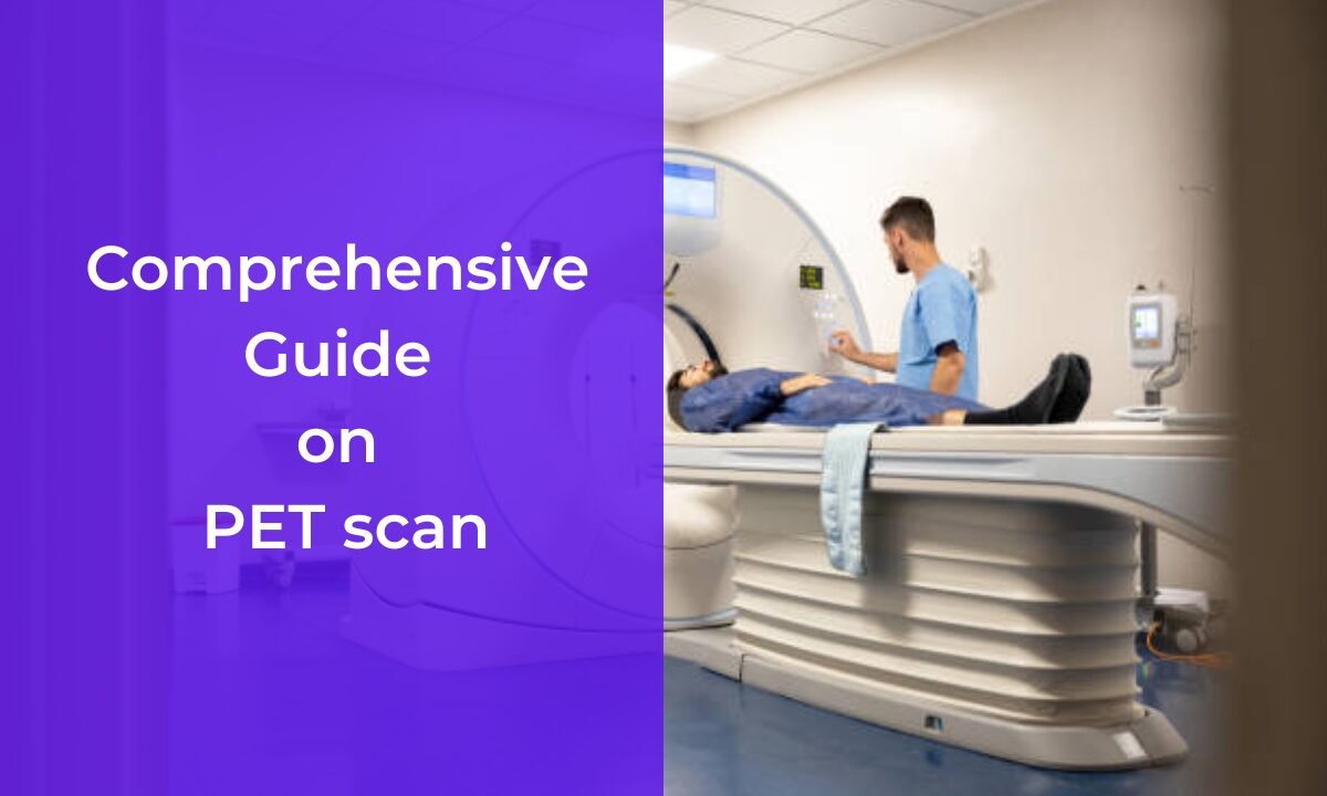
The PET Scan Process
Before receiving a PET scan you might inquire about what is happening during the “PET scan process”. Just because the process involves technical terminology it proves to be straightforward and not very discomforting to patients. To understand the PET scan procedure we need to analyze its simple components.
1. Pre-scan Preparation
The medical staff gives patients special instructions during the pre-scan period to enable precise image results. During the preparation phase, the team guiding the procedure requires you to remain without consuming any calories since empty stomach conditions improve test accuracy by reducing bloodstream sugar levels.
When doctors create eating restrictions for patients before their examination they might limit either high-carbohydrate drinks or foods before the test day.
2. Radiotracer Administration
After reaching the diagnostic center the medical staff administers a minor dose of the radiotracer to you. Clinical staff use a combination of intravenous (IV) injection with either liquid intake or gas inhalation based on the target region under evaluation. The quick procedure requires an injection which virtually causes no discomfort to patients. The injection procedure ends with a brief period of waiting from 30 to 60 minutes.
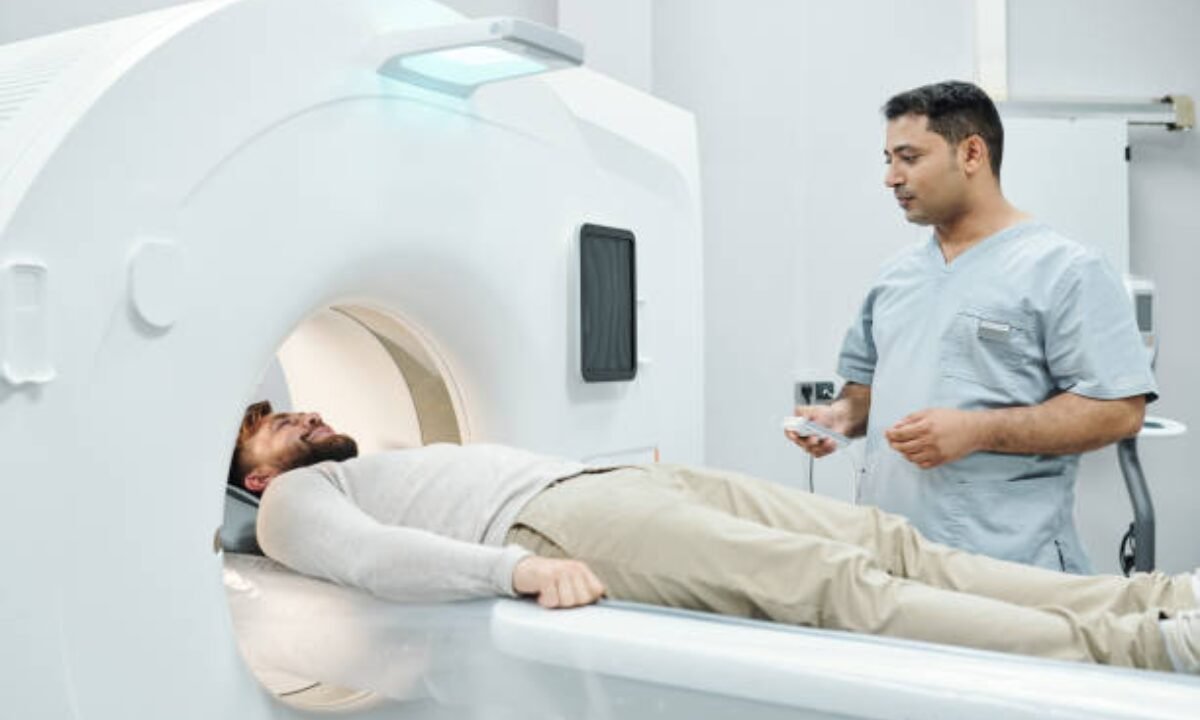
3. The Scanning Procedure
The examination process begins when you lie down on the scanning table while it moves toward the PET scanner which has a donut-shaped form. You need to stay motionless throughout the scan procedure to achieve clear images with proper precision. Patients undergoing a scan need between 15 to 60 minutes of total scanning time based on what region doctors assess.
The machine naturally produces soft clicking and humming noises that should be expected during operation. Operating inside the scanner will result in a fully painless experience.
4. Post-scan Guidance
The radiotracer will exit your body by passing through urine and stool after the scanning process is completed. Drinking fluids accelerates urine production for tracer elimination from the body. Pet scan data gets examined by radiologists together with doctors to determine the diagnosis before planning the next medical procedure.
Benefits of PET Scan
A PET scan offers advantages that conventional imaging modalities cannot provide. The modern healthcare world stands to gain significantly from this advanced diagnostic method because of the following features:
1. Early Detection of Diseases
The investigation via PET scan enables doctors to spot diseases before patients develop symptoms or structural abnormalities appear. Medical professionals detecting cancerous activity take place before the formation of tumors in cancer cases.
2. Monitoring Treatment Response
A PET scan provides real-time evaluation of treatment effectiveness to doctors who treat patients with cancer during their treatment. Treatment plans and medications can be adjusted when doctors assess the obtained test results.
3. Precision in Diagnosis and Staging
PET scans show metabolic processes to reveal the precise area, extent, and size of disease-related issues. The precise nature of this diagnostic agent makes it suitable for determining cancer staging along with heart condition assessment and brain disorder analysis.
4. Minimal Risk and Pain
Due to the very small volume of radiotracer injected risks associated with adverse effects become virtually nonexistent. A scanning operation causes no harm to patients through its completely painless non-invasive procedures.
5. Combination with CT Scans
PET scanners are commonly used alongside CT scanners to generate enhanced visualization of body structures. PET-CT scan technology merges metabolic PET results with CT structural data thus leading to better diagnostic precision.
What makes PET CT Scans so popular among Bangalore residents?
The diagnostic technique “pet CT scan in Bangalore” can be found by patients searching for diagnostic options in busy urban centers of India. PET CT scanning services are available at state-of-the-art hospitals and diagnostic facilities that operate in this city. The medical community of Bangalore connects high-level expertise to modern technology because they deliver smooth medical services to patients.
The standard of PET CT scanning in Bangalore remains competitive with major urban areas but comes at an accessible price for patients to seek advanced imaging. Patients seeking advanced medical imaging in need of oncology care and diagnostic expertise can find a beneficial setting in Bangalore because of its reasonable pricing structure and excellent care quality.
Precautions and Safety
The handling of radioactive tracers during PET scans produces an initially uncomfortable feeling but patients should understand these scans remain extremely safe. The small radiation amounts found in the radiotracer make the associated risks negligible. Doctors prescribe PET scans only when a correct medical cause exists or else they avoid these procedures.
Conclusion
In summary, a PET scan is more than just an imaging tool; it is a lifesaving resource that supports early diagnosis and guides treatments for critical medical conditions. Your understanding of pet scan basics including its definition, operational mechanism, and examination procedure will help you feel assured about the procedure.
The “pet CT scan in Bangalore” service provides patients with efficiency combined with affordable prices and modern technological capabilities. The exceptional advantages of advanced technology become apparent regardless of when patients need a PET scan for diagnosis or treatment monitoring.
The advanced imaging facilities at Koshikaa provide dependable PET scans in Bangalore service through advanced instruments and professional expertise for accurate diagnostic needs.