

Regular health check-ups perform an essential role in early problem detection because most people focus on sustaining good health in present-day society. High importance exists for early breast detection because it enables better outcomes of treatment. Women obtain different breast examination diagnostic tests for evaluation, but mammograms and ultrasounds are the most recommended choices. This article compares mammography vs ultrasound tests, which are crucial for breast health management through the identification of their unique characteristics.
What Is Mammography Screening?
The X-ray diagnostic procedure known as mammography helps healthcare providers detect abnormalities in breast tissues and detect different masses or irregular growths. Mammography Screening is the process of detecting early breast cancer signs using X-ray imaging techniques for the breast to generate visual results. Early signs of breast cancer become visible through mammograms at microscopic sizes before the cancer is detectable or could manifest noticeable symptoms.
The medical imaging technique known as mammography can operate within two separate domains.
1. Screening Mammograms: The screening method that doctors use on asymptomatic female patients to detect early cancerous breast cell changes.
2. Diagnostic Mammograms serve as advanced breast tests for women showing signs such as breast lumps or pain as well as unusual results from other breast screening tests.
The radiology technician uses a quick testing method, which remains simple to perform for both patients and professionals. During the X-ray process, your breast gets pressed between the two compression plates. The short compression period leads to slight annoyance. However, the information gained about breast health is very beneficial.
What Is an Ultrasound Scan for the Breast?
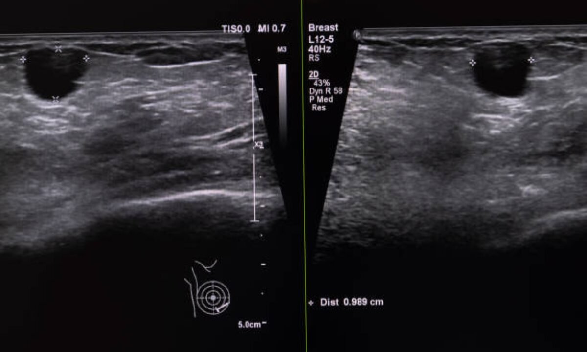
A breast tissue examination using sonography or an ultrasound scan represents another diagnostic procedure that medical professionals utilize. The method of imaging breast tissue differs between mammography and ultrasound because X-rays do not appear in ultrasound scans, where sound waves instead create real-time images.
The beneficial nature of an ultrasound scan arises from its ability to assess breast abnormalities detected by physical exams and mammograms.
Ultrasound provides the best evaluation tool for younger women who have dense breasts. The detection of abnormalities becomes less clear when dense breast tissue exists because mammograms have difficulty working in such cases, so ultrasound acts as a second screening method. The examination remains painless because a handheld transducer requires placement on the breast skin for imaging purposes.
Differences Between Mammography vs Ultrasound
Comfortable understanding between Mammography and Ultrasound remains crucial when patients need breast cancer screening. Every detection method possesses distinct features which make it suitable for particular diagnostic purposes. Let’s compare them in detail:
1. Nature of the Technology
Computers use low-dose X-rays to generate comprehensive breast images, known as mammography. The technology excels at detecting calcifications that represent potentially cancerous regions because of their tiny size.
The examination uses sound waves to display dynamic images of breasts. The imaging technique demonstrates value in differentiating fluid-filled cysts from solid breast masses because it does not include radiation exposure.
2. Diagnostic Accuracy
Detector systems in mammography excel at breast cancer stage detection as well as microcalcifications identification that ultrasounds rarely identify.
Images generated using ultrasound lack precision in detecting both very small tumors as well as microcalcifications. The testing method works together with mammography tests or functions as a supplemental measure when screening dense breasts.
3. Who Should Use It?
- Mammography Tests: Recommended for women above the age of 40 as part of routine breast cancer screenings. Doctors recommend earlier implementation of breast cancer screenings for females who have relatives who have experienced breast cancer.
- Ultrasound: The medical professional would recommend ultrasound inspections to adolescents or women with dense breasts because mammograms generate unclear results in these circumstances.
It is used as a dual test after an abnormality is found in a mammogram.
4. Time and Cost
- Mammography: Usually takes a bit longer and costs more as a result of the sophisticated technology and X-rays used. But with certainty in the health infrastructure now, affordable and quality check-ups through mammography tests are accessible since Bangalore has various cities.
- Ultrasound: Less time is needed, and it is usually affordable. It is available in most hospitals and diagnostic centers as a non-invasive and non-radiation procedure.
When to Choose Mammography or Ultrasound?
Having said that, which process is best for you, Mammography vs Ultrasound? The answer depends on your age, how dense your breasts are, your symptoms, and other risk factors.
For instance, regular mammography screening should be an essential part of every woman’s healthy yearly routine after her 40th birthday or at an earlier age if she has a family history of breast cancer. On the other hand, women under 40 and tissue with dense breasts may be helped by ultrasound scans.
In some cases, both tests may be suggested at the same time to give a better width picture of your breast health. No single test can assure 100% accuracy, but using multiple of them increases the chances of early and accurate detection of breast abnormality.
The Role of Breast Imaging in Cancer Screening
Early detection of breast cancer can also be facilitated with the help of breast imaging. Whether it is mammography or ultrasound, both are integral parts of a comprehensive cancer screening program. In a city like Bangalore, where there is a growing focus on healthcare and infrastructure, there are many hospitals and screening centers in Bangalore that offer advanced cancer screening Bangalore services. These programs are designed to make sure women have access to timely screenings, as well as the expertise that each one deserves. Also, there are many women’s extended health clinics that have advanced mammography in Bangalore.
In the prevention of breast cancer, mammography tests, and performing ultrasound, it is used for routine health check-ups or even for cancer awareness programs. They enable you to seize control of your health by identifying possible life-threatening conditions at a point that is often the most treatable stage.
Conclusion
It ultimately comes to a guess in the mammography vs ultrasound situation as your health circumstances determine the difference. However, everyone’s tests get their place in breast cancer detection. Mammography is used as a screening instrument at a much higher level than for detecting early-stage disease, making it vital in adjustments after the age of 40. On the other hand, ultrasound gives insight for women whose breast tissue is dense or for women who have abnormal findings during a physical exam.
The services offered by mammography tests and cancer screening in Bangalore enable patients to receive their diagnosis as early as possible with the highest level of accuracy.
The self-control of your health becomes possible when you understand the different diagnostic tools available to you. Early detection of potential concerns becomes more achievable by conducting regular breast imaging tests and routine check-ups.
Koshikaa serves as a trusted healthcare provider that delivers comprehensive & reliable services for cancer screening in Bangalore while offering early detection and individualized treatment care for effective medical intervention.