5 Things You Should Know About Radiation From Scans

Table of Contents

Radiation from scans is a subject that naturally raises many questions. With medical imaging becoming a routine part of healthcare today, more people wonder whether the radiation involved in procedures like X-rays, CT scans, or PET scans is safe.

At our trusted Health Screening Centre in Bangalore, we often meet individuals who want to know how much radiation they are exposed to, whether it carries risks, and why doctors sometimes recommend these scans over others. These are all valid concerns, and understanding the facts can help you feel more confident when a scan is advised.

In this blog, we will walk you through the five most important things you should know about radiation from scans. And stay with us till the end, you may be surprised at one of the comparisons we share.

1. Not All Imaging Scans Use Radiation

When we talk about imaging and diagnostics, it’s important to know that not every scan involves radiation.

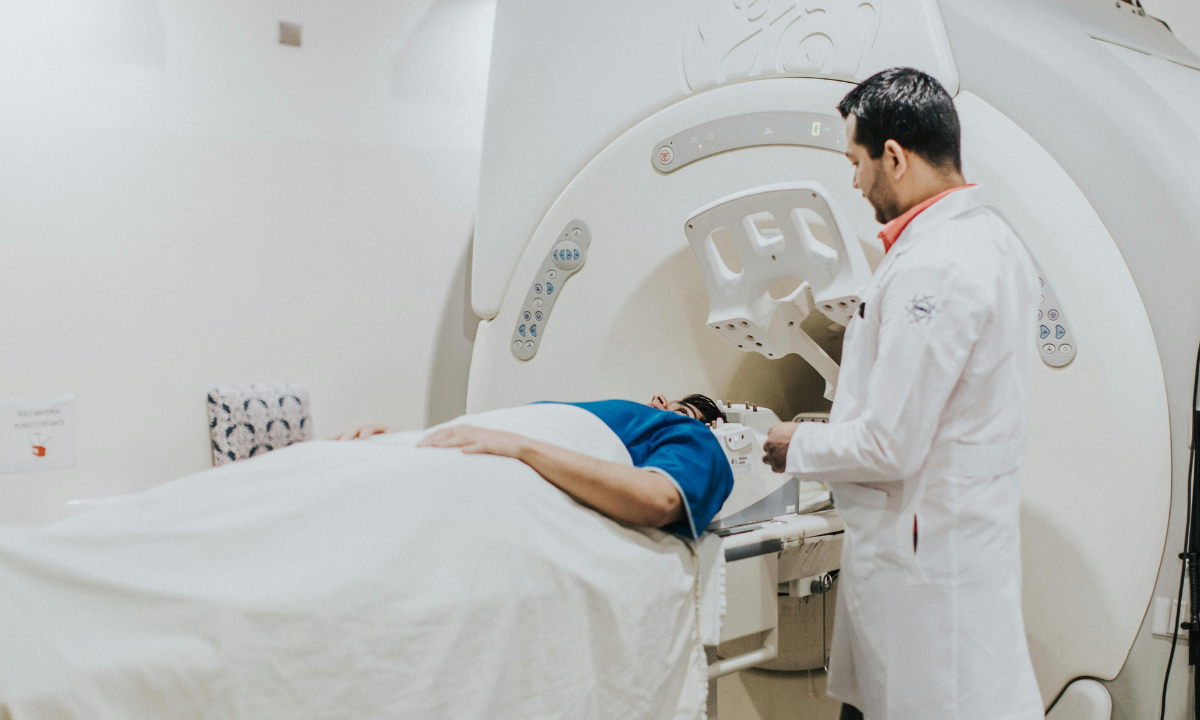
  • MRI (Magnetic Resonance Imaging) uses magnetic fields and radio waves, not radiation.
  • Ultrasound relies on sound waves to capture images of organs and tissues.
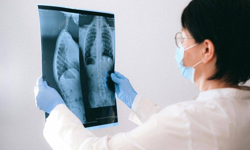
  • X-rays, CT scans, and PET scans, on the other hand, do use radiation.

This distinction is why your doctor carefully chooses the right type of scan for your situation. If radiation-free alternatives are suitable, they are often the first choice.

2. Radiation in X-rays is Extremely Low

X-rays are among the most common and oldest imaging tools in medicine. They work by passing a controlled amount of radiation through the body to capture images of bones, teeth, or the chest.

The amount of radiation in an X-ray is very small. For perspective:

  • A chest X-ray exposes you to about 0.1 mSv of radiation, which is roughly the same as the natural background radiation you are exposed to in just 10 days of daily life.

That means a single X-ray is considered safe, even for children, when used appropriately. Doctors usually advise them only when necessary, like to check fractures, lung infections, or dental issues.

3. How Much Radiation in a CT Scan Matters More

CT (Computed Tomography) scans provide detailed 3D images, which makes them incredibly useful in detecting internal injuries, tumors, or complex health conditions. But because they capture multiple X-ray images from different angles, they involve higher radiation compared to a single X-ray.

Here’s a simple comparison table to put things into perspective:

Scan TypeAverage Radiation Dose (in mSv)Equivalent Natural Exposure
Chest X-ray0.1 mSv10 days of natural exposure
Chest CT Scan7 mSv2 years of natural exposure
PET Scan25 mSv8 years of natural exposure

So yes, radiation exposure in CT scans is higher, but it’s still within medically acceptable safety limits. Doctors carefully weigh the diagnostic benefits against the small risks before recommending one.

Example to relate: Taking a chest CT scan is like getting the same radiation you’d receive if you flew around the world a couple of times. Air travel exposes you to natural cosmic radiation, yet millions of people fly safely every year.

4. Radiation Used in PET Scan Is Unique but Controlled

PET (Positron Emission Tomography) scans are slightly different. They use small amounts of radioactive tracers combined with imaging technology to study how organs and tissues function.

The radiation used in PET scan is higher than X-rays and CT scans, but the purpose is also more advanced, it helps detect cancer, track how treatments are working, and even identify early-stage diseases that might otherwise be missed.

Doctors carefully calculate the dosage of these tracers based on your age, health, and body size. The radiation disappears naturally from the body within hours or days, minimizing long-term risks.

5. Safety Measures Are Always a Priority

At any reputed health screening centre in Bangalore, strict safety protocols are followed to protect patients.

Here are some of the measures in place:

  • Low-dose technology: Modern machines are designed to use the smallest radiation dose needed.
  • Protective shielding: Sensitive areas, such as reproductive organs, are shielded whenever possible.
  • Avoiding repeat scans: Doctors ensure your scan history is reviewed so you don’t undergo unnecessary repetitions.
  • Special considerations for children and pregnant women: Extra caution is taken, and alternatives are often used first.

This means every scan you undergo is carefully justified for your health and safety.

Tips to Stay Safe and Informed

  • Always ask why a scan is recommended, it helps you understand its necessity.
  • Keep a personal record of your past scans to avoid duplication.
  • If you are pregnant or suspect pregnancy, inform your doctor before any radiation-based scan.
  • Don’t panic about one-time exposures, remember, the benefits usually outweigh the risks.

Wrapping Up

Radiation from scans is part of modern medical imaging, but it is carefully regulated to keep patients safe. Whether it’s radiation in X-ray, radiation exposure in CT scans, or radiation used in PET scan, the benefits of accurate diagnosis often outweigh the risks.

At a health screening centre in Bangalore like Koshikaa, our focus is on balancing advanced imaging and diagnostics with patient safety and awareness. By staying informed, you can approach your healthcare decisions with confidence.

FAQs on Radiation From Scans

1. Do all imaging scans use radiation?

No. MRI and Ultrasound use sound waves or magnetic fields. X-rays, CT scans, and PET scans involve radiation but at controlled and safe levels.

2. How much radiation in a CT scan is considered safe?

A chest CT scan exposes you to about 7 mSv, equal to roughly two years of natural background exposure, which is considered safe when medically required.

3. Is radiation exposure in CT scans dangerous if repeated?

Frequent CT scans can increase cumulative exposure, but doctors carefully track your medical history to avoid unnecessary repetition and reduce risks.

4. Why is radiation used in PET scan higher than others?

PET scans use radioactive tracers to study organ function, which requires higher radiation, but the insights gained often outweigh the minimal risks involved.

5. Can I refuse a scan if I’m concerned about radiation?

Yes, but speak with your doctor first. They may suggest an alternative like MRI or Ultrasound, or explain why the scan is crucial for diagnosis.


MRI of the uterus is primarily used for diagnosing and evaluating conditions such as uterine fibroids, adenomyosis, and malignancies. It is particularly useful in the planning of surgical procedures and assessing the extent of gynecological diseases. This imaging test is valuable for its ability to deliver detailed cross-sectional images of the uterus and surrounding structures.
An MRI of the spleen can help diagnose various disorders affecting the spleen including splenomegaly (enlarged spleen), tumors, and other abnormalities. It provides clear, detailed images that are crucial for assessing the health of the spleen and planning potential surgical interventions.
Prostate MRI is a non-invasive imaging technique that provides detailed pictures of the prostate gland and surrounding tissues. It helps in the detection, staging, and monitoring of prostate cancer. Prostate MRIs are increasingly used to guide biopsy procedures and assist in the planning of treatment strategies for prostate health.
An MRI of the pancreas is used to identify issues such as pancreatitis, pancreatic cancer, and other pancreatic disorders. It offers high-resolution images that help in detecting small lesions and assessing the pancreatic ducts. This imaging technique is valuable for guiding biopsies and planning surgeries involving the pancreas.
MRI scans of the liver, kidney, and bile ducts provide detailed images that help diagnose various conditions, such as tumors, cysts, and blockages in the bile ducts. They are crucial for assessing organ damage, staging cancer, and planning liver or kidney-related surgeries. This type of MRI can also evaluate the health and function of these organs comprehensively.
Breast MRI is highly sensitive for detecting breast cancer, especially in individuals with dense breast tissue or a high risk of cancer. It is often used in conjunction with mammography to provide a more comprehensive evaluation. Breast MRIs can also monitor the response to breast cancer treatments and provide detailed pre-surgical mapping.
Brain and spine MRIs are critical tools for diagnosing and monitoring neurological conditions, including tumors, spinal cord injuries, and multiple sclerosis. These MRIs can reveal abnormalities in brain tissue and spinal cord structure that might not be visible with other imaging methods. They are essential for planning surgeries and assessing the effectiveness of treatments in neurology.
PET-CT scans of transplant organs like the liver, kidney, or pancreas are performed to assess metabolic activity, detect complications, and monitor post-transplant function. These scans play a vital role in the early detection of issues such as organ rejection, recurrence of disease, or infection, guiding timely interventions and optimizing transplant outcomes for patients.
PET-CT examinations of organs such as the liver, kidney, or prostate are crucial for assessing metabolic activity, detecting abnormalities, and monitoring disease progression. These scans help diagnose conditions such as liver metastases, renal cell carcinoma, or prostate cancer, guiding treatment decisions and monitoring response to therapy over time.
PET-CT scans of soft-tissue masses are utilized to assess metabolic activity and detect malignant tumors or other abnormalities. By combining PET imaging with CT imaging, these scans provide detailed information about tumor location, size, and metabolic characteristics, aiding in accurate diagnosis and treatment planning for soft-tissue cancers.
PET-CT scans are valuable for detecting cysts or fluid collections within the body, including cysts in the liver, kidneys, or other organs. These scans help characterize the nature and metabolic activity of cysts, guiding diagnosis and treatment planning for conditions such as cystic liver disease, renal cysts, or pancreatic cysts.
PET-CT scans of muscles are performed to assess metabolic activity and detect abnormalities such as inflammation, infection, or tumors. These scans provide valuable information about muscle function and pathology, aiding in the diagnosis and management of conditions like myositis, muscular dystrophy, or soft-tissue sarcomas.
PET-CT scans of the brain are utilized to evaluate brain metabolism and detect abnormalities such as tumors, epilepsy, or neurodegenerative diseases. By combining functional PET imaging with anatomical CT imaging, these scans offer comprehensive information about brain function and structure, guiding neurosurgical interventions and treatment decisions for neurological disorders
PET-CT scans of bones are commonly used to detect bone metastases in cancer patients, assess bone health in conditions like osteoporosis, and evaluate response to treatment. By combining PET and CT imaging, these scans provide detailed information about metabolic activity and anatomical changes in bone structures, aiding in accurate diagnosis and treatment planning for various bone-related conditions.
CT scans of the spine are conducted to assess the vertebrae, spinal cord, and surrounding soft tissues for conditions like herniated discs, spinal fractures, or spinal stenosis, aiding in the diagnosis and management of spinal disorders with detailed imaging essential for surgical planning and intervention.
CT scans of the pelvis are utilized to visualize the pelvic bones, reproductive organs, bladder, and surrounding structures for conditions such as fractures, tumors, or pelvic inflammatory disease, guiding treatment plans and surgical interventions with precise anatomical information.
CT scans of the neck are performed to evaluate structures such as the throat, thyroid gland, lymph nodes, and blood vessels for abnormalities like tumors, infections, or injuries, assisting in diagnosis and management of head and neck disorders with detailed imaging.
CT scans of the extremities are employed to assess bones, joints, and soft tissues in the arms or legs, aiding in the diagnosis of fractures, arthritis, or other musculoskeletal conditions, guiding orthopedic treatment and rehabilitation plans for optimal recovery.
CT scans of the chest are utilized to examine the lungs, heart, blood vessels, and surrounding structures for conditions such as lung nodules, pneumonia, or pulmonary embolism, facilitating accurate diagnosis and management of respiratory and cardiovascular diseases.
CT scans of the brain are conducted to assess brain structures, identify abnormalities like tumors, hemorrhages, or strokes, and aid in diagnosing neurological conditions, guiding neurosurgical interventions and treatment strategies with precise imaging of the brain’s anatomy.
CT scans of the abdomen are performed to visualize and evaluate the internal organs such as the liver, kidneys, pancreas, and intestines for conditions like tumors, infections, or injuries, providing detailed images essential for diagnosis and treatment planning.
Transplant organ ultrasound scans, including those of the liver, kidney, or pancreas, are performed to assess organ viability, detect complications, and monitor post-transplant function. These scans play a vital role in the early detection of issues such as organ rejection, vascular complications, or fluid collections, guiding timely interventions and optimizing transplant outcomes for patients.
Ultrasound examinations of organs such as the liver, kidney, or prostate are crucial for assessing organ structure, detecting abnormalities, and monitoring disease progression. These scans help diagnose conditions such as liver cirrhosis, kidney stones, or prostate enlargement, guiding treatment decisions and monitoring response to therapy over time.
Soft-tissue masses can be evaluated using ultrasound to determine their size, location, and characteristics. Ultrasound imaging helps differentiate between benign and malignant masses, guiding further diagnostic evaluation and treatment planning. By providing real-time imaging, ultrasound assists in the precise localization of soft-tissue masses for biopsy or surgical removal if needed.
Ultrasound scans are valuable for detecting cysts or fluid collections within the body, including cysts in the liver, kidneys, or other organs. These scans help characterize the nature and size of cysts, aiding in diagnosis and treatment planning for conditions such as cystic liver disease or renal cysts, and guiding interventions such as cyst drainage or sclerotherapy.
Musculoskeletal ultrasound scans are used to assess the health and integrity of muscles, identifying issues such as strains, tears, or muscle inflammation. These scans provide detailed images of muscle structures, allowing healthcare providers to diagnose muscle injuries accurately and develop personalized rehabilitation plans to promote recovery and restore function.
Ultrasound examinations of joints are essential for evaluating joint health, detecting abnormalities such as inflammation, fluid accumulation, or cartilage damage. By visualizing the joint structures in real-time, ultrasound helps diagnose conditions like arthritis, bursitis, or ligament injuries, guiding treatment decisions and monitoring disease progression over time.
Ultrasound scans of tendons are performed to assess for injuries, inflammation, or tears, aiding in the diagnosis and management of conditions such as tendonitis or tendon tears. These scans provide detailed images of tendon structures, helping healthcare providers determine the extent of damage and plan appropriate treatment strategies, including physical therapy or surgical intervention if necessary.
Pelvis X-rays play a crucial role in diagnosing injuries, diseases, and developmental conditions affecting the pelvic region. They provide detailed images of the hip bones, sacrum, and coccyx, aiding in the assessment of fractures, dislocations, or signs of arthritis. Pelvic X-rays are instrumental in guiding surgical interventions, monitoring healing progress, and evaluating conditions such as osteoporosis or tumors. By providing precise anatomical information, these imaging studies enable healthcare professionals to deliver personalized care and improve patient outcomes.
Teeth X-rays, known as dental radiographs, are indispensable tools for dentists to assess oral health. They aid in diagnosing dental decay, gum disease, and abnormalities in tooth structure or alignment. These images provide valuable insights into hidden dental issues, allowing for early intervention and prevention of complications. Additionally, dental X-rays are essential for planning treatments such as orthodontics, root canals, or dental implants, ensuring optimal outcomes and patient satisfaction.
Spine X-rays are pivotal in diagnosing spinal fractures, degenerative conditions, and spinal misalignments, providing critical information for treatment planning and monitoring spinal health. They offer detailed images of the vertebrae and surrounding structures, enabling healthcare professionals to identify issues such as scoliosis, disc herniation, or spinal stenosis
Skull X-rays are valuable for detecting fractures, assessing head trauma, and evaluating the alignment of facial bones, crucial in diagnosing conditions like fractures, tumors, or developmental abnormalities in the skull. They provide detailed views of the cranial structure, aiding clinicians in determining appropriate treatment strategies and ensuring patient safety.
For the heart, X-ray solutions can aid in the detection of various cardiac problems, ranging from mild to severe, including – Heart failure, Cardiac congestion and other heart-related conditions. In addition to cardiac assessments, X-ray solutions are also used for breast examinations to identify tumors or signs of breast cancer.

Bone X-rays are essential for diagnosing fractures and assessing bone alignment, aiding doctors in developing treatment plans and monitoring healing progress. They provide detailed images of the skeletal system, helping identify abnormalities or injuries that may not be visible through other imaging techniques

Abdominal X-rays are performed when patients experience symptoms such as significant nausea, ongoing vomiting, abdominal pain, flank or back pain, or abdominal swelling. These X-rays can help diagnose various conditions.

Frequently Booked Tests

Frequently Booked Checkups