Why You Shouldn’t Ignore Your Lipid Profile Test

Table of Contents

As a doctor at Koshikaa Health Screening Centre, I often emphasize preventive health care, and one of the most underused yet powerful tools in our arsenal is the lipid profile test. 

When you consider that heart disease and stroke are leading causes of premature death, a simple blood screening can make a profound difference. The lipid profile test of blood offers us a clear window into your cholesterol and fat levels, so we can act early, optimize your risk, and help you live healthier.

Understanding What the Lipid Profile Test Is 

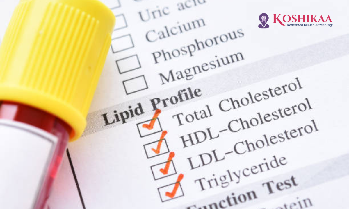
A lipid profile test (sometimes called a lipid panel) is a blood test that quantifies different types of fats in your bloodstream. In primary, cholesterol (total cholesterol, LDL “bad” cholesterol, and HDL “good” cholesterol) and triglycerides.

Since lipids travel in your blood and can accumulate in your arteries, the purpose of the lipid profile test becomes clear. This helps assess your cardiovascular health risk and guide prevention.

Why We Recommend the Lipid Profile Test and What It Detects

At Koshikaa, we ask: “What is the lipid profile test trying to tell us?” The answer lies in early detection and risk stratification. Conditions we evaluate using this blood test in Bangalore include:

  • Elevated cholesterol or triglycerides, even in asymptomatic individuals.
  • Screening those with a family history of heart disease, hypertension, diabetes, or obesity.
  • Monitoring therapy if you are already on lipid-lowering medications or lifestyle interventions.

Ignoring this test is akin to driving a car without checking your fuel gauge. You may be heading toward an unseen problem.

When to Undergo a Lipid Profile Test?

I frequently encounter patients who say, “Should I really take a lipid profile test when I feel fine?” 

The answer from our practice is yes, especially if you have risk factors. The lipid profile test, when to do it, often depends on your age, lifestyle, and comorbidities. Here’s our guideline at Koshikaa Health Screening Centre:

  • Healthy adults: first screening in their 20s or 30s, then repeat every 4–6 years if no risk factors.
  • Adults over 45 years old or with risk factors such as hypertension, diabetes, smoking, or obesity: annually or more frequently.
  • Patients with prior abnormal results: as recommended by the physician, often every 6–12 months to monitor trends. 

If you are in the Bangalore region and you seek a reliable blood test in Bangalore at a reputable health screening centre in Bangalore, selecting a facility tied to a recognized lab ensures accuracy and timely reporting.

What Happens During a Lipid Profile Test Procedure?

Here’s the step-by-step of how we perform the test at our clinic:

  1. You arrive, and we review your medications, fasting status, and any recent lifestyle changes.
  2. Typically, you fast for 9–12 hours (water allowed) before the test, unless we decide a non-fasting lipid profile is acceptable.
  3. A blood sample is drawn, usually from a vein in your arm. The process takes only a few minutes.
  4. The sample is sent for analysis, and the lipid profile test report is generated, listing total cholesterol, HDL, LDL, triglycerides, and sometimes VLDL.
  5. We review the results together and interpret your lipid profile test results in the context of your overall health, risk factors, and lifestyle.

How to Read Your Lipid Profile Test Report

When you receive your report, several numbers matter. Here’s how I guide patients at Koshikaa Health Screening Centre:

  • Total cholesterol: best if less than 200 mg/dL (for many healthy adults).
  • LDL cholesterol (“bad” cholesterol): lower is better; ideally under 100 mg/dL for most, under 70 mg/dL if you already have cardiovascular disease.
  • HDL cholesterol (“good” cholesterol): higher levels are protective; ideally above 40–60 mg/dL.
  • Triglycerides: best if under 150 mg/dL; elevated levels raise the risk of heart disease.
  • Additional ratios (like total cholesterol/HDL) and context (age, gender, medical history) guide how we customize action.

We emphasize a single abnormal reading is not a sentence; it’s a prompt. We use the report as a starting point to plan lifestyle changes, medication if needed, and repeat testing to track progress.

Why Ignoring the Lipid Profile Test is a Risk You Shouldn’t Take

There are three major reasons why skipping this test can be dangerous:

  • Silent Progression: High cholesterol and triglycerides often cause no symptoms until a major event happens (heart attack, stroke). The lipid profile test can alert us long before damage is done.
  • Intervention Window: Early abnormal results give us the chance to act with diet, exercise, and medications, reducing risk significantly.
  • Monitoring Effectiveness: If you undertake lifestyle changes or start therapy, repeat testing via the lipid profile test of blood helps us quantify improvement and fine-tune the strategy.

In Bangalore’s fast-paced environment, busy lifestyles, changing diets, and genetics are making dyslipidaemia more common, so preventive screening at a trusted health screening centre in Bangalore matters.

Wrapping Up 

At Koshikaa Health Screening Centre, I’ve cared for many patients whose illuminated lipid profile test acted as the wake-up call they needed. By addressing abnormal lipids early, we avoid the cascading complications of cardiovascular disease, stroke, and organ damage. The lipid profile test isn’t just another blood test; it’s a vital screening tool with the power to change your health trajectory.

If you’ve not had your lipid profile test yet, ask your physician about booking it at a trusted health screening centre in Bangalore. Once you have the results, we will review them together, interpret your risk profile, and guide you through diet, exercise, medications (if needed), and lifestyle habits appropriate for your profile. Your future self will thank you for taking this step.

Remember: good health isn’t only about fixing what’s broken; it’s about protecting what is working. The lipid profile test gives us insight. Let’s use that insight to ensure your heart stays strong and your life stays full.

FAQs

Q: What is the lipid profile test?

The lipid profile test is a blood screening that measures different types of fats (lipids) in your blood. cholesterol (total, HDL, LDL) and triglycerides, to help assess your risk of cardiovascular disease.

Q: Is fasting required for the Lipid profile test?

In most cases, yes. We often ask for 9–12 hours of fasting before the test to ensure accurate levels, especially for triglycerides. However, non-fasting tests are possible in certain settings; please follow your doctor’s instructions.

Q: What should I expect during a Lipid profile test?

You’ll have a simple blood draw, usually from a vein in your arm. The procedure takes only a few minutes. Beforehand, you may need to fast as instructed by your healthcare provider.

Q: When should I do a lipid profile test?

If you’re an adult with no risk factors, every 4–6 years may suffice. But if you have conditions like diabetes, high blood pressure, obesity, or a family history of heart disease or are over 45, then annual or more frequent testing is advisable.

MRI of the uterus is primarily used for diagnosing and evaluating conditions such as uterine fibroids, adenomyosis, and malignancies. It is particularly useful in the planning of surgical procedures and assessing the extent of gynecological diseases. This imaging test is valuable for its ability to deliver detailed cross-sectional images of the uterus and surrounding structures.
An MRI of the spleen can help diagnose various disorders affecting the spleen including splenomegaly (enlarged spleen), tumors, and other abnormalities. It provides clear, detailed images that are crucial for assessing the health of the spleen and planning potential surgical interventions.
Prostate MRI is a non-invasive imaging technique that provides detailed pictures of the prostate gland and surrounding tissues. It helps in the detection, staging, and monitoring of prostate cancer. Prostate MRIs are increasingly used to guide biopsy procedures and assist in the planning of treatment strategies for prostate health.
An MRI of the pancreas is used to identify issues such as pancreatitis, pancreatic cancer, and other pancreatic disorders. It offers high-resolution images that help in detecting small lesions and assessing the pancreatic ducts. This imaging technique is valuable for guiding biopsies and planning surgeries involving the pancreas.
MRI scans of the liver, kidney, and bile ducts provide detailed images that help diagnose various conditions, such as tumors, cysts, and blockages in the bile ducts. They are crucial for assessing organ damage, staging cancer, and planning liver or kidney-related surgeries. This type of MRI can also evaluate the health and function of these organs comprehensively.
Breast MRI is highly sensitive for detecting breast cancer, especially in individuals with dense breast tissue or a high risk of cancer. It is often used in conjunction with mammography to provide a more comprehensive evaluation. Breast MRIs can also monitor the response to breast cancer treatments and provide detailed pre-surgical mapping.
Brain and spine MRIs are critical tools for diagnosing and monitoring neurological conditions, including tumors, spinal cord injuries, and multiple sclerosis. These MRIs can reveal abnormalities in brain tissue and spinal cord structure that might not be visible with other imaging methods. They are essential for planning surgeries and assessing the effectiveness of treatments in neurology.
PET-CT scans of transplant organs like the liver, kidney, or pancreas are performed to assess metabolic activity, detect complications, and monitor post-transplant function. These scans play a vital role in the early detection of issues such as organ rejection, recurrence of disease, or infection, guiding timely interventions and optimizing transplant outcomes for patients.
PET-CT examinations of organs such as the liver, kidney, or prostate are crucial for assessing metabolic activity, detecting abnormalities, and monitoring disease progression. These scans help diagnose conditions such as liver metastases, renal cell carcinoma, or prostate cancer, guiding treatment decisions and monitoring response to therapy over time.
PET-CT scans of soft-tissue masses are utilized to assess metabolic activity and detect malignant tumors or other abnormalities. By combining PET imaging with CT imaging, these scans provide detailed information about tumor location, size, and metabolic characteristics, aiding in accurate diagnosis and treatment planning for soft-tissue cancers.
PET-CT scans are valuable for detecting cysts or fluid collections within the body, including cysts in the liver, kidneys, or other organs. These scans help characterize the nature and metabolic activity of cysts, guiding diagnosis and treatment planning for conditions such as cystic liver disease, renal cysts, or pancreatic cysts.
PET-CT scans of muscles are performed to assess metabolic activity and detect abnormalities such as inflammation, infection, or tumors. These scans provide valuable information about muscle function and pathology, aiding in the diagnosis and management of conditions like myositis, muscular dystrophy, or soft-tissue sarcomas.
PET-CT scans of the brain are utilized to evaluate brain metabolism and detect abnormalities such as tumors, epilepsy, or neurodegenerative diseases. By combining functional PET imaging with anatomical CT imaging, these scans offer comprehensive information about brain function and structure, guiding neurosurgical interventions and treatment decisions for neurological disorders
PET-CT scans of bones are commonly used to detect bone metastases in cancer patients, assess bone health in conditions like osteoporosis, and evaluate response to treatment. By combining PET and CT imaging, these scans provide detailed information about metabolic activity and anatomical changes in bone structures, aiding in accurate diagnosis and treatment planning for various bone-related conditions.
CT scans of the spine are conducted to assess the vertebrae, spinal cord, and surrounding soft tissues for conditions like herniated discs, spinal fractures, or spinal stenosis, aiding in the diagnosis and management of spinal disorders with detailed imaging essential for surgical planning and intervention.
CT scans of the pelvis are utilized to visualize the pelvic bones, reproductive organs, bladder, and surrounding structures for conditions such as fractures, tumors, or pelvic inflammatory disease, guiding treatment plans and surgical interventions with precise anatomical information.
CT scans of the neck are performed to evaluate structures such as the throat, thyroid gland, lymph nodes, and blood vessels for abnormalities like tumors, infections, or injuries, assisting in diagnosis and management of head and neck disorders with detailed imaging.
CT scans of the extremities are employed to assess bones, joints, and soft tissues in the arms or legs, aiding in the diagnosis of fractures, arthritis, or other musculoskeletal conditions, guiding orthopedic treatment and rehabilitation plans for optimal recovery.
CT scans of the chest are utilized to examine the lungs, heart, blood vessels, and surrounding structures for conditions such as lung nodules, pneumonia, or pulmonary embolism, facilitating accurate diagnosis and management of respiratory and cardiovascular diseases.
CT scans of the brain are conducted to assess brain structures, identify abnormalities like tumors, hemorrhages, or strokes, and aid in diagnosing neurological conditions, guiding neurosurgical interventions and treatment strategies with precise imaging of the brain’s anatomy.
CT scans of the abdomen are performed to visualize and evaluate the internal organs such as the liver, kidneys, pancreas, and intestines for conditions like tumors, infections, or injuries, providing detailed images essential for diagnosis and treatment planning.
Transplant organ ultrasound scans, including those of the liver, kidney, or pancreas, are performed to assess organ viability, detect complications, and monitor post-transplant function. These scans play a vital role in the early detection of issues such as organ rejection, vascular complications, or fluid collections, guiding timely interventions and optimizing transplant outcomes for patients.
Ultrasound examinations of organs such as the liver, kidney, or prostate are crucial for assessing organ structure, detecting abnormalities, and monitoring disease progression. These scans help diagnose conditions such as liver cirrhosis, kidney stones, or prostate enlargement, guiding treatment decisions and monitoring response to therapy over time.
Soft-tissue masses can be evaluated using ultrasound to determine their size, location, and characteristics. Ultrasound imaging helps differentiate between benign and malignant masses, guiding further diagnostic evaluation and treatment planning. By providing real-time imaging, ultrasound assists in the precise localization of soft-tissue masses for biopsy or surgical removal if needed.
Ultrasound scans are valuable for detecting cysts or fluid collections within the body, including cysts in the liver, kidneys, or other organs. These scans help characterize the nature and size of cysts, aiding in diagnosis and treatment planning for conditions such as cystic liver disease or renal cysts, and guiding interventions such as cyst drainage or sclerotherapy.
Musculoskeletal ultrasound scans are used to assess the health and integrity of muscles, identifying issues such as strains, tears, or muscle inflammation. These scans provide detailed images of muscle structures, allowing healthcare providers to diagnose muscle injuries accurately and develop personalized rehabilitation plans to promote recovery and restore function.
Ultrasound examinations of joints are essential for evaluating joint health, detecting abnormalities such as inflammation, fluid accumulation, or cartilage damage. By visualizing the joint structures in real-time, ultrasound helps diagnose conditions like arthritis, bursitis, or ligament injuries, guiding treatment decisions and monitoring disease progression over time.
Ultrasound scans of tendons are performed to assess for injuries, inflammation, or tears, aiding in the diagnosis and management of conditions such as tendonitis or tendon tears. These scans provide detailed images of tendon structures, helping healthcare providers determine the extent of damage and plan appropriate treatment strategies, including physical therapy or surgical intervention if necessary.
Pelvis X-rays play a crucial role in diagnosing injuries, diseases, and developmental conditions affecting the pelvic region. They provide detailed images of the hip bones, sacrum, and coccyx, aiding in the assessment of fractures, dislocations, or signs of arthritis. Pelvic X-rays are instrumental in guiding surgical interventions, monitoring healing progress, and evaluating conditions such as osteoporosis or tumors. By providing precise anatomical information, these imaging studies enable healthcare professionals to deliver personalized care and improve patient outcomes.
Teeth X-rays, known as dental radiographs, are indispensable tools for dentists to assess oral health. They aid in diagnosing dental decay, gum disease, and abnormalities in tooth structure or alignment. These images provide valuable insights into hidden dental issues, allowing for early intervention and prevention of complications. Additionally, dental X-rays are essential for planning treatments such as orthodontics, root canals, or dental implants, ensuring optimal outcomes and patient satisfaction.
Spine X-rays are pivotal in diagnosing spinal fractures, degenerative conditions, and spinal misalignments, providing critical information for treatment planning and monitoring spinal health. They offer detailed images of the vertebrae and surrounding structures, enabling healthcare professionals to identify issues such as scoliosis, disc herniation, or spinal stenosis
Skull X-rays are valuable for detecting fractures, assessing head trauma, and evaluating the alignment of facial bones, crucial in diagnosing conditions like fractures, tumors, or developmental abnormalities in the skull. They provide detailed views of the cranial structure, aiding clinicians in determining appropriate treatment strategies and ensuring patient safety.
For the heart, X-ray solutions can aid in the detection of various cardiac problems, ranging from mild to severe, including – Heart failure, Cardiac congestion and other heart-related conditions. In addition to cardiac assessments, X-ray solutions are also used for breast examinations to identify tumors or signs of breast cancer.

Bone X-rays are essential for diagnosing fractures and assessing bone alignment, aiding doctors in developing treatment plans and monitoring healing progress. They provide detailed images of the skeletal system, helping identify abnormalities or injuries that may not be visible through other imaging techniques

Abdominal X-rays are performed when patients experience symptoms such as significant nausea, ongoing vomiting, abdominal pain, flank or back pain, or abdominal swelling. These X-rays can help diagnose various conditions.

Frequently Booked Tests

Frequently Booked Checkups