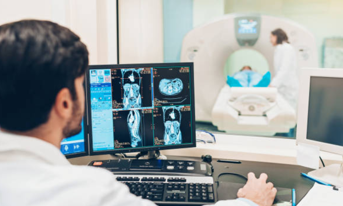
If you’re wondering whether an MRI scan is really necessary, the answer is simple: MRI is one of the most powerful and accurate tools in modern medicine. It helps doctors see what’s happening inside your body without any surgery or pain.
Why it’s so useful:
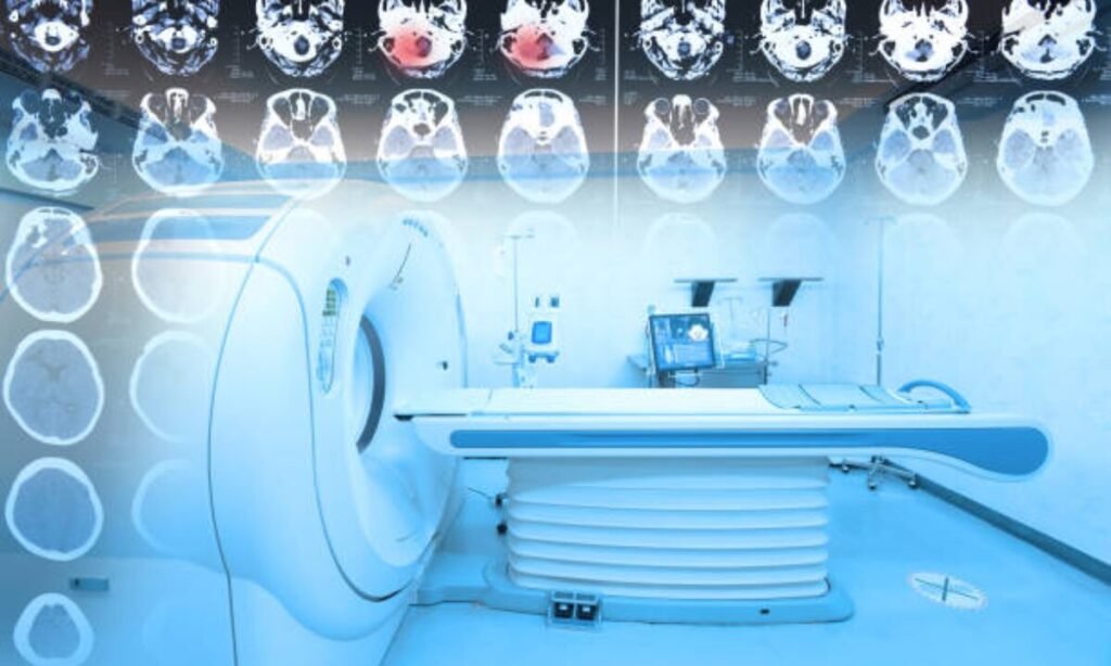
- Detailed Images: MRI creates high-quality images of your brain, spine, joints, organs, and soft tissues. This helps doctors catch problems that other scans might miss.
- No Radiation: Unlike CT scans or X-rays, MRIs use magnets and radio waves — making it safe, even for repeated use.
- Early Detection: MRI can spot diseases like tumors, infections, or nerve damage early, often before symptoms get worse.
- Helps Plan Treatment: Once your doctor knows exactly what’s wrong, they can choose the best treatment faster — whether it’s medicine, therapy, or surgery.
- Tracks Recovery: MRI is also used to check if your treatment is working and how well your body is healing.
In short, an MRI scan gives clear answers, saves time, and helps you and your doctor make smarter decisions about your health.
Need an MRI in Bangalore? At Koshikaa, we combine expert care with the latest MRI technology to give you accurate results and peace of mind.