Why Every Woman Over 40 Should Consider a Mammogram

MRI scan in Bangalore

Table of Contents

The health story of every woman is unique; nevertheless, there is one huge checkpoint that every woman is faced with at the age of 40: the mammogram. At this age, there is a possibility of changes in breast density, and other rates of risk might also shift. These alterations render the importance of regular screening. The understanding of the relevance of a mammogram allows women to make more confident decisions and take initiative. Have it done early, when catching a mammogram, small troubles can be noted before this mammogram, but after 40, it escalates. Fast treatment can make a difference between life and death.  

What is a Mammogram?  

Mammography

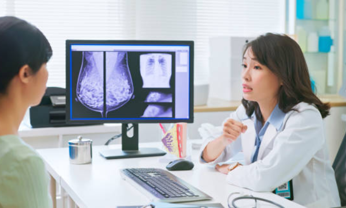
A mammogram refers to an X-ray image of the breast. It searches for alterations in the tissue structure. Modern applications of digital mammography involve the utilization of low doses of radiation and the generation of high-quality images that are examined by radiologists in search of minuscule indications of cancer. Such systems save the images in the electronic mode, unlike in ancient film-based machines, so the doctors can easily compare the new scan with the previous ones. This assists in improving the pace of review and, in many instances, alleviates the need to make additional visits.  

Why all women above the age of 40 need to be screened  

It has been observed that the risk of breast cancer increases as a person ages. The risk of an unhealthy tumor increases after 40. They are detected long before they show any signs through a simple X-ray scan. Having a mammogram for a person after the age of 40 is like taking a personal health initiative into one’s self, and it enables establishing a foundation, subsequent scans to more easily detect changes.  

The Role of Breast Cancer  Prevention

Periodic screening through mammography is recommended as a measure all over the world. Treatments are more effective and less invasive in the case of early detection.  In the case of early detection of mammograms, there are increased chances of recovery. You can even avoid harsh breast cancer treatments such as chemotherapy and proceed to more specific ones. The frequent checks also help alleviate pressure on the hospitals since fewer cases require sophisticated surgery.

Advantages of Early Detection: Your Screening Assistance

With the help of an early detection mammogram, the growth can be identified as early as it reaches a few millimeters in size, before pain is felt or a lump is detected. Once a physician suspects the presence of a problem, he/she can conduct biopsies or any other scans to definitively confirm the presence of cancer. Such a rapid response will aid in prevention and will provide patients with numerous treatment options. One of the finest behavioral patterns to achieve a good result is to go to regular yearly visits.

Getting ready for a mammogram: Preparation Tips  

This is made easy by planning. Going on with these mammogram preparation tips:  

  • Plan to get your screening a week after your period, when the breasts are not tender.  
  • Wear something that you can easily get undressed in quickly, such as a two-piece outfit.  
  • Do not apply underarm deodorant, lotion, or butter beneath your arms and your breasts, as they might reflect on the X-rays.  
  • Bring old scan images in case you have been tested at some other place.  
  • Report noticeable changes and/or implants of the breast to the technologist.
  • It makes the pictures clear and the exam less stressful.

Selection of the Right Facility

Ask a friend or a relative, or surf the net to identify a clinic or hospital nearby. Digital mammography test is now available in many centers, and it produces clearer images that result within a shorter duration. Do they also have ultrasounds or MRIs?

Affordability and Personal choice  

Others prefer being in the small clinics since they are more confidential and individualistic. Many select hospitals that offer comprehensive care for cancer. It may be an issue of costs, but most programs reduce the price:  

  • The government initiatives will provide free or subsidized checks  
  • Mobile units go to underserved communities  
  • Community health uses workshops and outreach to disseminate the word  
  • Volunteers assist in providing early screenings for women.

Community support  

The locally driven projects magnify cancer prevention. They are united with healthcare workers to have workshops, to provide preparation advice, and to demonstrate how modern images save lives. Mobile units go to isolated areas, and local collaborations with champions, activists, and survivors develop community trust and safer, healthy choices by women.  

Conclusion  

A first-time mammogram is a minor investment with a major payoff. It reduces early symptom concerns and retrains your brain to be on the move. All women beyond forty ought to have one. You will also make a difference when you educate yourself to better understand why mammogram matters, use the main prep suggestions, and embrace the digital opportunities available today, all when it comes to supporting the health of yourself and all people impacted by breast cancer in general. Early detection increases the chances of effective treatment. Make an appointment for your next early detection mammogram now.  

At Koshikaa, we provide advanced diagnostic care with compassion and expertise. Our state-of-the-art mammography services ensure accurate results, timely reporting, and a comfortable experience tailored to every woman’s needs. If you’re looking for trusted mammography in Bangalore, Koshikaa offers the perfect blend of medical precision and personalized care. Take charge of your health—schedule your mammogram with Koshikaa today and invest in a healthier tomorrow.

MRI of the uterus is primarily used for diagnosing and evaluating conditions such as uterine fibroids, adenomyosis, and malignancies. It is particularly useful in the planning of surgical procedures and assessing the extent of gynecological diseases. This imaging test is valuable for its ability to deliver detailed cross-sectional images of the uterus and surrounding structures.
An MRI of the spleen can help diagnose various disorders affecting the spleen including splenomegaly (enlarged spleen), tumors, and other abnormalities. It provides clear, detailed images that are crucial for assessing the health of the spleen and planning potential surgical interventions.
Prostate MRI is a non-invasive imaging technique that provides detailed pictures of the prostate gland and surrounding tissues. It helps in the detection, staging, and monitoring of prostate cancer. Prostate MRIs are increasingly used to guide biopsy procedures and assist in the planning of treatment strategies for prostate health.
An MRI of the pancreas is used to identify issues such as pancreatitis, pancreatic cancer, and other pancreatic disorders. It offers high-resolution images that help in detecting small lesions and assessing the pancreatic ducts. This imaging technique is valuable for guiding biopsies and planning surgeries involving the pancreas.
MRI scans of the liver, kidney, and bile ducts provide detailed images that help diagnose various conditions, such as tumors, cysts, and blockages in the bile ducts. They are crucial for assessing organ damage, staging cancer, and planning liver or kidney-related surgeries. This type of MRI can also evaluate the health and function of these organs comprehensively.
Breast MRI is highly sensitive for detecting breast cancer, especially in individuals with dense breast tissue or a high risk of cancer. It is often used in conjunction with mammography to provide a more comprehensive evaluation. Breast MRIs can also monitor the response to breast cancer treatments and provide detailed pre-surgical mapping.
Brain and spine MRIs are critical tools for diagnosing and monitoring neurological conditions, including tumors, spinal cord injuries, and multiple sclerosis. These MRIs can reveal abnormalities in brain tissue and spinal cord structure that might not be visible with other imaging methods. They are essential for planning surgeries and assessing the effectiveness of treatments in neurology.
PET-CT scans of transplant organs like the liver, kidney, or pancreas are performed to assess metabolic activity, detect complications, and monitor post-transplant function. These scans play a vital role in the early detection of issues such as organ rejection, recurrence of disease, or infection, guiding timely interventions and optimizing transplant outcomes for patients.
PET-CT examinations of organs such as the liver, kidney, or prostate are crucial for assessing metabolic activity, detecting abnormalities, and monitoring disease progression. These scans help diagnose conditions such as liver metastases, renal cell carcinoma, or prostate cancer, guiding treatment decisions and monitoring response to therapy over time.
PET-CT scans of soft-tissue masses are utilized to assess metabolic activity and detect malignant tumors or other abnormalities. By combining PET imaging with CT imaging, these scans provide detailed information about tumor location, size, and metabolic characteristics, aiding in accurate diagnosis and treatment planning for soft-tissue cancers.
PET-CT scans are valuable for detecting cysts or fluid collections within the body, including cysts in the liver, kidneys, or other organs. These scans help characterize the nature and metabolic activity of cysts, guiding diagnosis and treatment planning for conditions such as cystic liver disease, renal cysts, or pancreatic cysts.
PET-CT scans of muscles are performed to assess metabolic activity and detect abnormalities such as inflammation, infection, or tumors. These scans provide valuable information about muscle function and pathology, aiding in the diagnosis and management of conditions like myositis, muscular dystrophy, or soft-tissue sarcomas.
PET-CT scans of the brain are utilized to evaluate brain metabolism and detect abnormalities such as tumors, epilepsy, or neurodegenerative diseases. By combining functional PET imaging with anatomical CT imaging, these scans offer comprehensive information about brain function and structure, guiding neurosurgical interventions and treatment decisions for neurological disorders
PET-CT scans of bones are commonly used to detect bone metastases in cancer patients, assess bone health in conditions like osteoporosis, and evaluate response to treatment. By combining PET and CT imaging, these scans provide detailed information about metabolic activity and anatomical changes in bone structures, aiding in accurate diagnosis and treatment planning for various bone-related conditions.
CT scans of the spine are conducted to assess the vertebrae, spinal cord, and surrounding soft tissues for conditions like herniated discs, spinal fractures, or spinal stenosis, aiding in the diagnosis and management of spinal disorders with detailed imaging essential for surgical planning and intervention.
CT scans of the pelvis are utilized to visualize the pelvic bones, reproductive organs, bladder, and surrounding structures for conditions such as fractures, tumors, or pelvic inflammatory disease, guiding treatment plans and surgical interventions with precise anatomical information.
CT scans of the neck are performed to evaluate structures such as the throat, thyroid gland, lymph nodes, and blood vessels for abnormalities like tumors, infections, or injuries, assisting in diagnosis and management of head and neck disorders with detailed imaging.
CT scans of the extremities are employed to assess bones, joints, and soft tissues in the arms or legs, aiding in the diagnosis of fractures, arthritis, or other musculoskeletal conditions, guiding orthopedic treatment and rehabilitation plans for optimal recovery.
CT scans of the chest are utilized to examine the lungs, heart, blood vessels, and surrounding structures for conditions such as lung nodules, pneumonia, or pulmonary embolism, facilitating accurate diagnosis and management of respiratory and cardiovascular diseases.
CT scans of the brain are conducted to assess brain structures, identify abnormalities like tumors, hemorrhages, or strokes, and aid in diagnosing neurological conditions, guiding neurosurgical interventions and treatment strategies with precise imaging of the brain’s anatomy.
CT scans of the abdomen are performed to visualize and evaluate the internal organs such as the liver, kidneys, pancreas, and intestines for conditions like tumors, infections, or injuries, providing detailed images essential for diagnosis and treatment planning.
Transplant organ ultrasound scans, including those of the liver, kidney, or pancreas, are performed to assess organ viability, detect complications, and monitor post-transplant function. These scans play a vital role in the early detection of issues such as organ rejection, vascular complications, or fluid collections, guiding timely interventions and optimizing transplant outcomes for patients.
Ultrasound examinations of organs such as the liver, kidney, or prostate are crucial for assessing organ structure, detecting abnormalities, and monitoring disease progression. These scans help diagnose conditions such as liver cirrhosis, kidney stones, or prostate enlargement, guiding treatment decisions and monitoring response to therapy over time.
Soft-tissue masses can be evaluated using ultrasound to determine their size, location, and characteristics. Ultrasound imaging helps differentiate between benign and malignant masses, guiding further diagnostic evaluation and treatment planning. By providing real-time imaging, ultrasound assists in the precise localization of soft-tissue masses for biopsy or surgical removal if needed.
Ultrasound scans are valuable for detecting cysts or fluid collections within the body, including cysts in the liver, kidneys, or other organs. These scans help characterize the nature and size of cysts, aiding in diagnosis and treatment planning for conditions such as cystic liver disease or renal cysts, and guiding interventions such as cyst drainage or sclerotherapy.
Musculoskeletal ultrasound scans are used to assess the health and integrity of muscles, identifying issues such as strains, tears, or muscle inflammation. These scans provide detailed images of muscle structures, allowing healthcare providers to diagnose muscle injuries accurately and develop personalized rehabilitation plans to promote recovery and restore function.
Ultrasound examinations of joints are essential for evaluating joint health, detecting abnormalities such as inflammation, fluid accumulation, or cartilage damage. By visualizing the joint structures in real-time, ultrasound helps diagnose conditions like arthritis, bursitis, or ligament injuries, guiding treatment decisions and monitoring disease progression over time.
Ultrasound scans of tendons are performed to assess for injuries, inflammation, or tears, aiding in the diagnosis and management of conditions such as tendonitis or tendon tears. These scans provide detailed images of tendon structures, helping healthcare providers determine the extent of damage and plan appropriate treatment strategies, including physical therapy or surgical intervention if necessary.
Pelvis X-rays play a crucial role in diagnosing injuries, diseases, and developmental conditions affecting the pelvic region. They provide detailed images of the hip bones, sacrum, and coccyx, aiding in the assessment of fractures, dislocations, or signs of arthritis. Pelvic X-rays are instrumental in guiding surgical interventions, monitoring healing progress, and evaluating conditions such as osteoporosis or tumors. By providing precise anatomical information, these imaging studies enable healthcare professionals to deliver personalized care and improve patient outcomes.
Teeth X-rays, known as dental radiographs, are indispensable tools for dentists to assess oral health. They aid in diagnosing dental decay, gum disease, and abnormalities in tooth structure or alignment. These images provide valuable insights into hidden dental issues, allowing for early intervention and prevention of complications. Additionally, dental X-rays are essential for planning treatments such as orthodontics, root canals, or dental implants, ensuring optimal outcomes and patient satisfaction.
Spine X-rays are pivotal in diagnosing spinal fractures, degenerative conditions, and spinal misalignments, providing critical information for treatment planning and monitoring spinal health. They offer detailed images of the vertebrae and surrounding structures, enabling healthcare professionals to identify issues such as scoliosis, disc herniation, or spinal stenosis
Skull X-rays are valuable for detecting fractures, assessing head trauma, and evaluating the alignment of facial bones, crucial in diagnosing conditions like fractures, tumors, or developmental abnormalities in the skull. They provide detailed views of the cranial structure, aiding clinicians in determining appropriate treatment strategies and ensuring patient safety.
For the heart, X-ray solutions can aid in the detection of various cardiac problems, ranging from mild to severe, including – Heart failure, Cardiac congestion and other heart-related conditions. In addition to cardiac assessments, X-ray solutions are also used for breast examinations to identify tumors or signs of breast cancer.

Bone X-rays are essential for diagnosing fractures and assessing bone alignment, aiding doctors in developing treatment plans and monitoring healing progress. They provide detailed images of the skeletal system, helping identify abnormalities or injuries that may not be visible through other imaging techniques

Abdominal X-rays are performed when patients experience symptoms such as significant nausea, ongoing vomiting, abdominal pain, flank or back pain, or abdominal swelling. These X-rays can help diagnose various conditions.

Frequently Booked Tests

Frequently Booked Checkups