What to Expect During a Blood Cancer Detection Test

blood cancer detection

Table of Contents

Multiple people across the globe face serious complications from blood cancer as a medical illness. Receiving doctor advice to undergo a cancer detection test in Bangalore creates normal feelings of worry and confusion about what this procedure will be like. Learning the testing process allows you to reduce anxiety while gaining knowledge about blood cancer diagnosis procedures. This guide will explain in simple steps the complete process of a blood cancer detection test from preparation to final results.

What is Blood Cancer?  

blood test in Bangalore

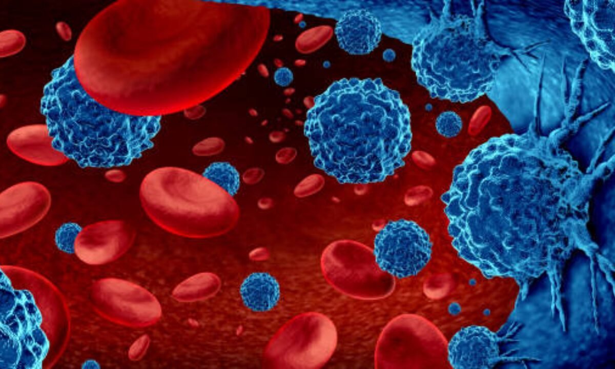
The test requires an initial clarification of blood cancer definitions. Blood cancer disrupts the structure and working procedure of blood cells, starting at the bone marrow where blood develops. The term describes cancerous growths that involve leukaemia along with lymphoma and multiple myeloma. Early medical identification of blood cancer remains essential because proper treatment planning requires it. A blood cancer detection test maintains its crucial position for detection purposes.

The significance of conducting blood cancer detection tests becomes essential to understand.

A blood cancer detection test will identify abnormalities in blood structures that need to be evaluated. You need this test based on specific signs including being tired, often having repeated infections or losing weight without reason and unusual bruises.

A health checkup sometimes incorporates a cancer detection test in Bangalore as its preventive aspect. Regular health screenings are a common aspect of healthy living practices for people residing in Bangalore and its neighbouring big cities. The detection tests are part of comprehensive health evaluations conducted by various individuals during full body checkup in Bangalore.

How to Prepare for the Test?

The process of preparation for blood cancer detection testing follows straightforward procedures that need no complex preparation steps. 

1. Fasting or Not?

A few blood tests need patients to follow a fasting timetable as one of their pre-test protocols. The medical staff performing the test will let you know if fasting is required as a part of your preparation.

2. Medication Check

You should notify both your doctor and technicians operating at the health screening centre in Bangalore whether you take medications or supplements before your test.

3. Stay Hydrated

You will benefit from high water intake before testing because it enhances the visibility of veins to blood drawing technicians.

The protocol for test preparation enables you to achieve a smoother and more pleasant experience during the testing procedure.

What Happens During the Test?

Medical facilities combine the blood cancer detection test with blood tests and imaging as well as biopsies as part of their diagnostic process. Let’s go step by step:  

The initial step requires you to sign up at the Health Center facilities.

All patients need to perform registration steps before entering the health centre. Any patient visiting a Bangalore health screening centre or any other facility needs to complete paperwork while sharing their medical records.

Blood Sample Collection

Doctors validate the presence of blood cancer by performing blood tests on collected blood samples. Here’s what typically happens:  

  • A healthcare worker cleans your arm with an antiseptic solution to stop the risk of infections that might occur during the treatment process.
  • A needle of small size enters your vein to extract blood samples. Most people experience only a light pinch sensation which becomes almost painless during the procedure.  
  • The technician will collect your blood sample into separate test tubes which receive labels before analysis begins.  

The Complete Blood Count test (CBC) is typically conducted at this time since it detects abnormal cell levels between white blood cells and platelets as well as red blood cells which might indicate blood cancer.

Conducting bone marrow aspiration

The doctor will order a bone marrow biopsy to get a better understanding of your condition if initial blood tests show unusual results. This medical procedure demands additional invasiveness through bone marrow sampling from your hip bone area. A local anaesthetic is used to reduce pain, but mild discomfort might be felt.

Bone marrow tests help determine whether the blood cells are being produced normally in your bone marrow or not. If your doctor recommends this, it’s to ensure a thorough evaluation. 

Additional Imaging Tests

The evaluation process might require medical imaging tests including X-rays, CT scans, or MRIs to confirm whether cancer cells have reached lymph nodes or other organs. A diagnosis testing package available in Bangalore includes cancer screening tests within its diagnostic package.

After the Test: What Happens Next?

The duration for receiving results depends on which tests were conducted. Basic blood test outcomes become available quickly while molecular tests combined with biopsies need one to two weeks for final results.  

You should visit your doctor when test results are prepared to explore the meaning behind the outcome. Abnormal results from testing will require your doctor to create a plan involving additional examination or therapy initiation.

Why Regular Screening is Important?

The majority of people schedule routine health examinations before diseases develop because they lack distinguishing symptoms. Blood testing during full body checkup in Bangalore provides an opportunity to identify blood cancer simultaneously with standard health screenings.   

Medical technology developments have made holistic health screening services convenient for residents of Bangalore and similar cities. Visiting a health screening centre in Bangalore for preventive or specific tests requires immediate action to improve your total health condition. 

Your Comfort Matters

Perceiving anxiety before a medical test is typical yet understanding what will happen during the examination helps decrease your stress levels. The detection of blood cancer through testing appears complicated but remains simple because these tests help you understand your health condition better.     

Your doctor and supporting staff at the health screening centre stand ready to help when you feel uncertain or overburdened. Health screening centre personnel and doctors will assist you with any questions you have about the process at their facility in Bangalore or any other location.

Final Thoughts

Getting a cancer detection test in Bangalore is an essential step toward better health, especially if you have symptoms or risk factors connected to blood cancer. The process, while slightly nerve-wracking at first, is generally simple and can provide critical insights into your health. 

Bangalore provides residents with many choices of high-quality medical facilities because it possesses a robust healthcare infrastructure. The healthcare facilities in Bangalore provide complete body exams together with detection tests for cancer that will secure your health. Prioritize your health because early detection stands as the key difference for your well-being.  

So, don’t hesitate. You should execute blood cancer detection tests exactly as your doctor prescribes since these assessments protect the course of your future health. 

Koshikaa is a well-respected health screening centre in Bangalore that provides comprehensive imaging services and preventive health care.

MRI of the uterus is primarily used for diagnosing and evaluating conditions such as uterine fibroids, adenomyosis, and malignancies. It is particularly useful in the planning of surgical procedures and assessing the extent of gynecological diseases. This imaging test is valuable for its ability to deliver detailed cross-sectional images of the uterus and surrounding structures.
An MRI of the spleen can help diagnose various disorders affecting the spleen including splenomegaly (enlarged spleen), tumors, and other abnormalities. It provides clear, detailed images that are crucial for assessing the health of the spleen and planning potential surgical interventions.
Prostate MRI is a non-invasive imaging technique that provides detailed pictures of the prostate gland and surrounding tissues. It helps in the detection, staging, and monitoring of prostate cancer. Prostate MRIs are increasingly used to guide biopsy procedures and assist in the planning of treatment strategies for prostate health.
An MRI of the pancreas is used to identify issues such as pancreatitis, pancreatic cancer, and other pancreatic disorders. It offers high-resolution images that help in detecting small lesions and assessing the pancreatic ducts. This imaging technique is valuable for guiding biopsies and planning surgeries involving the pancreas.
MRI scans of the liver, kidney, and bile ducts provide detailed images that help diagnose various conditions, such as tumors, cysts, and blockages in the bile ducts. They are crucial for assessing organ damage, staging cancer, and planning liver or kidney-related surgeries. This type of MRI can also evaluate the health and function of these organs comprehensively.
Breast MRI is highly sensitive for detecting breast cancer, especially in individuals with dense breast tissue or a high risk of cancer. It is often used in conjunction with mammography to provide a more comprehensive evaluation. Breast MRIs can also monitor the response to breast cancer treatments and provide detailed pre-surgical mapping.
Brain and spine MRIs are critical tools for diagnosing and monitoring neurological conditions, including tumors, spinal cord injuries, and multiple sclerosis. These MRIs can reveal abnormalities in brain tissue and spinal cord structure that might not be visible with other imaging methods. They are essential for planning surgeries and assessing the effectiveness of treatments in neurology.
PET-CT scans of transplant organs like the liver, kidney, or pancreas are performed to assess metabolic activity, detect complications, and monitor post-transplant function. These scans play a vital role in the early detection of issues such as organ rejection, recurrence of disease, or infection, guiding timely interventions and optimizing transplant outcomes for patients.
PET-CT examinations of organs such as the liver, kidney, or prostate are crucial for assessing metabolic activity, detecting abnormalities, and monitoring disease progression. These scans help diagnose conditions such as liver metastases, renal cell carcinoma, or prostate cancer, guiding treatment decisions and monitoring response to therapy over time.
PET-CT scans of soft-tissue masses are utilized to assess metabolic activity and detect malignant tumors or other abnormalities. By combining PET imaging with CT imaging, these scans provide detailed information about tumor location, size, and metabolic characteristics, aiding in accurate diagnosis and treatment planning for soft-tissue cancers.
PET-CT scans are valuable for detecting cysts or fluid collections within the body, including cysts in the liver, kidneys, or other organs. These scans help characterize the nature and metabolic activity of cysts, guiding diagnosis and treatment planning for conditions such as cystic liver disease, renal cysts, or pancreatic cysts.
PET-CT scans of muscles are performed to assess metabolic activity and detect abnormalities such as inflammation, infection, or tumors. These scans provide valuable information about muscle function and pathology, aiding in the diagnosis and management of conditions like myositis, muscular dystrophy, or soft-tissue sarcomas.
PET-CT scans of the brain are utilized to evaluate brain metabolism and detect abnormalities such as tumors, epilepsy, or neurodegenerative diseases. By combining functional PET imaging with anatomical CT imaging, these scans offer comprehensive information about brain function and structure, guiding neurosurgical interventions and treatment decisions for neurological disorders
PET-CT scans of bones are commonly used to detect bone metastases in cancer patients, assess bone health in conditions like osteoporosis, and evaluate response to treatment. By combining PET and CT imaging, these scans provide detailed information about metabolic activity and anatomical changes in bone structures, aiding in accurate diagnosis and treatment planning for various bone-related conditions.
CT scans of the spine are conducted to assess the vertebrae, spinal cord, and surrounding soft tissues for conditions like herniated discs, spinal fractures, or spinal stenosis, aiding in the diagnosis and management of spinal disorders with detailed imaging essential for surgical planning and intervention.
CT scans of the pelvis are utilized to visualize the pelvic bones, reproductive organs, bladder, and surrounding structures for conditions such as fractures, tumors, or pelvic inflammatory disease, guiding treatment plans and surgical interventions with precise anatomical information.
CT scans of the neck are performed to evaluate structures such as the throat, thyroid gland, lymph nodes, and blood vessels for abnormalities like tumors, infections, or injuries, assisting in diagnosis and management of head and neck disorders with detailed imaging.
CT scans of the extremities are employed to assess bones, joints, and soft tissues in the arms or legs, aiding in the diagnosis of fractures, arthritis, or other musculoskeletal conditions, guiding orthopedic treatment and rehabilitation plans for optimal recovery.
CT scans of the chest are utilized to examine the lungs, heart, blood vessels, and surrounding structures for conditions such as lung nodules, pneumonia, or pulmonary embolism, facilitating accurate diagnosis and management of respiratory and cardiovascular diseases.
CT scans of the brain are conducted to assess brain structures, identify abnormalities like tumors, hemorrhages, or strokes, and aid in diagnosing neurological conditions, guiding neurosurgical interventions and treatment strategies with precise imaging of the brain’s anatomy.
CT scans of the abdomen are performed to visualize and evaluate the internal organs such as the liver, kidneys, pancreas, and intestines for conditions like tumors, infections, or injuries, providing detailed images essential for diagnosis and treatment planning.
Transplant organ ultrasound scans, including those of the liver, kidney, or pancreas, are performed to assess organ viability, detect complications, and monitor post-transplant function. These scans play a vital role in the early detection of issues such as organ rejection, vascular complications, or fluid collections, guiding timely interventions and optimizing transplant outcomes for patients.
Ultrasound examinations of organs such as the liver, kidney, or prostate are crucial for assessing organ structure, detecting abnormalities, and monitoring disease progression. These scans help diagnose conditions such as liver cirrhosis, kidney stones, or prostate enlargement, guiding treatment decisions and monitoring response to therapy over time.
Soft-tissue masses can be evaluated using ultrasound to determine their size, location, and characteristics. Ultrasound imaging helps differentiate between benign and malignant masses, guiding further diagnostic evaluation and treatment planning. By providing real-time imaging, ultrasound assists in the precise localization of soft-tissue masses for biopsy or surgical removal if needed.
Ultrasound scans are valuable for detecting cysts or fluid collections within the body, including cysts in the liver, kidneys, or other organs. These scans help characterize the nature and size of cysts, aiding in diagnosis and treatment planning for conditions such as cystic liver disease or renal cysts, and guiding interventions such as cyst drainage or sclerotherapy.
Musculoskeletal ultrasound scans are used to assess the health and integrity of muscles, identifying issues such as strains, tears, or muscle inflammation. These scans provide detailed images of muscle structures, allowing healthcare providers to diagnose muscle injuries accurately and develop personalized rehabilitation plans to promote recovery and restore function.
Ultrasound examinations of joints are essential for evaluating joint health, detecting abnormalities such as inflammation, fluid accumulation, or cartilage damage. By visualizing the joint structures in real-time, ultrasound helps diagnose conditions like arthritis, bursitis, or ligament injuries, guiding treatment decisions and monitoring disease progression over time.
Ultrasound scans of tendons are performed to assess for injuries, inflammation, or tears, aiding in the diagnosis and management of conditions such as tendonitis or tendon tears. These scans provide detailed images of tendon structures, helping healthcare providers determine the extent of damage and plan appropriate treatment strategies, including physical therapy or surgical intervention if necessary.
Pelvis X-rays play a crucial role in diagnosing injuries, diseases, and developmental conditions affecting the pelvic region. They provide detailed images of the hip bones, sacrum, and coccyx, aiding in the assessment of fractures, dislocations, or signs of arthritis. Pelvic X-rays are instrumental in guiding surgical interventions, monitoring healing progress, and evaluating conditions such as osteoporosis or tumors. By providing precise anatomical information, these imaging studies enable healthcare professionals to deliver personalized care and improve patient outcomes.
Teeth X-rays, known as dental radiographs, are indispensable tools for dentists to assess oral health. They aid in diagnosing dental decay, gum disease, and abnormalities in tooth structure or alignment. These images provide valuable insights into hidden dental issues, allowing for early intervention and prevention of complications. Additionally, dental X-rays are essential for planning treatments such as orthodontics, root canals, or dental implants, ensuring optimal outcomes and patient satisfaction.
Spine X-rays are pivotal in diagnosing spinal fractures, degenerative conditions, and spinal misalignments, providing critical information for treatment planning and monitoring spinal health. They offer detailed images of the vertebrae and surrounding structures, enabling healthcare professionals to identify issues such as scoliosis, disc herniation, or spinal stenosis
Skull X-rays are valuable for detecting fractures, assessing head trauma, and evaluating the alignment of facial bones, crucial in diagnosing conditions like fractures, tumors, or developmental abnormalities in the skull. They provide detailed views of the cranial structure, aiding clinicians in determining appropriate treatment strategies and ensuring patient safety.
For the heart, X-ray solutions can aid in the detection of various cardiac problems, ranging from mild to severe, including – Heart failure, Cardiac congestion and other heart-related conditions. In addition to cardiac assessments, X-ray solutions are also used for breast examinations to identify tumors or signs of breast cancer.

Bone X-rays are essential for diagnosing fractures and assessing bone alignment, aiding doctors in developing treatment plans and monitoring healing progress. They provide detailed images of the skeletal system, helping identify abnormalities or injuries that may not be visible through other imaging techniques

Abdominal X-rays are performed when patients experience symptoms such as significant nausea, ongoing vomiting, abdominal pain, flank or back pain, or abdominal swelling. These X-rays can help diagnose various conditions.
Search Bar

Frequently Booked Tests

Frequently Booked Checkups