
Diagnostic X-rays have emerged as one of the most fundamental distinctive procedures for medical diagnosis as the healthcare industry continues its technological advancement. This detailed explanation starts by defining x-rays before explaining their operational principles, different variations, and diagnostic capabilities throughout several varied applications. We will maintain a basic approach while making everything simple to understand.
What is an X-ray?
X-Ray operates as electromagnetic radiation that reaches through various substances to display human tissue images and material structures because of its brief wavelength. X-ray technology generates images to display internal body conditions that doctors cannot see through surface examination.
X-Ray technology operates by measuring the varying absorption levels of different body tissues to X-ray energy flow. This penetrates bones more effectively than they do soft tissues allowing bones to appear white whilst soft tissues show only gray hues on imaging results.
How Does an X-Ray Work?
The x-ray machine projects small focused x-ray radiation through the examined body region. The body movement allows x-rays to transmit through various tissues until they get absorbed by bones. A detector located behind the body collects the X-rays which have passed through.
The processed results show bones appearing as bright white with lung air displayed in absolute black and muscular or adipose tissue appearing as variations of grey.
The x-ray digital images serve medical purposes because they present easy storage options and flexible transfer capabilities and image enhancement possibilities.
Types of X-Rays
X-rays function effectively based on multiple distinctive requirements. Medical X-ray equipment exists in multiple variations to treat different medical conditions. Numerous popular X-ray types exist as shown below.
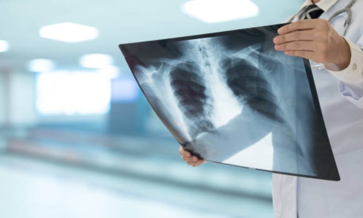
1. Chest X-ray
X-Rays of the chest rank as among the most regular radiological procedures utilized in medical settings. The diagnostic method helps doctors identify pneumonia along with lung infections and find indications of rib fractures along with tuberculosis and other diseases.
2. Bone X-Ray
X-Ray images of bones reveal breaks as well as fractures, joint dislocations and all abnormal pathways in the skeletal system. Orthopedists depend on bone X-rays when diagnosing injuries together with bone-related conditions that lead to arthritis.
3. Abdominal X-ray
Abdominal X-ray serves mainly to reveal problems in stomach and intestinal structures and abdominal organs. Ultrasound imaging shows doctors where kidney stones exist alongside bowel obstructions and swallowed items in the stomach.
4. Dental X-ray
During dental examinations dentists depend on X-rays to see all components of the teeth and jaws and adjacent tissues. Healthcare providers use X-rays to detect tooth decay, infections together with alignment problems in dental structures.
5. Mammography (Breast X-ray)
The detection of breast tissue requires special X-ray technology known as mammography. Normal breast cancer detection at an early stage heavily depends on the performance of X-ray-based screening procedures.
The Role of X-Rays in Diagnosis
Patients consider X-ray technology as a reliable and rapid diagnostic instrument. X-rays play significant roles in five distinct medical scenarios which include dental diagnosis and bone examination processes.
1. Identifying Fractures and Injuries
Medical professionals commonly use X-ray imaging to detect bone fractures together with joint dislocations and internal injuries resulting from accidents and trauma.
2. Detecting Lung Issues
The diagnostic process for pulmonary conditions including pneumonia, bronchitis and lung cancer primarily depends on Chest X-ray exams.
3. Examining Dental Problems
The teeth require Dental X-rays to show signs of cavities along with gum disease and impacted teeth which cannot be detected by standard eye observation.
4. Screening for Cancer
Early cancer detection is possible through X-ray examinations for both breast cancer and bone cancer. The identification of cancerous growths becomes more effective when medical personnel utilize specialized imaging such as mammograms.
5. Monitoring the Progress of Diseases
X-ray examinations help doctors monitor diseases in treated patients by determining how treatments have been performed.
When Should You Get an X-Ray?
Doctor recommendations along with particular symptoms might indicate doing an X-ray to proceed with medical investigations. One standard procedure for doctors to endorse this imaging strategy includes several specific scenarios.
- Continuous pain symptoms along with swelling and unknown discomfort warrant an X-ray interpretation.
- One gets X-ray tests after experiencing major traumatic events following a fall or accident.
- Routine screenings for conditions like cancer (mammography).
- The health care provider may request additional X-ray tests to check medical conditions which already exist or follow current treatments.
Complete health analysis needs can be met through a combination of X-rays with a full body checkup in Bangalore that will generate a comprehensive medical picture.
Advantages of Using X-Rays
Healthcare facilities use X-rays as a standard diagnostic tool because of multiple useful advantages.
- Customers obtain X-ray results in a matter of minutes when this technology yields quick outcomes during most examinations.
- The technological method performs body examinations without requiring incisions or inflicting any pain during the procedures.
- X-ray examinations are less expensive than MRI or CT scan diagnostic options thus making them more affordable for patients.
- Early cancer detection becomes possible through X-rays for diseases including cancer since this technique can result in life-saving outcomes.
The majority of medical professionals consider X-rays safe for human use yet these procedures require exposing patients to some radiation amounts.
The Importance of X Rays and Early Cancer Detection
X-ray services primarily serve to detect cancers at their earliest possible stages. The identification of lung and breast cancer at their earliest stages depends on X-ray scanning procedures. Patients who receive early diagnoses get better treatment choices alongside higher survival statistics and their treatments become more manageable and time-effective.
Facilities providing services for X Ray in Bangalore focus on illustrating the preventive healthcare advantages of the X-ray examination. The correct course of action requires screening participation before symptoms intensify especially for patients in high-risk status categories.
X-Rays as Part of Routine Health Checks
X-Rays have started appearing regularly during standard health examination procedures. Health checkup packages in Bangalore incorporate X-rays jointly with blood tests and ultrasounds besides additional diagnostic procedures. The combination of comprehensive health checks enables an early identification of hidden conditions for better health results.
How to Prepare for an X-Ray?
The process of preparing for X-ray tests is simple and does not involve strenuous work. The X-ray preparation process requires you to consider several key points as follows:
- Inform your physician about possible pregnancy status and all medical implants you have since these could affect your X-ray results.
- All patients need to eliminate metal accessories such as jewellery watches before undergoing an X-ray.
- Because different X-ray procedures need various preparations, patients should follow the exact instructions which might include changing into a hospital gown and fasting for gastrointestinal tract X-ray exams.
The X-ray procedure requires only a short duration causes no discomfort to the patient and lasts for a few minutes normally.
Conclusion
Modern healthcare benefits tremendously from X-ray technology since they became essential diagnostic tools over a hundred years ago. In cities like Bangalore, access to quality diagnostic centres for services such as X Ray in Bangalore is now more convenient and affordable than ever. Whether you’re going for a targeted X-ray or a full body checkup in Bangalore, do it in consultation with a qualified healthcare provider for the best outcomes.
Your understanding now includes complete knowledge about X-ray operations together with a clear awareness of diagnostic needs and multiple critical applications which save lives.
Koshikaa operates as a respected healthcare facility that showcases full body checkup in Bangalore through advanced testing equipment and expert medical care to detect health risks early.