What Is a Hemoglobin Blood Test and Why Is It Important?

Full body checkup in Bangalore

Table of Contents

In your red blood cells, hemoglobin helps move oxygen from your lungs to all the other parts of your body. Every part of your body needs oxygen for it to function normally. Some of the carbon dioxide your body makes attaches to your blood’s hemoglobin, and then you breathe it out. Lack of hemoglobin in your body can lead to your cells not getting enough oxygen, so you feel tired and might become short of breath.

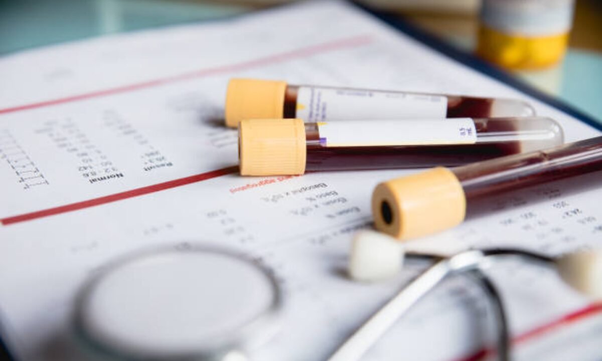
Looking for changes in your oxygen-carrying blood is as simple as getting a hemoglobin test. Most of the time, you’ll get a complete blood test during a normal checkup or if you feel extremely tired or weak.

Why Does a Blood Test Hemoglobin Matter?

Blood test in Bangalore

Many health problems are diagnosed and monitored using the results of blood tests for hemoglobin. Whether your body has a good supply of oxygen can be seen by your hemoglobin levels. 

You should check hemoglobin to keep track of your health.

Spotting anemia (a lack of red blood cells in the blood)

  • Finding out if too many red blood cells are seen
  • Keeping an eye on illnesses that are hard to cure, such as kidney disease and different cancers
  • Examining the body’s capacity to move oxygen throughout itself

When Should You Get a Blood Test for Hemoglobin?

For many reasons, you might find that this test is useful. These situations can be seen when someone:

  • Having more weakness or tiredness than usual
  • Feeling like you’re struggling to breathe, faint, or turn pale
  • Suffering from a chronic illness, such as having kidney disease
  • As one step in a panel of tests during a regular health checkup

Doctors could order a blood test for hemoglobin before an operation just to be certain your blood is suitable for the procedure.

What is Involved in Having a Blood Test for Hemoglobin?

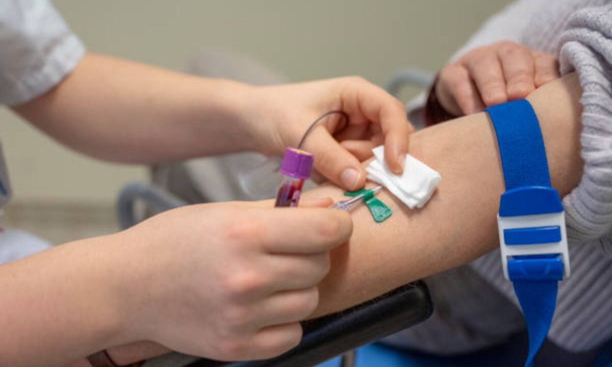
Hemoglobin blood tests are simple to do. Your blood will be taken by a nurse or lab technician and sent to a laboratory, where it is tested. Under such conditions, carrying out a Blood test in Bangalore is very straightforward and pleasant for many. You typically don’t have to prepare differently for diagnostic tests, unless your doctor tells you to.

Ways to Test Hemoglobin

Your hemoglobin level is measured based on grams per deciliter of blood. This test shows doctors if you have enough protein in your system for good health.

A blood test for hemoglobin is included in the tests that make up a complete blood count, or CBC. While looking at white and red blood cells and platelets, a CBC test depends on hemoglobin to tell if your red blood cells are in good health.

What Do Healthy Hemoglobin Levels Tell You?

Learning what your data means can benefit you. Hemoglobin’s healthy ranges are influenced by a person’s age, sex, and location. Usually, it is organized something like this:

  • The recommended range for men who are adults is from 13.8 to 17.2 g/dL
  • Women up to 19 years: 12.1-15.1 g/dL

Both children’s and adults’ hemoglobin levels are different and newborns generally have even more hemoglobin in their blood.

On average, the hemoglobin level for women’s blood is below what is normal for men. During menstruation, a woman’s blood usually contains a lower level of hemoglobin.

Why Should Hemoglobin Levels Matter to Women’s Health?

Hemoglobin levels for women should be frequently monitored. A condition called iron deficiency anemia is found more frequently in women because of menstruation, pregnancy, and often a diet lacking in iron.

  • When hemoglobin levels in the blood are low, you might feel exhausted, regular headaches and difficulties focusing on things.
  • Pregnant women are watched more closely by their doctors. Fortunately, there are signs that you should be aware of if you are pregnant and have a low hemoglobin level. 
  • As a result, doctors suggest taking more iron or folic acid if they see hemoglobin levels go down.

What the Red Blood Cells Normal Range Looks Like

Hemoglobin needs help to do its job. It goes hand in hand with red blood cells. Red blood cells normal range is measured by counting millions per microliter of blood.

  • 4.7 to 6.1 million men cells/ml
  • Women: between 4.2 and 5.4 million cells are found per milliliter of plasma

A red blood cell range that is not normal can affect both your hemoglobin and the oxygen that tissues receive.

What it Means When a Person Has Abnormal Levels of Hemoglobin

Every so often, the level of hemoglobin in your blood can be outside of its normal range. Some potential reasons are:

  • Hemoglobin deficiency (Anemia).
  • Loss of blood (caused by an injury, surgery, menstruation, or bleeding inside the body)
  • Deficiencies in iron, one of the B vitamins (B12) or folate

For example, chronic diseases like kidney disease or cancer.

  • Problems with bone marrow
  • Too much hemoglobin.
  • The low amount of oxygen at high altitudes leads your body to make more red blood cells. 
  • Problems in the lungs (such as COPD)
  • Smoking
  • Some people carry blood diseases that are not very common (examples include polycythemia vera).

What You Should Do to Keep Hemoglobin at the Optimal Level

Maintaining healthy blood contains a large amount of Vitamin C. Including meat, beans, spinach and fortified cereals in your diet helps produce hemoglobin. The folate and vitamin B12 in leafy greens, dairy and eggs can help as well. If someone’s hemoglobin level is low, doctors may recommend taking supplements or using medicines.

Certain health conditions may need different treatments to correct the hemoglobin problem.

Choosing a quick and easy way to get tested.

Getting a blood test to check your hemoglobin is now done quickly and easily. You can receive services at clinics, diagnostic centers, and hospitals in major cities. Fortunately, several centers in Bangalore let you book your blood test from home and schedule a nurse to collect your samples there.

How Do You Know When to Talk to Your Doctor

If you find that you are always tired, can’t breathe easily, or begin to bruise easily for no reason, mention this to your doctor. Sometimes, such symptoms may mean that the hemoglobin in your blood falls below the healthy limit. Your doctor can quickly order a blood test for hemoglobin to help figure out what’s going on.

If you have long-term illnesses that increase your risk of anemia—such as kidney disease or chronic inflammatory problems—regular monitoring of your hemoglobin and red blood cells normal range is essential. Blood test Bangalore centers provide patients with fast and accurate outcomes which help determine any required treatment.

Summary

Testing hemoglobin in blood is used to assess your oxygen levels and is a valuable method for finding and tracking different health problems. Learning about your hemoglobin levels, particularly those for women, helps you spot early signs of anemia. Both your red blood cells’ normal range and hemoglobin levels can give you important information about your health. Luckily, several cities, including Bangalore, now make it very easy to have blood tests done. Sticking to regular Doctor visits, eating healthy foods, and paying attention to any symptoms can help you stay healthy and full of energy.

If you need accurate results for Blood test in Bangalore, Koshikaa will offer you a full range of services and provide you with results in little time. So in case you or any family member have an emergency, then directly connect with our team.

MRI of the uterus is primarily used for diagnosing and evaluating conditions such as uterine fibroids, adenomyosis, and malignancies. It is particularly useful in the planning of surgical procedures and assessing the extent of gynecological diseases. This imaging test is valuable for its ability to deliver detailed cross-sectional images of the uterus and surrounding structures.
An MRI of the spleen can help diagnose various disorders affecting the spleen including splenomegaly (enlarged spleen), tumors, and other abnormalities. It provides clear, detailed images that are crucial for assessing the health of the spleen and planning potential surgical interventions.
Prostate MRI is a non-invasive imaging technique that provides detailed pictures of the prostate gland and surrounding tissues. It helps in the detection, staging, and monitoring of prostate cancer. Prostate MRIs are increasingly used to guide biopsy procedures and assist in the planning of treatment strategies for prostate health.
An MRI of the pancreas is used to identify issues such as pancreatitis, pancreatic cancer, and other pancreatic disorders. It offers high-resolution images that help in detecting small lesions and assessing the pancreatic ducts. This imaging technique is valuable for guiding biopsies and planning surgeries involving the pancreas.
MRI scans of the liver, kidney, and bile ducts provide detailed images that help diagnose various conditions, such as tumors, cysts, and blockages in the bile ducts. They are crucial for assessing organ damage, staging cancer, and planning liver or kidney-related surgeries. This type of MRI can also evaluate the health and function of these organs comprehensively.
Breast MRI is highly sensitive for detecting breast cancer, especially in individuals with dense breast tissue or a high risk of cancer. It is often used in conjunction with mammography to provide a more comprehensive evaluation. Breast MRIs can also monitor the response to breast cancer treatments and provide detailed pre-surgical mapping.
Brain and spine MRIs are critical tools for diagnosing and monitoring neurological conditions, including tumors, spinal cord injuries, and multiple sclerosis. These MRIs can reveal abnormalities in brain tissue and spinal cord structure that might not be visible with other imaging methods. They are essential for planning surgeries and assessing the effectiveness of treatments in neurology.
PET-CT scans of transplant organs like the liver, kidney, or pancreas are performed to assess metabolic activity, detect complications, and monitor post-transplant function. These scans play a vital role in the early detection of issues such as organ rejection, recurrence of disease, or infection, guiding timely interventions and optimizing transplant outcomes for patients.
PET-CT examinations of organs such as the liver, kidney, or prostate are crucial for assessing metabolic activity, detecting abnormalities, and monitoring disease progression. These scans help diagnose conditions such as liver metastases, renal cell carcinoma, or prostate cancer, guiding treatment decisions and monitoring response to therapy over time.
PET-CT scans of soft-tissue masses are utilized to assess metabolic activity and detect malignant tumors or other abnormalities. By combining PET imaging with CT imaging, these scans provide detailed information about tumor location, size, and metabolic characteristics, aiding in accurate diagnosis and treatment planning for soft-tissue cancers.
PET-CT scans are valuable for detecting cysts or fluid collections within the body, including cysts in the liver, kidneys, or other organs. These scans help characterize the nature and metabolic activity of cysts, guiding diagnosis and treatment planning for conditions such as cystic liver disease, renal cysts, or pancreatic cysts.
PET-CT scans of muscles are performed to assess metabolic activity and detect abnormalities such as inflammation, infection, or tumors. These scans provide valuable information about muscle function and pathology, aiding in the diagnosis and management of conditions like myositis, muscular dystrophy, or soft-tissue sarcomas.
PET-CT scans of the brain are utilized to evaluate brain metabolism and detect abnormalities such as tumors, epilepsy, or neurodegenerative diseases. By combining functional PET imaging with anatomical CT imaging, these scans offer comprehensive information about brain function and structure, guiding neurosurgical interventions and treatment decisions for neurological disorders
PET-CT scans of bones are commonly used to detect bone metastases in cancer patients, assess bone health in conditions like osteoporosis, and evaluate response to treatment. By combining PET and CT imaging, these scans provide detailed information about metabolic activity and anatomical changes in bone structures, aiding in accurate diagnosis and treatment planning for various bone-related conditions.
CT scans of the spine are conducted to assess the vertebrae, spinal cord, and surrounding soft tissues for conditions like herniated discs, spinal fractures, or spinal stenosis, aiding in the diagnosis and management of spinal disorders with detailed imaging essential for surgical planning and intervention.
CT scans of the pelvis are utilized to visualize the pelvic bones, reproductive organs, bladder, and surrounding structures for conditions such as fractures, tumors, or pelvic inflammatory disease, guiding treatment plans and surgical interventions with precise anatomical information.
CT scans of the neck are performed to evaluate structures such as the throat, thyroid gland, lymph nodes, and blood vessels for abnormalities like tumors, infections, or injuries, assisting in diagnosis and management of head and neck disorders with detailed imaging.
CT scans of the extremities are employed to assess bones, joints, and soft tissues in the arms or legs, aiding in the diagnosis of fractures, arthritis, or other musculoskeletal conditions, guiding orthopedic treatment and rehabilitation plans for optimal recovery.
CT scans of the chest are utilized to examine the lungs, heart, blood vessels, and surrounding structures for conditions such as lung nodules, pneumonia, or pulmonary embolism, facilitating accurate diagnosis and management of respiratory and cardiovascular diseases.
CT scans of the brain are conducted to assess brain structures, identify abnormalities like tumors, hemorrhages, or strokes, and aid in diagnosing neurological conditions, guiding neurosurgical interventions and treatment strategies with precise imaging of the brain’s anatomy.
CT scans of the abdomen are performed to visualize and evaluate the internal organs such as the liver, kidneys, pancreas, and intestines for conditions like tumors, infections, or injuries, providing detailed images essential for diagnosis and treatment planning.
Transplant organ ultrasound scans, including those of the liver, kidney, or pancreas, are performed to assess organ viability, detect complications, and monitor post-transplant function. These scans play a vital role in the early detection of issues such as organ rejection, vascular complications, or fluid collections, guiding timely interventions and optimizing transplant outcomes for patients.
Ultrasound examinations of organs such as the liver, kidney, or prostate are crucial for assessing organ structure, detecting abnormalities, and monitoring disease progression. These scans help diagnose conditions such as liver cirrhosis, kidney stones, or prostate enlargement, guiding treatment decisions and monitoring response to therapy over time.
Soft-tissue masses can be evaluated using ultrasound to determine their size, location, and characteristics. Ultrasound imaging helps differentiate between benign and malignant masses, guiding further diagnostic evaluation and treatment planning. By providing real-time imaging, ultrasound assists in the precise localization of soft-tissue masses for biopsy or surgical removal if needed.
Ultrasound scans are valuable for detecting cysts or fluid collections within the body, including cysts in the liver, kidneys, or other organs. These scans help characterize the nature and size of cysts, aiding in diagnosis and treatment planning for conditions such as cystic liver disease or renal cysts, and guiding interventions such as cyst drainage or sclerotherapy.
Musculoskeletal ultrasound scans are used to assess the health and integrity of muscles, identifying issues such as strains, tears, or muscle inflammation. These scans provide detailed images of muscle structures, allowing healthcare providers to diagnose muscle injuries accurately and develop personalized rehabilitation plans to promote recovery and restore function.
Ultrasound examinations of joints are essential for evaluating joint health, detecting abnormalities such as inflammation, fluid accumulation, or cartilage damage. By visualizing the joint structures in real-time, ultrasound helps diagnose conditions like arthritis, bursitis, or ligament injuries, guiding treatment decisions and monitoring disease progression over time.
Ultrasound scans of tendons are performed to assess for injuries, inflammation, or tears, aiding in the diagnosis and management of conditions such as tendonitis or tendon tears. These scans provide detailed images of tendon structures, helping healthcare providers determine the extent of damage and plan appropriate treatment strategies, including physical therapy or surgical intervention if necessary.
Pelvis X-rays play a crucial role in diagnosing injuries, diseases, and developmental conditions affecting the pelvic region. They provide detailed images of the hip bones, sacrum, and coccyx, aiding in the assessment of fractures, dislocations, or signs of arthritis. Pelvic X-rays are instrumental in guiding surgical interventions, monitoring healing progress, and evaluating conditions such as osteoporosis or tumors. By providing precise anatomical information, these imaging studies enable healthcare professionals to deliver personalized care and improve patient outcomes.
Teeth X-rays, known as dental radiographs, are indispensable tools for dentists to assess oral health. They aid in diagnosing dental decay, gum disease, and abnormalities in tooth structure or alignment. These images provide valuable insights into hidden dental issues, allowing for early intervention and prevention of complications. Additionally, dental X-rays are essential for planning treatments such as orthodontics, root canals, or dental implants, ensuring optimal outcomes and patient satisfaction.
Spine X-rays are pivotal in diagnosing spinal fractures, degenerative conditions, and spinal misalignments, providing critical information for treatment planning and monitoring spinal health. They offer detailed images of the vertebrae and surrounding structures, enabling healthcare professionals to identify issues such as scoliosis, disc herniation, or spinal stenosis
Skull X-rays are valuable for detecting fractures, assessing head trauma, and evaluating the alignment of facial bones, crucial in diagnosing conditions like fractures, tumors, or developmental abnormalities in the skull. They provide detailed views of the cranial structure, aiding clinicians in determining appropriate treatment strategies and ensuring patient safety.
For the heart, X-ray solutions can aid in the detection of various cardiac problems, ranging from mild to severe, including – Heart failure, Cardiac congestion and other heart-related conditions. In addition to cardiac assessments, X-ray solutions are also used for breast examinations to identify tumors or signs of breast cancer.

Bone X-rays are essential for diagnosing fractures and assessing bone alignment, aiding doctors in developing treatment plans and monitoring healing progress. They provide detailed images of the skeletal system, helping identify abnormalities or injuries that may not be visible through other imaging techniques

Abdominal X-rays are performed when patients experience symptoms such as significant nausea, ongoing vomiting, abdominal pain, flank or back pain, or abdominal swelling. These X-rays can help diagnose various conditions.

Frequently Booked Tests

Frequently Booked Checkups