
Imagine driving your car with the dashboard covered up. You wouldn’t know if you are low on fuel. You wouldn’t see the “Check Engine” light blinking and would only stop when the engine actually smokes.
Most of us treat our bodies exactly like that. We wait for pain, fever, or dizziness. We wait for the “smoke” before visiting a doctor. But your body was sending signals long before that.
Blood Tests are your body’s dashboard. Your blood visits every cell, organ, and tissue. It carries secret messages about your health. It warns you about sugar spikes or silent infections.
In this guide, we stop the guessing game. We decode those confusing terms like Complete blood count and HbA1c. We break down the Blood test Price in Bangalore. And we show you why a simple prick can save your life.
Let’s uncover the story flowing in your veins.
Disclaimer
Medical Disclaimer: The information provided in this blog, including test descriptions, reference ranges, and interpretation guides, is for educational and informational purposes only. It should not be considered as professional medical advice, diagnosis, or treatment. Blood test results can be influenced by various factors such as age, gender, medical history, and medications. Always consult a qualified doctor or pathologist to interpret your specific reports before making any health decisions.
Financial Disclosure: Prices mentioned for Koshikaa services are accurate as per the official website at the time of writing. All other “Market Average” costs listed in this article are estimates derived from independent online research and public data from various diagnostic centres in Bangalore. Actual prices may vary depending on the laboratory, location, and specific technology used.
The 4 BIG Tests: Decoding the Jargon
Doctors often speak in code language. They say “CBC” or “Lipid Profile,” and you nod silently. But knowing what these tests look for is your right.
Here are the four most common tests translated into simple English.
1. Complete blood count
This is the most common test in the world. Think of your blood as an army defending a castle (your body). The Complete blood count (CBC) counts your soldiers.
| Cells | What are they | Normal Range (Men) | Normal Range (Women) | What “High” Indicates | What “Low” Indicates |
|---|---|---|---|---|---|
RBCs (Red Blood Cells) | The Oxygen Trucks of our body. | 13.5 – 17.5 g/dL | 12.0 – 15.5 g/dL | Dehydration, Lung Disease | Anemia, Iron Deficiency |
WBCs (White Blood Cells) | The Soldiers of our body who fight foreign cells (bacteria, viruses) | 4,500 – 11,000 /mcL | 4,500 – 11,000 /mcL | Infection, Inflammation | Weak Immunity, Bone Marrow Issue |
Platelets | The “Bandages.” They clot your blood and form scabs when wounded. | 1.5 – 4.5 Lakh /mcL | 1.5 – 4.5 Lakh /mcL | Inflammation, Cancer (Rare) | Viral Fevers (Dengue), Clotting risk |
2. Lipid profile test
Lipid profile test is not just about “fat”; it is about Cardiovascular Risk.
It measures the specific types of fats (lipids) circulating in your blood.
The ratio between these fats is often more important than the total number.
- Low-Density Lipoprotein (LDL): The “Bad” Cholesterol. It deposits plaque on artery walls, leading to blockages.
- High-Density Lipoprotein (HDL): The “Good” Cholesterol. It acts as a scavenger, carrying LDL away from arteries to the liver.
- Triglycerides: The most common type of fat. High levels are directly linked to eating too many simple carbs (sugar, Maida, Alcohol).
Target Levels (Indian Heart Association Standards)
| Parameter | Desirable / Safe | Borderline Risk | High Risk |
|---|---|---|---|
| Total Cholesterol | < 200 mg/dL | 200 – 239 mg/dL | > 240 mg/dL |
| LDL (Bad) | < 100 mg/dL | 130 – 159 mg/dL | > 160 mg/dL |
| HDL (Good) | > 40 mg/dL (Men)> 50 mg/dL (Women) | 40 – 50 mg/dL | < 40 mg/dL (Too Low) |
| Triglycerides | < 150 mg/dL | 150 – 199 mg/dL | > 200 mg/dL |
Logic behind the tests: High LDL + Low HDL → Plaque Buildup → Narrow Arteries → High BP → Heart Attack Risk.
3. HbA1c
Most people cheat on their fasting sugar test. They eat healthy for 2 days before the test to get a good result. HbA1c catches that lie.
It doesn’t measure your sugar today. It measures the average sugar level of the last 3 months.
Sometimes HbA1c is also called glycated hemoglobin, hemoglobin A1c or just A1c.
The Hb part of it refers to the hemoglobin and the A1c part suggests a molecule where the sugar attaches to the RBCs.
The more sugar is attached to your blood, the higher the number will be shown; it is directly proportional. That makes cheating the blood sugar levels almost impossible without following a proper diet.
It is the gold standard for diagnosing Diabetes. If your score is above 6.5%, you are likely diabetic.
Here is a table explaining to you how the values are expressed in your tests (confusingly) in mmol/mol, but we have converted them into percentages for you to understand them better.
| Diagnosis | HbA1c Score | Magnitude in mmol/mol | Action Plan |
|---|---|---|---|
| Normal (safe level) | Below 6.0% | Below 42 mmol/mol | Maintain a healthy lifestyle. |
| Pre-diabetes (book consultation) | 6.0% to 6.4% | 42 to 47 mmol/mol | Crucial Window. Reversible with diet & exercise. |
| Diabetes (visit a doctor immediately) | 6.5% or above | 48 mmol/mol or above | Needs medical management & lifestyle overhaul. |
This is the HbA1c glucose attachment diagram for you to understand the difference between them.
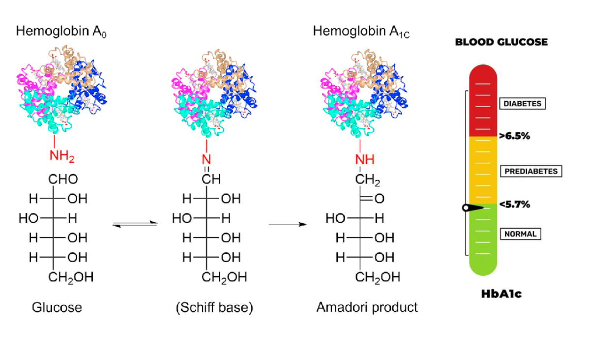
Image source: ResearchGate
4. Tumor marker blood test
This sounds scary, but it is a lifesaver. Cancer cells often release specific proteins into the blood. A Tumor marker blood test looks for these proteins.
These tests will help you the doctors identify if you have cancer and if so, whether it is spreading in your body. It also helps in determining the growth rate of the cancer cells.
If caught early, doctors will be able to choose the best medical treatment to cure you without any complications.
These tests mainly comprise the following three parts:
| Tumor Maker Test | Cancer Type | Tested to see what? |
|---|---|---|
| CA (Cancer Antigen) 125 | Ovarian Cancer | If the given treatment is working, or to see if cancer is coming back anytime soon, even after finishing the treatment. |
| CA 15-3 and CA 27-29 | Breast Cancer | This test is to monitor treatment for advanced breast cancer. |
| PSA (Prostate Specific Cancer) | Prostate Cancer | To see if you have prostate cancer and monitor the treatment if diagnosed already. |
| CEA (carcinoembryonic antigen) | Colorectal Cancer. (Also for the lung, stomach, thyroid, and pancreas) | Usually, to monitor the cancer treatment. |
| B2M (Beta 2 macroglobulin) | multiple myeloma, some lymphomas, and leukemias | To predict chances for recovery. |
Mainly, there are three ways to complete the Tumor maker test.
- Blood Test
- Urine Test
- Biopsy (Not as scary as you think): a small part of your tissue is removed and diagnosed.
Pro Tip for Readers:
Never interpret a Tumor Marker report alone. It must always be correlated with imaging (MRI/CT Scans) and a doctor’s clinical assessment.
How Does a Blood Test Work?
We have all been there—sitting in the chair, looking away while the nurse preps the needle. But understanding the science behind that pinch removes the fear.
Here is exactly what happens during a Blood Test Sample Collection at Home in Bengaluru.
The Vacuum Way (it doesn’t hurt!)
You might notice the nurse changes multiple tubes while the needle stays in your arm. This is possible because of the Vacutainer System.
- The needle is double-ended. One end goes into your vein, the other pierces the tube.
- The tubes have a pre-set vacuum inside. They automatically suck out the exact amount of blood needed (e.g., 3ml) and then stop.
- Result: One prick, multiple samples.
The Colorful Caps Decoder
Ever wonder why they use Purple, Red, or Yellow/Gray caps?
It’s not for decoration. Each color contains a different “chemical agent” to keep your blood ready for specific tests.
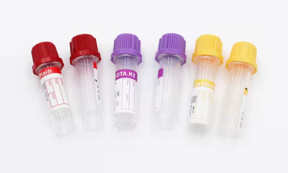
| Tube Cap Color | What’s Inside? | Used For |
|---|---|---|
| Lavender / Purple | EDTA (Anti-clotting agent) | CBC, HbA1c. It keeps blood liquid so cells can be counted. |
| Yellow / Gold | Gel Separator | Thyroid, Lipid, Vitamin B12. The gel separates cells from serum (the gold liquid). |
| Grey | Fluoride | Glucose (Sugar) Test. It freezes the sugar level instantly, so it doesn’t drop during transport. |
| Blue | Citrate | Clotting Tests (PT/INR). Used if you are on blood thinners. |
Do you need to fast for the blood test?
This is the #1 question people ask: “Can I have my morning chai?” The shortest answer to this question is, IT DEPENDS.
Getting this wrong is the main reason for inaccurate blood test results.
For example, if you eat, your blood turns “milky” (Lipemia), which confuses the lab machines.
Save this table before your next booking. Here we have simplified which test you are going for and if you need to fast or not.
| Test Name | Fasting Required? | Duration | Can I Drink Water? | Can I Drink Tea/Coffee? |
|---|---|---|---|---|
| Lipid Profile | MANDATORY | 10–12 Hours | YES (Plain water only) | NO (Not even without sugar) |
| Fasting Blood Sugar | MANDATORY | 8–10 Hours | YES | NO (Caffeine alters sugar) |
| Thyroid (TSH) | Preferred | Morning Sample | YES | NO (Tea can suppress TSH) |
| CBC / Platelets | NO | 0 Hours | YES | YES |
| HbA1c | NO | 0 Hours | YES | YES |
| Vitamin D / B12 | NO | 0 Hours | YES | YES |
Tip for “Smart” Fasting: Book your slot for 7:00 AM. Stop eating by 8:00 PM the previous night. You sleep through most of the fasting window and wake up ready for the test!
Does the Needle Hurt? (Honest Answer)
It feels like a sharp mosquito bite for 2 seconds.
How to reduce pain:
- Drink Water: Hydrated veins are “plump” and easier to find. Dry veins roll away, causing multiple pricks.
- Don’t Look: Watching the needle tenses your muscles, making it hurt more.
- Ask for a “Butterfly Needle”: If you have thin veins, ask Koshikaa’s phlebotomist for a butterfly needle (it’s thinner and gentler).
We believe knowing about what you are going through prepares you mentally and may even reduce the stress, which can affect the blood tests at some point, possibly.
Be as relaxed as possible for the honest results.
Blood Test Price in Bangalore
Bangalore has thousands of labs, from small single-room centres to giant national chains.
The price variation can be confusing. Why does one lab charge ₹300 while another charges ₹800 for the same test?
The variation in prices of the same lab may happen due to the type of service you are choosing, for such as, Koshikaa charges ₹349 for the blood test at home, whereas the same test can be completed for ₹299 by visiting their laboratory in Bangalore.
Don’t overpay. This is what you should be paying in Bangalore for high-quality labs (NABL Accredited).
| Test Name | Market Price Range (Bangalore) | Koshikaa Standard Rate for Home Sample | Koshikaa Standard Rate for Lab Visit | Report Time |
|---|---|---|---|---|
| CBC (Haemogram) | ₹250 – ₹450 | ₹349 | ₹299 | 6 – 8 Hours |
| Lipid Profile | ₹500 – ₹800 | ₹399 | ₹349 | 8 – 12 Hours |
| HbA1c (Sugar Average) | ₹450 – ₹700 | ₹349 | ₹299 | Same Day |
| Thyroid Profile (T3, T4, TSH) | ₹400 – ₹650 | ₹349 | ₹299 | Same Day |
| Vitamin D Total | ₹1,200 – ₹2,000 | ₹749 | ₹649 | 24 Hours |
| Vitamin B12 | ₹800 – ₹1,500 | ₹749 | ₹649 | 24 Hours |
If you need multiple tests, never book them individually.
Example: Booking CBC + Lipid + Thyroid separately costs ₹1,400. But a “Full Body Checkup” package often includes all these plus Liver & Kidney tests for just ₹1,099.
Always check for packages first.
You must be wondering where your money goes. Even if not, knowing this would make you wiser.
You aren’t just paying for a printout. Here is the hidden engineering behind the fee.
- The Cold Chain (20% of Cost): Your blood is alive. If it gets too hot in Bangalore traffic, the cells die. Premium services use Temperature-Controlled Boxes to keep samples at exactly 2°C–8°C during transport. Cheap labs use normal ice packs, which melt.
- The Technology (40% of Cost): Automated machines (like Roche or Abbott) cost crores. They give results with 99.9% accuracy. Manual microscope checking is cheaper but prone to human error.
- The Phlebotomist (15% of Cost): An expert phlebotomist finds the vein in one prick. An untrained one might need three. You pay for that skill and painless experience.
Is it safe to get a Blood Test Sample Collection at Home in Bengaluru?
Absolutely. In fact, it is often better because you are relaxed in your own chair (stress changes some blood markers!).
The 3-Step Process:
- The Booking: You select a slot (e.g., 7:00 AM) via the Koshikaa app/website.
- The Visit: A vaccinated phlebotomist arrives. They bring a sealed, sterile kit (never reused).
- Check: Ensure they open the needle packet in front of your eyes.
- The Transport: The sample is barcoded instantly (so no mix-ups happen) and placed in the cool box.
Understanding Your Report
My Report is Here. Now what?
So, the email from Koshikaa has arrived. You open the PDF, and you see some numbers in Bold or Red. You may jump to the question “Is something wrong?”
Before you panic-Google your symptoms (and convince yourself you have 3 days to live, which is sarcasm, absolutely.), follow this 3-Step Action Plan.
Step 1: The Assessment
Not all “Abnormal” results are dangerous. Classify your report into these three zones.
| Zone | What it looks like on the Report | The Action Plan |
|---|---|---|
| GREEN | All values are within the “Reference Range”. | Action: Smile! Your current lifestyle is working. Keep it up. Plan your next checkup in 12 months. |
| YELLOW | The value is slightly high/low. (e.g., Cholesterol is 210, limit is 200). | Action: This is a “Wake Up Call.” You don’t need medicine yet, but you DO need lifestyle changes. Retest in 3 months. |
| RED | Action: Do not wait. Book a doctor’s appointment immediately. |
Never look at a single number in isolation.
Example: High Total Cholesterol isn’t bad if your HDL (Good Cholesterol) is also very high. The ratio matters more.
Step 2: The “Lifestyle” Prescription
If you are in the Yellow Zone, start these changes TODAY.
- For High Cholesterol:
- Cut: Deep-fried foods, biscuits, and red meat.
- Add: Flaxseeds, walnuts, and oats (Soluble fiber cleans your pipes).
- Move: Walk 30 minutes daily to boost HDL.
- For High Blood Sugar (Pre-Diabetes):
- Cut: Fruit juices, white rice, and maida.
- Add: Methi water (morning) and raw veggies before every meal.
- Rule: “No carbs after 8 PM.”
- For Low Iron/Hemoglobin:
- Add: Dates, jaggery, and spinach.
- Tip: Squeeze lemon on your food (Vitamin C helps absorb Iron).
Step 3: The Doctor Discussion Checklist
Don’t go to the clinic empty-handed. Ask these 3 specific questions.
- “Is this ‘abnormal’ result a temporary spike?” (Stress, a recent cold, or a heavy dinner last night can skew results like WBC or Glucose).
- “Do I need medicine, or can I fix this with diet?” (Most doctors prefer diet changes for Borderline cases if you ask).
- “When should I re-test to check progress?” (Usually: Sugar = 3 months, Lipid = 6 weeks, Thyroid = 6 weeks).
If you ever feel any doubt about your blood health, test results or need any type of related consultation, feel free to contact the 24/7 support team of Koshikaa.
Conclusion
We spend thousands on car servicing and new mobile phones. But we often ignore the only machine that cannot be replaced—our body.
A blood test isn’t just about finding a disease; it is about Peace of Mind.
Knowing your blood test results are “Green” gives you confidence that no vitamin pill can buy. And if they are “Yellow,” you have the golden opportunity to fix it before it becomes “Red.”
In a city like Bangalore, where traffic makes everything hard, Koshikaa makes health easy. You don’t need to drive to a lab or wait in queues.
With our Blood Test Sample Collection at Home in Bengaluru, the lab comes to your doorstep. Whether you need a simple sugar check or a full preventive package, we ensure clinical precision at a fair Blood test Price in Bangalore.
Don’t wait for a symptom to force you into a hospital. Take charge today. Book a Blood test in Bengaluru with Koshikaa and secure your future, one drop at a time.
Frequently Asked Questions (FAQs)
Q1: Which Blood Test Requires Fasting?
Fasting is crucial for accuracy.
- Must Fast (10–12 Hours): Lipid Profile, Fasting Blood Sugar, Insulin.
- No Fasting Needed: CBC, HbA1c, Thyroid (TSH), Vitamin D/B12.
- Tip: Water is allowed, but strictly no tea, coffee, or biscuits.
Q2: How Does a Blood Test Work?
It is a simple 3-step science.
- Collection: A phlebotomist draws 3–5ml of blood using a vacuum tube system (painless).
- Processing: The blood is spun in a machine (centrifuge) to separate red cells from the liquid serum.
- Analysis: Automated analyzers count cells and measure chemicals to generate your report.
Q3: Can A Blood Test Detect Cancer?
Not directly, but it raises “Red Flags.” Tests like CBC can show abnormal white blood cells (Leukemia). Tumor Markers (like PSA for prostate or CA-125 for ovaries) indicate risk. However, a biopsy is always needed for a 100% confirmation.
Q4: How Long Does a Blood Test Take, and When Will I Receive the Results?
The sample collection takes just 5 to 10 minutes. For most routine tests (CBC, Sugar, Lipid), you will receive the digital report within 6 to 12 hours. Specialized tests (Vitamins, Hormones) may take 24 hours.
Q5: How Much Does A Blood Test Cost In Bangalore?
Prices vary by lab, but at Koshikaa, we ensure NABL-quality at honest prices.
- CBC: ~₹299
- Lipid Profile: ~₹349
- HbA1c: ~₹299
- Full Body Package: Starts from ₹1,099 (Best Value).
Sources:
For Test Parameters (RBC, WBC, Tumor Markers): Medlineplus.gov
For Lipid Profile Standards: National Heart Lung and Blood Institute
For Bangalore Market Pricing & Packages: Koshikaa
Images: (Google Images, Linked below each Image)