Prices As Low As

₹1500

Urine Test Near You In Bangalore

Elevate your well-being at our advanced Health Screening Centre in Bangalore, where you'll experience personalized care, precision screening, and peace of mind for a healthier tomorrow.

Cutting-edge Ultrasound Technology

We use the latest ultrasound equipment to ensure the highest quality images for precise diagnoses.

Expert Medical Team Members

Our experienced doctors and sonographers interpret the ultrasound results with great accuracy

Patient-Focused Approach

Your well-being is our priority, and we ensure a comfortable and stress-free experience during the ultrasound examination.

Urine Test in Bangalore

At Koshikaa, we offer advanced and comprehensive Urine testing services to provide you with valuable insights into your health. Our state-of-the-art laboratory and experienced medical professionals ensure accurate and reliable results, empowering you to make informed decisions about your well-being.
Blood Test

State-of-the-art laboratory

Accessible and affordable X-Ray In Bangalore

Accessible and affordable

Patient-Focused Approach

100% Reliable and accurate reports

Easy and convenient X Ray booking in Bangalore

Easy and convenient booking

PET CT scan in Bangalore with comprehensive screening

Comprehensive Screening

Request a Call Back

How Urine / Stool Test Can Save Lives

Experience reliable and accurate Urine/Stool Tests at Koshikaa. Our state-of-the-art facilities ensure precise results for comprehensive health assessment. Trust Koshikaa for your diagnostic needs.
We at Koshikaa recognize the significance of comprehensive health screening, which is why we provide a variety of diagnostic tests, such as analysis of stools and urine. These tests aid in the early detection of potential health issues and provide valuable insights into your overall health.
Our non-invasive urine test, also known as a urinalysis, involves analysing a sample of your urine. This straightforward yet potent test gives you a lot of information about how your body works and can help you find various health conditions. The presence of bacteria, red and white blood cell counts, protein levels, glucose levels, and pH balance are all evaluated by the test. Our skilled medical professionals are able to identify diabetes, urinary tract infections, kidney disorders, and other abnormalities of the urinary system by examining these markers. Effective treatment and prevention of complications require prompt diagnosis of these conditions.

What is Urine Test cost in Bangalore?

Bone marrow₹5999

Bone marrow is a soft, spongy tissue found inside certain bones, primarily in the center of large bones like the hip bones, breastbone (sternum), ribs, and vertebrae. It is responsible for producing blood cells, including red blood cells (erythrocytes), white blood cells (leukocytes), and platelets (thrombocytes).

Urine Analysis/Routine Examination₹199

A urine analysis, also known as a urinalysis or routine urine examination, is a common diagnostic test that evaluates the physical, chemical, and microscopic properties of urine.

Urine Culture ₹599

A urine culture is a laboratory test that examines urine for the presence of bacteria or fungi that can cause urinary tract infections (UTIs).

Urine Protein Test ₹299

A urine protein test, also known as a urine protein-to-creatinine ratio (UPCR) or urine albumin test, measures the amount of protein present in urine.

Urine Glucose Test ₹199

A urine glucose test is a diagnostic test that measures the level of glucose (sugar) present in urine.

Urine Ketone Test ₹199

A urine ketone test is a diagnostic test that measures the level of ketones in the urine.

Urine Pregnancy Test ₹99

A urine pregnancy test is a common diagnostic test used to detect human chorionic gonadotropin (hCG), a hormone produced by the placenta during pregnancy.

Urine Microscopy₹299

Urine microscopy is a laboratory test that examines urine under a microscope to detect and analyze the presence of cells, crystals, casts, bacteria, and other substances.

24-Hour Urine Protein Test ₹799

A 24-hour urine protein test, also known as a 24-hour urine protein excretion or proteinuria test, measures the total amount of protein excreted in urine over a 24-hour period.

What is Stool Test cost in Bangalore?

Stool Occult Blood (Benzidine) ₹119

Stool occult blood testing, using benzidine or other similar chemical reagents, is a diagnostic test used to detect the presence of small amounts of blood in the stool that are not visibly apparent (occult).

Stool Routine Examination ₹299

A stool routine examination, also known as a stool analysis or stool culture, is a diagnostic test used to evaluate the physical, chemical, and microbiological characteristics of stool.

Stool Culture ₹599

A stool culture is a diagnostic test used to detect and identify bacterial pathogens present in the stool.

Occult Blood Test (FOBT) ₹399

An Occult Blood Test, specifically the Fecal Occult Blood Test (FOBT), is a diagnostic screening tool used to detect hidden (occult) blood in the stool that is not visibly apparent.

Ova and Parasites (O&P) Test₹499

The Ova and Parasites (O&P) test is a diagnostic examination used to detect the presence of parasites or their eggs (ova) in stool samples.

Fecal Fat Test ₹999

The Fecal Fat Test, also known as the Quantitative Fecal Fat Test (QFFT) or Stool Fat Analysis, is a diagnostic test used to measure the amount of fat in stool samples.

Clostridium difficile (C. diff) Test ₹1299

The Clostridium difficile (C. diff) test is a diagnostic examination used to detect the presence of Clostridium difficile bacteria or their toxins in stool samples.

Calprotectin Test ₹1499

The Calprotectin Test is a diagnostic tool used to measure the levels of calprotectin in stool samples.

Helicobacter pylori (H. pylori) Antigen Test ₹899

The Helicobacter pylori (H. pylori) Antigen Test is a diagnostic tool used to detect the presence of H. pylori antigens in stool samples.

Where We Use Urine Test

Utilizing Urine test in Bangalore enables diagnosis of various conditions within the body,

Urine Culture Test

Gastrointestinal Tract

Heart

Urine Culture Test

Kidneys

Urine Culture Test

Liver

Urine Culture test

Pancreas

Urine Culture test

RBC

Urine Culture Test

Thyroid

How Urine test works?

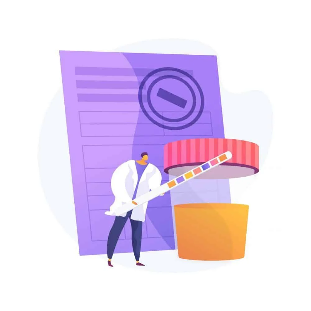
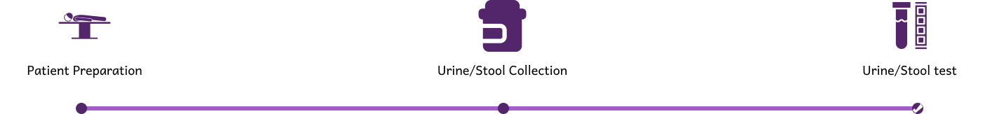
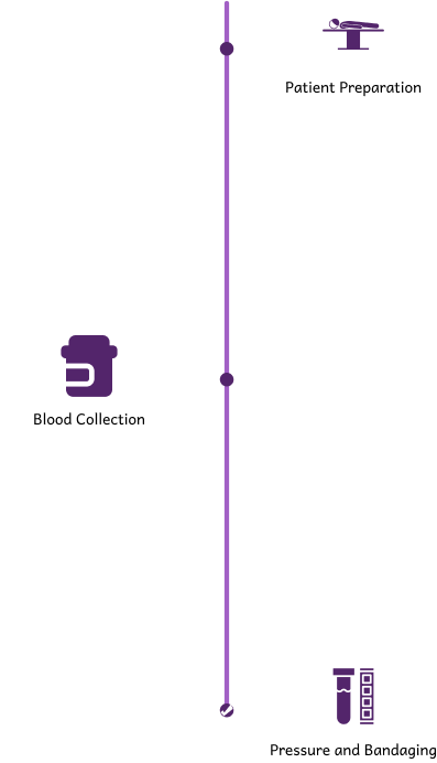
Patient Preparation: During blood test in Bangalore, you will sit on a comfortable table. Lab technician will clean the skin surface by application of an antiseptic liquid.

Blood Collection: Wrap an elastic band around patient's upper arm to apply pressure and blood is drawn.

Pressure and Bandaging: The elastic band and needle is removed. Apply cotton or a bandage to stop the bleeding.

Reviews from Koshikaa Customers

How Urine test works?

Urine Test in Bangalore

How does Urine test in Bangalore help in early cancer detection?

Frequently Asked Questions

What is a urine test and why is it performed?
  • A urine test, or urinalysis, is a diagnostic procedure used to examine the content of urine. It helps detect and manage a wide range of disorders, such as urinary tract infections, kidney disease, diabetes, and more. It evaluates various aspects like color, clarity, concentration, and content of the urine.
How should I prepare for a urine test?
  • Generally, no special preparation is required for a routine urine test. However, you may be advised to avoid certain foods, beverages, or medications that can affect test results. It’s best to follow any specific instructions given by your healthcare provider.
What types of urine tests are available?
  • There are several types of urine tests, including routine urinalysis, urine culture, 24-hour urine collection, and specific tests for drugs, pregnancy, and kidney function. Each type assesses different aspects of health and bodily function.
How is a urine sample collected?
  • For most urine tests, a “clean-catch” midstream sample is collected. This involves cleaning the genital area, starting to urinate into the toilet, then collecting the urine midstream into a sterile container. For certain tests, a 24-hour urine collection or a catheterized urine sample might be required.
What can a urine test detect?
  • A urine test can detect a variety of substances and conditions, including glucose (sugar), protein, blood, bacteria, ketones, and pH levels. It can indicate infections, kidney disease, diabetes, liver problems, and other metabolic disorders.
Are there any risks associated with urine tests?
  • Urine tests are generally safe and non-invasive with no significant risks. However, if a catheterized sample is required, there may be a slight risk of discomfort or infection.
How long does it take to get urine test results?
  • The time it takes to get results can vary. Routine urinalysis results are often available within a few hours, while more detailed tests like urine culture might take a few days to allow for bacterial growth.
What do abnormal urine test results mean?
  • Abnormal results can indicate various health issues. For example, high glucose levels may suggest diabetes, presence of white blood cells may indicate a urinary tract infection, and elevated protein levels could point to kidney disease. It’s important to discuss abnormal results with your healthcare provider to understand the implications and next steps.
MRI of the uterus is primarily used for diagnosing and evaluating conditions such as uterine fibroids, adenomyosis, and malignancies. It is particularly useful in the planning of surgical procedures and assessing the extent of gynecological diseases. This imaging test is valuable for its ability to deliver detailed cross-sectional images of the uterus and surrounding structures.
An MRI of the spleen can help diagnose various disorders affecting the spleen including splenomegaly (enlarged spleen), tumors, and other abnormalities. It provides clear, detailed images that are crucial for assessing the health of the spleen and planning potential surgical interventions.
Prostate MRI is a non-invasive imaging technique that provides detailed pictures of the prostate gland and surrounding tissues. It helps in the detection, staging, and monitoring of prostate cancer. Prostate MRIs are increasingly used to guide biopsy procedures and assist in the planning of treatment strategies for prostate health.
An MRI of the pancreas is used to identify issues such as pancreatitis, pancreatic cancer, and other pancreatic disorders. It offers high-resolution images that help in detecting small lesions and assessing the pancreatic ducts. This imaging technique is valuable for guiding biopsies and planning surgeries involving the pancreas.
MRI scans of the liver, kidney, and bile ducts provide detailed images that help diagnose various conditions, such as tumors, cysts, and blockages in the bile ducts. They are crucial for assessing organ damage, staging cancer, and planning liver or kidney-related surgeries. This type of MRI can also evaluate the health and function of these organs comprehensively.
Breast MRI is highly sensitive for detecting breast cancer, especially in individuals with dense breast tissue or a high risk of cancer. It is often used in conjunction with mammography to provide a more comprehensive evaluation. Breast MRIs can also monitor the response to breast cancer treatments and provide detailed pre-surgical mapping.
Brain and spine MRIs are critical tools for diagnosing and monitoring neurological conditions, including tumors, spinal cord injuries, and multiple sclerosis. These MRIs can reveal abnormalities in brain tissue and spinal cord structure that might not be visible with other imaging methods. They are essential for planning surgeries and assessing the effectiveness of treatments in neurology.
PET-CT scans of transplant organs like the liver, kidney, or pancreas are performed to assess metabolic activity, detect complications, and monitor post-transplant function. These scans play a vital role in the early detection of issues such as organ rejection, recurrence of disease, or infection, guiding timely interventions and optimizing transplant outcomes for patients.
PET-CT examinations of organs such as the liver, kidney, or prostate are crucial for assessing metabolic activity, detecting abnormalities, and monitoring disease progression. These scans help diagnose conditions such as liver metastases, renal cell carcinoma, or prostate cancer, guiding treatment decisions and monitoring response to therapy over time.
PET-CT scans of soft-tissue masses are utilized to assess metabolic activity and detect malignant tumors or other abnormalities. By combining PET imaging with CT imaging, these scans provide detailed information about tumor location, size, and metabolic characteristics, aiding in accurate diagnosis and treatment planning for soft-tissue cancers.
PET-CT scans are valuable for detecting cysts or fluid collections within the body, including cysts in the liver, kidneys, or other organs. These scans help characterize the nature and metabolic activity of cysts, guiding diagnosis and treatment planning for conditions such as cystic liver disease, renal cysts, or pancreatic cysts.
PET-CT scans of muscles are performed to assess metabolic activity and detect abnormalities such as inflammation, infection, or tumors. These scans provide valuable information about muscle function and pathology, aiding in the diagnosis and management of conditions like myositis, muscular dystrophy, or soft-tissue sarcomas.
PET-CT scans of the brain are utilized to evaluate brain metabolism and detect abnormalities such as tumors, epilepsy, or neurodegenerative diseases. By combining functional PET imaging with anatomical CT imaging, these scans offer comprehensive information about brain function and structure, guiding neurosurgical interventions and treatment decisions for neurological disorders
PET-CT scans of bones are commonly used to detect bone metastases in cancer patients, assess bone health in conditions like osteoporosis, and evaluate response to treatment. By combining PET and CT imaging, these scans provide detailed information about metabolic activity and anatomical changes in bone structures, aiding in accurate diagnosis and treatment planning for various bone-related conditions.
CT scans of the spine are conducted to assess the vertebrae, spinal cord, and surrounding soft tissues for conditions like herniated discs, spinal fractures, or spinal stenosis, aiding in the diagnosis and management of spinal disorders with detailed imaging essential for surgical planning and intervention.
CT scans of the pelvis are utilized to visualize the pelvic bones, reproductive organs, bladder, and surrounding structures for conditions such as fractures, tumors, or pelvic inflammatory disease, guiding treatment plans and surgical interventions with precise anatomical information.
CT scans of the neck are performed to evaluate structures such as the throat, thyroid gland, lymph nodes, and blood vessels for abnormalities like tumors, infections, or injuries, assisting in diagnosis and management of head and neck disorders with detailed imaging.
CT scans of the extremities are employed to assess bones, joints, and soft tissues in the arms or legs, aiding in the diagnosis of fractures, arthritis, or other musculoskeletal conditions, guiding orthopedic treatment and rehabilitation plans for optimal recovery.
CT scans of the chest are utilized to examine the lungs, heart, blood vessels, and surrounding structures for conditions such as lung nodules, pneumonia, or pulmonary embolism, facilitating accurate diagnosis and management of respiratory and cardiovascular diseases.
CT scans of the brain are conducted to assess brain structures, identify abnormalities like tumors, hemorrhages, or strokes, and aid in diagnosing neurological conditions, guiding neurosurgical interventions and treatment strategies with precise imaging of the brain’s anatomy.
CT scans of the abdomen are performed to visualize and evaluate the internal organs such as the liver, kidneys, pancreas, and intestines for conditions like tumors, infections, or injuries, providing detailed images essential for diagnosis and treatment planning.
Transplant organ ultrasound scans, including those of the liver, kidney, or pancreas, are performed to assess organ viability, detect complications, and monitor post-transplant function. These scans play a vital role in the early detection of issues such as organ rejection, vascular complications, or fluid collections, guiding timely interventions and optimizing transplant outcomes for patients.
Ultrasound examinations of organs such as the liver, kidney, or prostate are crucial for assessing organ structure, detecting abnormalities, and monitoring disease progression. These scans help diagnose conditions such as liver cirrhosis, kidney stones, or prostate enlargement, guiding treatment decisions and monitoring response to therapy over time.
Soft-tissue masses can be evaluated using ultrasound to determine their size, location, and characteristics. Ultrasound imaging helps differentiate between benign and malignant masses, guiding further diagnostic evaluation and treatment planning. By providing real-time imaging, ultrasound assists in the precise localization of soft-tissue masses for biopsy or surgical removal if needed.
Ultrasound scans are valuable for detecting cysts or fluid collections within the body, including cysts in the liver, kidneys, or other organs. These scans help characterize the nature and size of cysts, aiding in diagnosis and treatment planning for conditions such as cystic liver disease or renal cysts, and guiding interventions such as cyst drainage or sclerotherapy.
Musculoskeletal ultrasound scans are used to assess the health and integrity of muscles, identifying issues such as strains, tears, or muscle inflammation. These scans provide detailed images of muscle structures, allowing healthcare providers to diagnose muscle injuries accurately and develop personalized rehabilitation plans to promote recovery and restore function.
Ultrasound examinations of joints are essential for evaluating joint health, detecting abnormalities such as inflammation, fluid accumulation, or cartilage damage. By visualizing the joint structures in real-time, ultrasound helps diagnose conditions like arthritis, bursitis, or ligament injuries, guiding treatment decisions and monitoring disease progression over time.
Ultrasound scans of tendons are performed to assess for injuries, inflammation, or tears, aiding in the diagnosis and management of conditions such as tendonitis or tendon tears. These scans provide detailed images of tendon structures, helping healthcare providers determine the extent of damage and plan appropriate treatment strategies, including physical therapy or surgical intervention if necessary.
Pelvis X-rays play a crucial role in diagnosing injuries, diseases, and developmental conditions affecting the pelvic region. They provide detailed images of the hip bones, sacrum, and coccyx, aiding in the assessment of fractures, dislocations, or signs of arthritis. Pelvic X-rays are instrumental in guiding surgical interventions, monitoring healing progress, and evaluating conditions such as osteoporosis or tumors. By providing precise anatomical information, these imaging studies enable healthcare professionals to deliver personalized care and improve patient outcomes.
Teeth X-rays, known as dental radiographs, are indispensable tools for dentists to assess oral health. They aid in diagnosing dental decay, gum disease, and abnormalities in tooth structure or alignment. These images provide valuable insights into hidden dental issues, allowing for early intervention and prevention of complications. Additionally, dental X-rays are essential for planning treatments such as orthodontics, root canals, or dental implants, ensuring optimal outcomes and patient satisfaction.
Spine X-rays are pivotal in diagnosing spinal fractures, degenerative conditions, and spinal misalignments, providing critical information for treatment planning and monitoring spinal health. They offer detailed images of the vertebrae and surrounding structures, enabling healthcare professionals to identify issues such as scoliosis, disc herniation, or spinal stenosis
Skull X-rays are valuable for detecting fractures, assessing head trauma, and evaluating the alignment of facial bones, crucial in diagnosing conditions like fractures, tumors, or developmental abnormalities in the skull. They provide detailed views of the cranial structure, aiding clinicians in determining appropriate treatment strategies and ensuring patient safety.
For the heart, X-ray solutions can aid in the detection of various cardiac problems, ranging from mild to severe, including – Heart failure, Cardiac congestion and other heart-related conditions. In addition to cardiac assessments, X-ray solutions are also used for breast examinations to identify tumors or signs of breast cancer.
Bone X-rays are essential for diagnosing fractures and assessing bone alignment, aiding doctors in developing treatment plans and monitoring healing progress. They provide detailed images of the skeletal system, helping identify abnormalities or injuries that may not be visible through other imaging techniques
Abdominal X-rays are performed when patients experience symptoms such as significant nausea, ongoing vomiting, abdominal pain, flank or back pain, or abdominal swelling. These X-rays can help diagnose various conditions.
Search Bar

Frequently Booked Tests

Frequently Booked Checkups