Understanding the Importance of Routine Health Screenings

Full body checkup in Bangalore

Table of Contents

Regimen-based disease screening methods serve two essential functions which include disease prevention through early detection at treatable stages. Preventative medical attention stands equally important with sick patient care although most individuals wait until sickness to seek medical attention. The combination of regular health screenings protects lives while offering an improved quality of life to people. This article examines why health screening matters and reveals methods for achieving better life health.

What Are Health Screenings?

Health screenings involve performing multiple medical tests to detect possible health dangers and disease manifestations during a time before symptoms become apparent. Preventive measures can detect potential major illnesses during their early stages through proactive methods. Health screening programs use identification factors from three sectors: age, gender, and familial medical background.

Preview health screenings combine tests of blood pressure measurements with cholesterol levels and blood sugar checkups to detect developing chronic medical conditions including diabetes and heart disease. People can use screening results as a guide when creating programs to protect themselves from significant medical illnesses.

Why Health Screening is Important
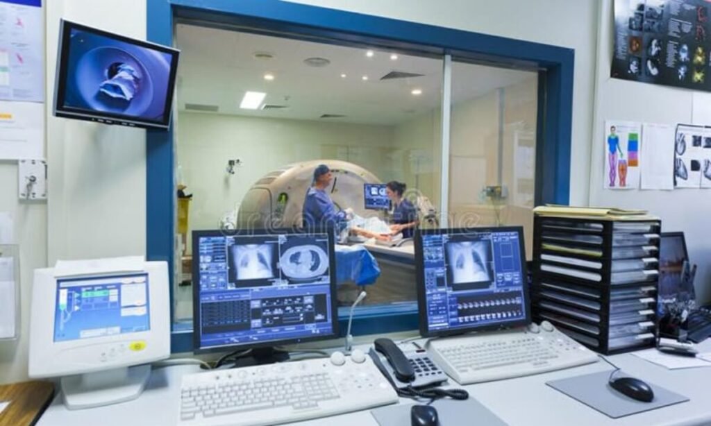
Pet CT Scan

Everyone should perform screening for various important reasons. Multiple advantages exist from using health screenings.

1. Early Detection Saves Lives

Health screenings help doctors discover symptoms that remain hidden or minor until they become noticeable. Detecting diseases early provides better treatment options together with reduced possibilities of medical complications.

2. Prevention of Chronic Diseases

The common practice of  full body checkup in Bangalore enables medical professionals to detect lifestyle diseases that include diabetes, obesity and high blood pressure. The early identification of such conditions allows healthcare professionals to treat patients through medication along with lifestyle modifications to stop complications from developing.

3. Reduce Healthcare Costs

Preventive measures adopt benefits for personal wellness and they make financial sense. Moving forward with preventive health checkup or scheduled tests proves much less expensive than confronting advanced medical conditions when their diagnosis remains undetected.

Key Screenings Everyone Should Consider

The following is a list of fundamental tests that healthcare providers recommend for every individual.

  • The monitoring of cardiovascular health through blood pressure and cholesterol tests should be performed by everyone, regardless of age.
  • Personal factors and family medical data determine the necessity of screenings that include mammograms, colonoscopies, or Pap smears.
  • Diabetes Screening: Detecting insulin resistance or elevated sugar levels.
  • Treatment of bone density requires testing specifically for people aged 50 years old and above.
  • Booster shots with immunizations protect against diseases that can be prevented including tetanus and flu.

Participate with a certified health screening centre in Bangalore because their personalized packages cater to individual health requirements leading to full assessments.

The Role of Preventive Health Checkups

Health screening tests provide medical protection for people’s physical wellness. People with diverse medical situations and risk levels should receive their checks either once each year or twice each year as per their needs. Here’s why they’re integral:

  • Preventive health checkups reveal comprehensive health information to doctors who use these results in making customized treatment plans for individuals.
  • Regular evaluations of your health condition bring peace of mind while bouncing your troubles in the form of worry.
  • Your knowledge of health matters leads you to practice beneficial healthy habits including eating a balanced diet together exercising regularly and managing stress effectively.
  • Visiting a health screening centre within Bangalore ensures residents will find both advanced diagnostic services combined with qualified medical personnel.

The Search for the Right Health Screening Facility

Regular screenings need people to pick a trustworthy health screening centre for maximum benefits. Use the following pointers for reference:

Reputation

People should select health screening centres that hold proper accreditation and maintain positive feedback. The name of a reliable testing facility ensures both accurate information and quick reporting.

Comprehensive Packages

Select medical centres that offer multiple testing packages to include full body checkups in Bangalore as their complete health assessment method.

Access to Specialists

Health screening centres should have medical specialists along with doctors who both explain your test results and provide appropriate guidance.

Convenience and Facilities

The centre selection should focus on facilities with comfortable state-of-the-art equipment and a convenient location within Bangalore city.

Making Health Screenings a Priority

The combination of work responsibilities and family obligations together with a dangerous belief in being invulnerable leads numerous individuals toward health neglect. Health represents the highest form of wealth so taking little steps now prevents future health emergencies. Various health examination methods which include annual physical checks and preventive screenings lead to substantial enhancements in your overall health condition.

People who live in cities with busy environments can lower their vulnerability to stress combined with pollution and lack of physical activity through routine medical checks. You should select a  health screening centre in Bangalore that provides state-of-the-art technology and specialized healthcare directly accessible to city residents.

The Road Ahead

The knowledge of health screening importance becomes essential for everyone who desires to live healthfully. Scheduled medical examinations help you find previously unseen medical conditions that can evolve into serious medical conditions. Preventing health problems always proves superior to healing from them after they occur. 

All patients should always prioritize their health because they can access simple cholesterol testing or services for full body checkup in Bangalore. A well-being-centered preventive strategy brings two distinct benefits by both extending life expectancy and adding life energy that enhances daily health.

In Conclusion

Having routine health screening tests represents affordable spending to achieve better health outcomes tomorrow. The combination of technological progress in healthcare makes both early diagnosis detection and prevention accessible and highly accurate to patients. Medical science offers effective ways to control lifestyle diseases together with genetic conditions when health professionals detect them at an early stage.

Every person of all ages should put preventive medical care at the top of their health priorities. A dependable health screening centre in Bangalore requires you to confirm both diagnostic capabilities and exceptional healthcare service delivery. Screening procedures provide you with the prevention of major health threats and enable both early diagnosis and quality preventative care for lifelong wellness.

Koshikaa operates as a respected health screening centre in Bangalore offering extensive and customized preventive care programs for all diagnostic requirements.

MRI of the uterus is primarily used for diagnosing and evaluating conditions such as uterine fibroids, adenomyosis, and malignancies. It is particularly useful in the planning of surgical procedures and assessing the extent of gynecological diseases. This imaging test is valuable for its ability to deliver detailed cross-sectional images of the uterus and surrounding structures.
An MRI of the spleen can help diagnose various disorders affecting the spleen including splenomegaly (enlarged spleen), tumors, and other abnormalities. It provides clear, detailed images that are crucial for assessing the health of the spleen and planning potential surgical interventions.
Prostate MRI is a non-invasive imaging technique that provides detailed pictures of the prostate gland and surrounding tissues. It helps in the detection, staging, and monitoring of prostate cancer. Prostate MRIs are increasingly used to guide biopsy procedures and assist in the planning of treatment strategies for prostate health.
An MRI of the pancreas is used to identify issues such as pancreatitis, pancreatic cancer, and other pancreatic disorders. It offers high-resolution images that help in detecting small lesions and assessing the pancreatic ducts. This imaging technique is valuable for guiding biopsies and planning surgeries involving the pancreas.
MRI scans of the liver, kidney, and bile ducts provide detailed images that help diagnose various conditions, such as tumors, cysts, and blockages in the bile ducts. They are crucial for assessing organ damage, staging cancer, and planning liver or kidney-related surgeries. This type of MRI can also evaluate the health and function of these organs comprehensively.
Breast MRI is highly sensitive for detecting breast cancer, especially in individuals with dense breast tissue or a high risk of cancer. It is often used in conjunction with mammography to provide a more comprehensive evaluation. Breast MRIs can also monitor the response to breast cancer treatments and provide detailed pre-surgical mapping.
Brain and spine MRIs are critical tools for diagnosing and monitoring neurological conditions, including tumors, spinal cord injuries, and multiple sclerosis. These MRIs can reveal abnormalities in brain tissue and spinal cord structure that might not be visible with other imaging methods. They are essential for planning surgeries and assessing the effectiveness of treatments in neurology.
PET-CT scans of transplant organs like the liver, kidney, or pancreas are performed to assess metabolic activity, detect complications, and monitor post-transplant function. These scans play a vital role in the early detection of issues such as organ rejection, recurrence of disease, or infection, guiding timely interventions and optimizing transplant outcomes for patients.
PET-CT examinations of organs such as the liver, kidney, or prostate are crucial for assessing metabolic activity, detecting abnormalities, and monitoring disease progression. These scans help diagnose conditions such as liver metastases, renal cell carcinoma, or prostate cancer, guiding treatment decisions and monitoring response to therapy over time.
PET-CT scans of soft-tissue masses are utilized to assess metabolic activity and detect malignant tumors or other abnormalities. By combining PET imaging with CT imaging, these scans provide detailed information about tumor location, size, and metabolic characteristics, aiding in accurate diagnosis and treatment planning for soft-tissue cancers.
PET-CT scans are valuable for detecting cysts or fluid collections within the body, including cysts in the liver, kidneys, or other organs. These scans help characterize the nature and metabolic activity of cysts, guiding diagnosis and treatment planning for conditions such as cystic liver disease, renal cysts, or pancreatic cysts.
PET-CT scans of muscles are performed to assess metabolic activity and detect abnormalities such as inflammation, infection, or tumors. These scans provide valuable information about muscle function and pathology, aiding in the diagnosis and management of conditions like myositis, muscular dystrophy, or soft-tissue sarcomas.
PET-CT scans of the brain are utilized to evaluate brain metabolism and detect abnormalities such as tumors, epilepsy, or neurodegenerative diseases. By combining functional PET imaging with anatomical CT imaging, these scans offer comprehensive information about brain function and structure, guiding neurosurgical interventions and treatment decisions for neurological disorders
PET-CT scans of bones are commonly used to detect bone metastases in cancer patients, assess bone health in conditions like osteoporosis, and evaluate response to treatment. By combining PET and CT imaging, these scans provide detailed information about metabolic activity and anatomical changes in bone structures, aiding in accurate diagnosis and treatment planning for various bone-related conditions.
CT scans of the spine are conducted to assess the vertebrae, spinal cord, and surrounding soft tissues for conditions like herniated discs, spinal fractures, or spinal stenosis, aiding in the diagnosis and management of spinal disorders with detailed imaging essential for surgical planning and intervention.
CT scans of the pelvis are utilized to visualize the pelvic bones, reproductive organs, bladder, and surrounding structures for conditions such as fractures, tumors, or pelvic inflammatory disease, guiding treatment plans and surgical interventions with precise anatomical information.
CT scans of the neck are performed to evaluate structures such as the throat, thyroid gland, lymph nodes, and blood vessels for abnormalities like tumors, infections, or injuries, assisting in diagnosis and management of head and neck disorders with detailed imaging.
CT scans of the extremities are employed to assess bones, joints, and soft tissues in the arms or legs, aiding in the diagnosis of fractures, arthritis, or other musculoskeletal conditions, guiding orthopedic treatment and rehabilitation plans for optimal recovery.
CT scans of the chest are utilized to examine the lungs, heart, blood vessels, and surrounding structures for conditions such as lung nodules, pneumonia, or pulmonary embolism, facilitating accurate diagnosis and management of respiratory and cardiovascular diseases.
CT scans of the brain are conducted to assess brain structures, identify abnormalities like tumors, hemorrhages, or strokes, and aid in diagnosing neurological conditions, guiding neurosurgical interventions and treatment strategies with precise imaging of the brain’s anatomy.
CT scans of the abdomen are performed to visualize and evaluate the internal organs such as the liver, kidneys, pancreas, and intestines for conditions like tumors, infections, or injuries, providing detailed images essential for diagnosis and treatment planning.
Transplant organ ultrasound scans, including those of the liver, kidney, or pancreas, are performed to assess organ viability, detect complications, and monitor post-transplant function. These scans play a vital role in the early detection of issues such as organ rejection, vascular complications, or fluid collections, guiding timely interventions and optimizing transplant outcomes for patients.
Ultrasound examinations of organs such as the liver, kidney, or prostate are crucial for assessing organ structure, detecting abnormalities, and monitoring disease progression. These scans help diagnose conditions such as liver cirrhosis, kidney stones, or prostate enlargement, guiding treatment decisions and monitoring response to therapy over time.
Soft-tissue masses can be evaluated using ultrasound to determine their size, location, and characteristics. Ultrasound imaging helps differentiate between benign and malignant masses, guiding further diagnostic evaluation and treatment planning. By providing real-time imaging, ultrasound assists in the precise localization of soft-tissue masses for biopsy or surgical removal if needed.
Ultrasound scans are valuable for detecting cysts or fluid collections within the body, including cysts in the liver, kidneys, or other organs. These scans help characterize the nature and size of cysts, aiding in diagnosis and treatment planning for conditions such as cystic liver disease or renal cysts, and guiding interventions such as cyst drainage or sclerotherapy.
Musculoskeletal ultrasound scans are used to assess the health and integrity of muscles, identifying issues such as strains, tears, or muscle inflammation. These scans provide detailed images of muscle structures, allowing healthcare providers to diagnose muscle injuries accurately and develop personalized rehabilitation plans to promote recovery and restore function.
Ultrasound examinations of joints are essential for evaluating joint health, detecting abnormalities such as inflammation, fluid accumulation, or cartilage damage. By visualizing the joint structures in real-time, ultrasound helps diagnose conditions like arthritis, bursitis, or ligament injuries, guiding treatment decisions and monitoring disease progression over time.
Ultrasound scans of tendons are performed to assess for injuries, inflammation, or tears, aiding in the diagnosis and management of conditions such as tendonitis or tendon tears. These scans provide detailed images of tendon structures, helping healthcare providers determine the extent of damage and plan appropriate treatment strategies, including physical therapy or surgical intervention if necessary.
Pelvis X-rays play a crucial role in diagnosing injuries, diseases, and developmental conditions affecting the pelvic region. They provide detailed images of the hip bones, sacrum, and coccyx, aiding in the assessment of fractures, dislocations, or signs of arthritis. Pelvic X-rays are instrumental in guiding surgical interventions, monitoring healing progress, and evaluating conditions such as osteoporosis or tumors. By providing precise anatomical information, these imaging studies enable healthcare professionals to deliver personalized care and improve patient outcomes.
Teeth X-rays, known as dental radiographs, are indispensable tools for dentists to assess oral health. They aid in diagnosing dental decay, gum disease, and abnormalities in tooth structure or alignment. These images provide valuable insights into hidden dental issues, allowing for early intervention and prevention of complications. Additionally, dental X-rays are essential for planning treatments such as orthodontics, root canals, or dental implants, ensuring optimal outcomes and patient satisfaction.
Spine X-rays are pivotal in diagnosing spinal fractures, degenerative conditions, and spinal misalignments, providing critical information for treatment planning and monitoring spinal health. They offer detailed images of the vertebrae and surrounding structures, enabling healthcare professionals to identify issues such as scoliosis, disc herniation, or spinal stenosis
Skull X-rays are valuable for detecting fractures, assessing head trauma, and evaluating the alignment of facial bones, crucial in diagnosing conditions like fractures, tumors, or developmental abnormalities in the skull. They provide detailed views of the cranial structure, aiding clinicians in determining appropriate treatment strategies and ensuring patient safety.
For the heart, X-ray solutions can aid in the detection of various cardiac problems, ranging from mild to severe, including – Heart failure, Cardiac congestion and other heart-related conditions. In addition to cardiac assessments, X-ray solutions are also used for breast examinations to identify tumors or signs of breast cancer.

Bone X-rays are essential for diagnosing fractures and assessing bone alignment, aiding doctors in developing treatment plans and monitoring healing progress. They provide detailed images of the skeletal system, helping identify abnormalities or injuries that may not be visible through other imaging techniques

Abdominal X-rays are performed when patients experience symptoms such as significant nausea, ongoing vomiting, abdominal pain, flank or back pain, or abdominal swelling. These X-rays can help diagnose various conditions.
Search Bar

Frequently Booked Tests

Frequently Booked Checkups