
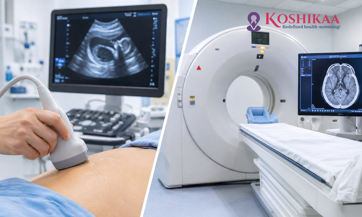
Ultrasound and CT scans are two of the most commonly used imaging tests, and both allow doctors to see inside the body without surgery. Yet they work in completely different ways, and each is better suited to certain conditions. If you have been referred for one of these scans, or wondered why your doctor chose one over the other, understanding the difference can help you feel more confident about the process.
In short, ultrasound uses sound waves, involves no radiation and shows organs in real time, while CT uses X-rays to produce highly detailed cross-sectional images. Neither test is better in every situation. The right choice depends on the part of the body being examined, the suspected condition, how urgent the situation is and individual factors such as pregnancy.
This guide explains how each test works, the key differences between them, which conditions each is commonly used for and how doctors decide which scan is appropriate. Whether you visit a trusted health screening centre in Bangalore for a routine checkup or need a CT scan in Bangalore after a doctor’s referral, knowing what to expect from each test will help you prepare better.
What Is the Difference Between Ultrasound and CT?
The fundamental difference is the technology. Ultrasound uses high-frequency sound waves that bounce off tissues to create images, while a CT (computed tomography) scan uses X-rays taken from many angles, which a computer combines into detailed slices of the body.
This means ultrasound is radiation-free, portable and excellent for looking at soft organs and moving structures, such as blood flow or a developing baby. CT provides a broader, more detailed view of the whole area being examined, including bones, lungs and deeper structures that ultrasound cannot see clearly.
How Does Ultrasound Imaging Work?
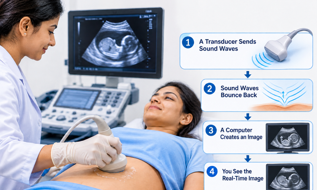
During an ultrasound, a sonographer or radiologist applies a water-based gel to your skin and moves a small handheld device called a transducer over the area being examined. The transducer sends sound waves into the body and picks up the echoes, which are turned into live images on a screen.
Because the images are produced in real time, ultrasound can show movement, such as blood flowing through vessels (Doppler ultrasound) or a baby moving in the womb. The scan is painless, and most examinations take about 15 to 30 minutes. Some scans need preparation, such as fasting for an abdominal ultrasound or a full bladder for a pelvic or KUB scan.
Learn more about our ultrasound scan in Bangalore services.
How Does CT Imaging Work?
During a CT scan, you lie on a table that moves through a wide, ring-shaped scanner. An X-ray tube rotates around you, capturing images from many angles. A computer then creates detailed cross-sectional images that can also be combined into 3D views.
The scanning itself is very quick, often just seconds for a single area, and the whole appointment usually takes 15 to 30 minutes. For some scans, a contrast agent containing iodine is injected into a vein or taken by mouth to make blood vessels and organs stand out more clearly.
Find out more about our CT scan in Bangalore services.
Ultrasound vs CT Scan: Key Differences
| Feature | Ultrasound | CT Scan |
| Technology | High-frequency sound waves | X-rays and computer processing |
| Ionising radiation | None | Yes, a small, controlled dose |
| Type of images | Real-time, moving images | Detailed cross-sectional and 3D images |
| Best at showing | Soft organs, fluid, blood flow, pregnancy | Bone, lungs, bleeding, deep structures, wider areas |
| Limitations | Cannot see through bone or air; image quality varies with body size and bowel gas | Less suited to repeated use; contrast not suitable for everyone |
| Use in pregnancy | Safe and routinely used | Generally avoided unless medically necessary |
| Typical cost | Usually lower | Usually higher |
| Preparation | Sometimes fasting or a full bladder | Sometimes fasting, especially with contrast |
Neither test is universally more accurate. Each is more accurate for certain questions and less useful for others.
Which Test Is Used for Different Conditions?
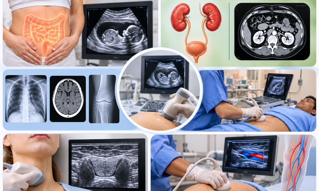
Abdominal and Pelvic Conditions
Ultrasound is often the first test for abdominal pain, particularly when gallstones, liver changes, fatty liver or problems with the uterus and ovaries are suspected. It is also widely used to investigate conditions such as polycystic ovaries and fibroids.
CT may be recommended when ultrasound does not give a clear answer, when a wider view of the abdomen is needed or in emergencies. For suspected appendicitis, ultrasound is often preferred first in children and pregnant women, while CT is commonly used in adults, although the choice also depends on the clinical pathway, availability and severity. Starting with ultrasound and moving to CT if needed is normal practice.
Kidney and Urinary Tract Concerns
For kidney and bladder problems, ultrasound is a safe first test that can show swelling of the kidneys, cysts, larger stones and how well the bladder empties. A non-contrast CT KUB scan is more accurate for detecting and measuring kidney and ureteric stones, especially small ones, and is often used before planning stone treatment. Read our KUB ultrasound guide to learn more about this scan.
Chest, Head and Bone Conditions
Ultrasound cannot see through air or bone, so it is of limited use for the lungs, brain and skeleton. For chest symptoms, a chest X-ray is often the first test, with CT used when more detail is needed. For many fractures, an X-ray is also the first test, while CT gives more detail in complex injuries. CT is commonly used to investigate head injuries and detect bleeding in the brain.
In a suspected stroke, a non-contrast CT scan is usually the first test because it is fast and effective at showing bleeding in the brain. However, it may not show an early ischaemic stroke caused by a blocked artery. Depending on the clinical situation, doctors may also use CT angiography, CT perfusion or MRI to assess blood vessels and brain tissue.
Injuries and Urgent Medical Conditions
In major trauma, a bedside ultrasound may be used first to look quickly for free fluid or bleeding in the abdomen or chest. If the patient is stable, a CT scan can provide a more detailed assessment of internal injuries. The choice depends on the person’s condition and the emergency team’s assessment, and in some situations immediate surgery or resuscitation takes priority over any scan.
Neck, Thyroid and Blood Vessels
Ultrasound is commonly the first test for examining thyroid nodules, neck lumps and many blood vessel problems, such as checking for clots in the leg veins using Doppler ultrasound. CT or CT angiography may be used for more complex vascular problems. It can also guide needles during biopsies and other procedures. Because it shows images in real time, the doctor can watch the needle reach the right spot, which makes these procedures safer and more accurate.
Radiation, Safety and Limitations
Ultrasound Safety
Ultrasound does not use ionising radiation and is considered safe for people of all ages, including during pregnancy and for repeated monitoring. Its main limitations are that it cannot see through bone or air, and image quality can be affected by body size, bowel gas and the skill of the person performing the scan.
CT Safety
CT uses ionising radiation. The dose from a single scan is carefully controlled, and the individual risk is small, but doctors avoid unnecessary repeat scans, particularly in children. CT is generally avoided during pregnancy unless it is medically necessary, in which case steps are taken to minimise exposure. Our article on CT radiation exposure and safety measures explains this in more detail.
Contrast Considerations
Some CT scans use an iodine-based contrast agent. Tell the team if you have kidney problems, diabetes, asthma or have reacted to contrast before. Ultrasound rarely needs contrast, and when it does, the contrast agent is different and does not involve radiation.
How to Prepare for Each Test
Preparing for an Ultrasound
Preparation depends on the area being scanned:
• Abdominal ultrasound: you will usually be asked to fast for six to eight hours, so that gas in the bowel does not block the view of organs such as the gallbladder and pancreas
• Pelvic or KUB ultrasound: you will usually be asked to drink several glasses of water beforehand and avoid passing urine until the scan, as a full bladder improves the images
• Thyroid, neck, breast and Doppler scans: usually need no special preparation
Preparing for a CT Scan
• Plain CT scans: often need no special preparation apart from removing metal items such as jewellery and belts
• CT scans with contrast: you may be asked to fast for four to six hours, and a recent kidney function blood test may be requested if you have diabetes, kidney disease or other risk factors
• Abdominal CT: you may be given a drink of oral contrast to take before the scan
For both tests, wear loose, comfortable clothing, bring your prescription and previous reports, and tell the team if you are or might be pregnant. Always follow the specific instructions you are given when you book.
How Do Doctors Decide Which Scan Is Appropriate?
Your doctor considers several factors when choosing between ultrasound and CT:
• The organ or symptom involved: some organs are best seen with ultrasound, others with CT
• How urgent the situation is: emergencies may call for the speed and detail of CT
• Radiation sensitivity: children and pregnant women are usually offered ultrasound first where possible
• Previous test results: an unclear ultrasound may be followed by CT, or a CT finding may be checked with ultrasound
• Individual factors: such as kidney function, allergies, body size and the ability to lie still
It is common to need both tests. They provide complementary information, and using them together often leads to a clearer diagnosis.
Ultrasound and CT Scans at Koshikaa
Koshikaa offers both ultrasound and CT scans at our centre in Banashankari, Bengaluru. Ultrasound is generally the more affordable option. As an indicative guide, an abdomen and pelvis ultrasound is currently listed at ₹1,000, and some plain CT scans start from around ₹4,000. Prices depend on the area scanned, whether contrast is used and any current offers, so please confirm when you book.Reports are usually shared within 24 to 48 hours.
Frequently Asked Questions
Which is better, ultrasound or CT?
Neither is universally better. Doctors choose based on the body area, symptoms, urgency, pregnancy status and the clinical question being asked.
Is a CT scan more accurate than an ultrasound?
It depends on what is being examined. CT is more accurate for bones, lungs, bleeding and small kidney stones, while ultrasound is better for many soft organs, blood flow, the thyroid and pregnancy.
Is ultrasound always the first test?
Often, but not always. Ultrasound is commonly used first for abdominal, pelvic, kidney and thyroid concerns, but CT may be chosen first in emergencies, for head injuries or for lung problems.
Is the radiation from a CT scan dangerous?
The radiation dose from a single CT scan is small and carefully controlled, and the individual risk is low. Doctors recommend CT only when the expected benefit outweighs the risk.
Can I have both an ultrasound and a CT scan?
Yes. Many people have both, as the tests provide different information. An ultrasound is often done first, with CT used if more detail is needed.
Which is cheaper, ultrasound or CT?
Ultrasound is usually cheaper than CT. Exact costs depend on the body part being scanned and whether contrast is needed.
Is a CT scan safe during pregnancy?
CT is generally avoided in pregnancy unless it is medically necessary. Ultrasound, and sometimes MRI, are preferred because they do not use ionising radiation.
Conclusion
Ultrasound and CT are both valuable imaging tests, but they answer different questions. Ultrasound is safe, radiation-free and ideal for many soft organs, pregnancy and blood flow, while CT offers fast, detailed images of bones, lungs, bleeding and complex conditions. Your doctor will recommend the test, or combination of tests, that gives the clearest answer for your situation.
To book an ultrasound or CT scan, contact Koshikaa in Banashankari, Bengaluru, on +91 7996666106.