Ultrasound Scan in JP Nagar

Get a safe and accurate ultrasound scan in JP Nagar at our trusted health screening centre in Bangalore, where we offer advanced imaging, quick reports, and patient-friendly diagnostic care for every need.

About Ultrasound Scan

An ultrasound scan is a non-invasive imaging test that uses sound waves to create real-time body images. It helps diagnose medical conditions safely and is widely preferred for routine evaluations.
When choosing an Ultrasound Scan in Bangalore, accuracy and expertise matter. Modern centres provide detailed imaging for abdominal, pelvic, and pregnancy assessments, ensuring timely diagnosis and improved clinical decision-making.
Our reliable health screening centre in Bangalore offers advanced ultrasound technology, skilled radiologists, and quick report delivery. These scans support preventive healthcare by detecting issues early, guiding treatments, and helping patients understand their health status with clarity and confidence.

Request a Call Back

Types Of Ultrasound Scan

Using an ultrasound scan helps doctors clearly see different parts of the body. Moreover, it helps detect various conditions effectively.

Abdomen Ultrasound Scan

Abdomen

An abdominal ultrasound provides clear images of organs like the liver, pancreas, and gallbladder, helping diagnose pain, infections, or digestive system concerns effectively.
Pelvic Ultrasound Scan

Pelvic

A pelvic ultrasound evaluates reproductive organs, including the uterus and ovaries, assisting in diagnosing menstrual issues, pelvic pain, fertility concerns, and pregnancy.
Ultrasound Scan For Joints

Joints

A Joint ultrasound helps detect inflammation, ligament injuries, fluid accumulation, and arthritis-related changes, offering accurate diagnosis and treatment planning.
Ultrasound For Kidney

Kidney

A Kidney ultrasound identifies stones, cysts, structural abnormalities, and infections, supporting early detection and effective management of various renal health concerns.
KUB Ultrasound Scan

KUB Ultrasound

A KUB ultrasound scans the kidneys, ureters, and bladder to detect stones, obstructions, or infections, providing essential insights into urinary tract health.

Cost of Ultrasound Scan in JP Nagar

Abdominal Ultrasound ₹1250

An abdominal ultrasound captures detailed images of internal organs, helping diagnose pain, infections, stones, and digestive issues with safe, non-invasive real-time imaging.

USG Abdomen and Pelvis ₹1000

A USG abdomen and pelvis scan provides detailed images of abdominal and pelvic organs, helping detect pain causes, infections, stones, and reproductive health issues.

Kidney Ultrasound ₹850

A kidney ultrasound produces clear images of the kidneys to detect stones, infections, cysts, or structural issues, supporting early diagnosis and effective treatment planning.

Breast Ultrasound ₹1000

A breast ultrasound uses sound waves to examine breast tissue, helping detect lumps, cysts, or abnormalities and supporting accurate, early diagnosis without radiation exposure.

Pelvis Ultrasound ₹850

A pelvis ultrasound captures detailed images of reproductive organs, helping diagnose pelvic pain, menstrual issues, fertility concerns, and other abnormalities with safe, non-invasive evaluation.

Transvaginal Ultrasound ₹1000

A transvaginal ultrasound provides close-up imaging of pelvic organs using an internal probe, helping detect menstrual issues, fertility concerns, early pregnancy details, and pelvic abnormalities accurately.

Thyroid Ultrasound ₹1000

A thyroid ultrasound produces clear images of the thyroid gland, helping detect nodules, swelling, cysts, or structural abnormalities for accurate diagnosis and treatment planning.

When An Ultrasound Scan Is Recommended

Why Choose Koshikaa's Ultrasound Scan in JP Nagar?

Advanced Scan Technology

Advanced Imaging Technology

Uses modern ultrasound machines that deliver clear, precise images for accurate diagnosis and improved patient outcomes.

Expert Radiologists

Expert Radiologists

Skilled radiologists interpret scans accurately, offering reliable insights for timely and effective medical decisions.

Reliable Service

Maintains consistent, dependable diagnostic services with a strong focus on accuracy and patient satisfaction.

Hygiene & Comfortable Atmosphere

Follows strict hygiene protocols and offers a clean, welcoming environment to ensure patient comfort.

Fast Reports & Easy Booking

Ensures quick report delivery and hassle-free appointment scheduling for a smooth patient experience.

Affordable Scan Price

Affordable Pricing

Provides high-quality ultrasound scans at budget-friendly rates, ensuring accessible healthcare for every patient.

How to Book Ultrasound Scan

Booking an Affordable Ultrasound scan at the Best Health Screening Centre is easy and takes less than a minute. Just follow these 4 simple steps: 

Fill Out the Booking Form

Complete the online booking form with your details and preferred time, allowing the centre to schedule your ultrasound scan quickly and conveniently.

Receive a Confirmation Call

A team member will call to confirm your appointment, verify details, and answer any questions regarding your ultrasound scan.

Get Details

You will receive complete information about the scan, including preparation guidelines, reporting time, location, and necessary documents to carry.

Walk In for Ultrasound Scan

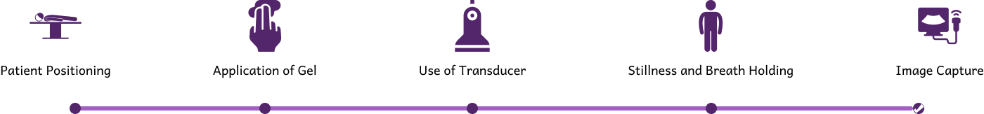
Visit the centre at your scheduled time, complete the simple check-in process, and proceed with your ultrasound scan without delay.

How To Prepare For An Ultrasound Scan?

Fasting Instructions

Follow the fasting instructions given in clinic.

Arrive Early

Arrive early for smooth registration.

Drink Water

Drink water if bladder filling is required.

Wear comfortable, easily removable clothing.

Wear comfortable, easily removable clothing.

Previous Reports

Bring previous reports for comparison.

How Ultrasound Scan Works?

process of ultrasound scan
Best ultrasound scan in Bangalore

Benefits Of Ultrasound Scanning

Safe Scanning Process

Safe Scanning Process

Ultrasound scanning offers safe, radiation-free imaging, making it ideal for pregnancy, routine health checks, and diagnosing various medical conditions accurately.
Better Treatment Outcomes

Better Treatment Outcomes

It provides real-time visuals of internal organs, helping doctors assess abnormalities, monitor changes, and make timely clinical decisions for effective treatment.

Non-invasive Procedure

Ultrasound scans are quick, non-invasive, and painless, ensuring patient comfort while delivering detailed diagnostic information for various health concerns.
Improved Visibility of Abnormality

Improved Visibility

They help detect stones, cysts, infections, and structural issues early, supporting preventive healthcare and reducing future complications significantly.

accurate imaging

Accurate Imaging

Ultrasound scans guide medical procedures like biopsies, ensuring precision, reducing risks, and improving overall diagnostic accuracy for patients.

Reviews

Our Recent Blogs

Frequency Asked Questions

How Much Does An Ultrasound Cost In JP Nagar?

Ultrasound costs in JP Nagar vary depending on the scan type. Koshikaa offers budget-friendly, transparent pricing with high-quality imaging, ensuring patients receive accurate diagnostic results without financial burden.

What Is A Type 2 Ultrasound For Pregnancy?

A Type 2 ultrasound, also known as the anomaly scan, is done around 18–22 weeks to assess fetal organs, growth, and development and detect any abnormalities for safer pregnancy monitoring.

Can I Get Same-Day Ultrasound Reports?

Yes, most ultrasound scans at Koshikaa offer same-day reports. Radiologists review images promptly to ensure quick, accurate results for timely medical decision-making.

Can I Eat Or Drink Before An Abdominal Ultrasound?

For abdominal ultrasounds, fasting is generally required for clearer imaging. Follow the centre’s specific instructions regarding food, water, and preparation to ensure accurate diagnostic results.

Do You Offer Pregnancy And Fetal Ultrasound Scans?

Yes, Koshikaa provides a complete range of pregnancy and fetal scans, including early pregnancy, NT, anomaly, growth, and 3D/4D ultrasounds for comprehensive fetal monitoring.

How Early Should I Arrive For My Ultrasound Appointment?

Arrive 10–15 minutes early to complete basic registration, receive instructions, and ensure a smooth, stress-free start to your ultrasound appointment.

Where Can I Get A Reliable Ultrasound Scan In JP Nagar?

Koshikaa offers reliable ultrasound services in JP Nagar with advanced imaging technology, experienced radiologists, fast reporting, and patient-focused care for all types of scans.

What Are The Services Offered By Koshikaa?

Koshikaa provides ultrasound scans, X-rays, CTs, MRIs, PET-CTs, blood tests, health checkups, and specialized diagnostic services, offering complete imaging and screening under one roof.

How Do I Reach Your Ultrasound Scan Centre In JP Nagar?

The centre is conveniently located in JP Nagar with easy access by public and private transport. After booking, you’ll receive exact directions and location details for a smooth visit.

What Types Of Ultrasound Scans Are Available In JP Nagar?

Available scans include abdomen, pelvis, pregnancy, breast, thyroid, kidney, KUB, joint, and soft-tissue ultrasounds, all performed using advanced equipment for clear and accurate results.

Are There Affordable Ultrasound Scan Options In JP Nagar For Routine And Pregnancy Scans?

Yes, Koshikaa offers cost-effective ultrasound services for routine, diagnostic, and pregnancy scans, ensuring affordability without compromising on accuracy or care quality.

How Do I Book An Appointment For An Ultrasound Scan At Koshikaa?

You can book by filling out the online form, calling the centre +91 7996666104, or walking in directly. The team will confirm your appointment and provide preparation details.

What Are The Clinic Hours For Ultrasound Scans At Koshikaa?

Koshikaa offers convenient morning and evening slots for ultrasound scans. You can check the exact hours online or contact the centre for updated scheduling details.
MRI of the uterus is primarily used for diagnosing and evaluating conditions such as uterine fibroids, adenomyosis, and malignancies. It is particularly useful in the planning of surgical procedures and assessing the extent of gynecological diseases. This imaging test is valuable for its ability to deliver detailed cross-sectional images of the uterus and surrounding structures.
An MRI of the spleen can help diagnose various disorders affecting the spleen including splenomegaly (enlarged spleen), tumors, and other abnormalities. It provides clear, detailed images that are crucial for assessing the health of the spleen and planning potential surgical interventions.
Prostate MRI is a non-invasive imaging technique that provides detailed pictures of the prostate gland and surrounding tissues. It helps in the detection, staging, and monitoring of prostate cancer. Prostate MRIs are increasingly used to guide biopsy procedures and assist in the planning of treatment strategies for prostate health.
An MRI of the pancreas is used to identify issues such as pancreatitis, pancreatic cancer, and other pancreatic disorders. It offers high-resolution images that help in detecting small lesions and assessing the pancreatic ducts. This imaging technique is valuable for guiding biopsies and planning surgeries involving the pancreas.
MRI scans of the liver, kidney, and bile ducts provide detailed images that help diagnose various conditions, such as tumors, cysts, and blockages in the bile ducts. They are crucial for assessing organ damage, staging cancer, and planning liver or kidney-related surgeries. This type of MRI can also evaluate the health and function of these organs comprehensively.
Breast MRI is highly sensitive for detecting breast cancer, especially in individuals with dense breast tissue or a high risk of cancer. It is often used in conjunction with mammography to provide a more comprehensive evaluation. Breast MRIs can also monitor the response to breast cancer treatments and provide detailed pre-surgical mapping.
Brain and spine MRIs are critical tools for diagnosing and monitoring neurological conditions, including tumors, spinal cord injuries, and multiple sclerosis. These MRIs can reveal abnormalities in brain tissue and spinal cord structure that might not be visible with other imaging methods. They are essential for planning surgeries and assessing the effectiveness of treatments in neurology.
PET-CT scans of transplant organs like the liver, kidney, or pancreas are performed to assess metabolic activity, detect complications, and monitor post-transplant function. These scans play a vital role in the early detection of issues such as organ rejection, recurrence of disease, or infection, guiding timely interventions and optimizing transplant outcomes for patients.
PET-CT examinations of organs such as the liver, kidney, or prostate are crucial for assessing metabolic activity, detecting abnormalities, and monitoring disease progression. These scans help diagnose conditions such as liver metastases, renal cell carcinoma, or prostate cancer, guiding treatment decisions and monitoring response to therapy over time.
PET-CT scans of soft-tissue masses are utilized to assess metabolic activity and detect malignant tumors or other abnormalities. By combining PET imaging with CT imaging, these scans provide detailed information about tumor location, size, and metabolic characteristics, aiding in accurate diagnosis and treatment planning for soft-tissue cancers.
PET-CT scans are valuable for detecting cysts or fluid collections within the body, including cysts in the liver, kidneys, or other organs. These scans help characterize the nature and metabolic activity of cysts, guiding diagnosis and treatment planning for conditions such as cystic liver disease, renal cysts, or pancreatic cysts.
PET-CT scans of muscles are performed to assess metabolic activity and detect abnormalities such as inflammation, infection, or tumors. These scans provide valuable information about muscle function and pathology, aiding in the diagnosis and management of conditions like myositis, muscular dystrophy, or soft-tissue sarcomas.
PET-CT scans of the brain are utilized to evaluate brain metabolism and detect abnormalities such as tumors, epilepsy, or neurodegenerative diseases. By combining functional PET imaging with anatomical CT imaging, these scans offer comprehensive information about brain function and structure, guiding neurosurgical interventions and treatment decisions for neurological disorders
PET-CT scans of bones are commonly used to detect bone metastases in cancer patients, assess bone health in conditions like osteoporosis, and evaluate response to treatment. By combining PET and CT imaging, these scans provide detailed information about metabolic activity and anatomical changes in bone structures, aiding in accurate diagnosis and treatment planning for various bone-related conditions.
CT scans of the spine are conducted to assess the vertebrae, spinal cord, and surrounding soft tissues for conditions like herniated discs, spinal fractures, or spinal stenosis, aiding in the diagnosis and management of spinal disorders with detailed imaging essential for surgical planning and intervention.
CT scans of the pelvis are utilized to visualize the pelvic bones, reproductive organs, bladder, and surrounding structures for conditions such as fractures, tumors, or pelvic inflammatory disease, guiding treatment plans and surgical interventions with precise anatomical information.
CT scans of the neck are performed to evaluate structures such as the throat, thyroid gland, lymph nodes, and blood vessels for abnormalities like tumors, infections, or injuries, assisting in diagnosis and management of head and neck disorders with detailed imaging.
CT scans of the extremities are employed to assess bones, joints, and soft tissues in the arms or legs, aiding in the diagnosis of fractures, arthritis, or other musculoskeletal conditions, guiding orthopedic treatment and rehabilitation plans for optimal recovery.
CT scans of the chest are utilized to examine the lungs, heart, blood vessels, and surrounding structures for conditions such as lung nodules, pneumonia, or pulmonary embolism, facilitating accurate diagnosis and management of respiratory and cardiovascular diseases.
CT scans of the brain are conducted to assess brain structures, identify abnormalities like tumors, hemorrhages, or strokes, and aid in diagnosing neurological conditions, guiding neurosurgical interventions and treatment strategies with precise imaging of the brain’s anatomy.
CT scans of the abdomen are performed to visualize and evaluate the internal organs such as the liver, kidneys, pancreas, and intestines for conditions like tumors, infections, or injuries, providing detailed images essential for diagnosis and treatment planning.
Transplant organ ultrasound scans, including those of the liver, kidney, or pancreas, are performed to assess organ viability, detect complications, and monitor post-transplant function. These scans play a vital role in the early detection of issues such as organ rejection, vascular complications, or fluid collections, guiding timely interventions and optimizing transplant outcomes for patients.
Ultrasound examinations of organs such as the liver, kidney, or prostate are crucial for assessing organ structure, detecting abnormalities, and monitoring disease progression. These scans help diagnose conditions such as liver cirrhosis, kidney stones, or prostate enlargement, guiding treatment decisions and monitoring response to therapy over time.
Soft-tissue masses can be evaluated using ultrasound to determine their size, location, and characteristics. Ultrasound imaging helps differentiate between benign and malignant masses, guiding further diagnostic evaluation and treatment planning. By providing real-time imaging, ultrasound assists in the precise localization of soft-tissue masses for biopsy or surgical removal if needed.
Ultrasound scans are valuable for detecting cysts or fluid collections within the body, including cysts in the liver, kidneys, or other organs. These scans help characterize the nature and size of cysts, aiding in diagnosis and treatment planning for conditions such as cystic liver disease or renal cysts, and guiding interventions such as cyst drainage or sclerotherapy.
Musculoskeletal ultrasound scans are used to assess the health and integrity of muscles, identifying issues such as strains, tears, or muscle inflammation. These scans provide detailed images of muscle structures, allowing healthcare providers to diagnose muscle injuries accurately and develop personalized rehabilitation plans to promote recovery and restore function.
Ultrasound examinations of joints are essential for evaluating joint health, detecting abnormalities such as inflammation, fluid accumulation, or cartilage damage. By visualizing the joint structures in real-time, ultrasound helps diagnose conditions like arthritis, bursitis, or ligament injuries, guiding treatment decisions and monitoring disease progression over time.
Ultrasound scans of tendons are performed to assess for injuries, inflammation, or tears, aiding in the diagnosis and management of conditions such as tendonitis or tendon tears. These scans provide detailed images of tendon structures, helping healthcare providers determine the extent of damage and plan appropriate treatment strategies, including physical therapy or surgical intervention if necessary.
Pelvis X-rays play a crucial role in diagnosing injuries, diseases, and developmental conditions affecting the pelvic region. They provide detailed images of the hip bones, sacrum, and coccyx, aiding in the assessment of fractures, dislocations, or signs of arthritis. Pelvic X-rays are instrumental in guiding surgical interventions, monitoring healing progress, and evaluating conditions such as osteoporosis or tumors. By providing precise anatomical information, these imaging studies enable healthcare professionals to deliver personalized care and improve patient outcomes.
Teeth X-rays, known as dental radiographs, are indispensable tools for dentists to assess oral health. They aid in diagnosing dental decay, gum disease, and abnormalities in tooth structure or alignment. These images provide valuable insights into hidden dental issues, allowing for early intervention and prevention of complications. Additionally, dental X-rays are essential for planning treatments such as orthodontics, root canals, or dental implants, ensuring optimal outcomes and patient satisfaction.
Spine X-rays are pivotal in diagnosing spinal fractures, degenerative conditions, and spinal misalignments, providing critical information for treatment planning and monitoring spinal health. They offer detailed images of the vertebrae and surrounding structures, enabling healthcare professionals to identify issues such as scoliosis, disc herniation, or spinal stenosis
Skull X-rays are valuable for detecting fractures, assessing head trauma, and evaluating the alignment of facial bones, crucial in diagnosing conditions like fractures, tumors, or developmental abnormalities in the skull. They provide detailed views of the cranial structure, aiding clinicians in determining appropriate treatment strategies and ensuring patient safety.
For the heart, X-ray solutions can aid in the detection of various cardiac problems, ranging from mild to severe, including – Heart failure, Cardiac congestion and other heart-related conditions. In addition to cardiac assessments, X-ray solutions are also used for breast examinations to identify tumors or signs of breast cancer.

Bone X-rays are essential for diagnosing fractures and assessing bone alignment, aiding doctors in developing treatment plans and monitoring healing progress. They provide detailed images of the skeletal system, helping identify abnormalities or injuries that may not be visible through other imaging techniques

Abdominal X-rays are performed when patients experience symptoms such as significant nausea, ongoing vomiting, abdominal pain, flank or back pain, or abdominal swelling. These X-rays can help diagnose various conditions.

Frequently Booked Tests

Frequently Booked Checkups