Transvaginal Ultrasound for Early Ovarian Cyst Detection

Table of Contents

Transvaginal Ultrasound is one of the most effective tools for early detection of ovarian cysts. This non-invasive imaging technique helps doctors evaluate the ovaries, detect abnormalities, and support timely intervention for better reproductive and overall health.

Ovarian cysts can develop quietly, sometimes causing few or no symptoms initially. With a transvaginal ultrasound, you can get precise insights about your ovarian health. At our Health Screening Centre in Bangalore, we ensure that every scan is comfortable, accurate, and interpreted by experienced radiologists.

Curious about what your ultrasound might reveal about ovarian cysts and reproductive health? Keep reading to learn more.

What is a transvaginal ultrasound?

Transvaginal ultrasound

A transvaginal ultrasound uses a specially designed transvaginal probe to create high-resolution images of the ovaries, uterus, and surrounding pelvic structures.

Unlike abdominal ultrasounds, this method provides a closer and clearer view of the ovaries, making it particularly effective for detecting small cysts or early changes.

It is safe, radiation-free, and takes only about 15–20 minutes to complete.

Purpose of Transvaginal Ultrasound

A transvaginal ultrasound purpose includes:

  • Detecting ovarian cysts, growths, or abnormal structures
  • Evaluating uterine health and fertility-related concerns
  • Guiding doctors for treatment planning or surgery if needed
  • Monitoring existing ovarian cysts over time

This scan is often recommended at our Health Screening Centre in Bangalore for women with symptoms, a family history of ovarian issues, or routine reproductive health check-ups.

Understanding Ovarian Cysts

Ovarian cysts refer to fluid-filled sacs that develop in or on the ovaries. They are common in women of all ages and often disappear on their own, but some cysts may require treatment.

Ovarian cysts symptoms can include:

  • Lower abdominal pain or bloating
  • Irregular periods or spotting
  • Pain during intercourse
  • Frequent urination or pressure in the pelvic area

Many cysts are asymptomatic, which is why a transvaginal ultrasound is crucial for early detection of cancer.

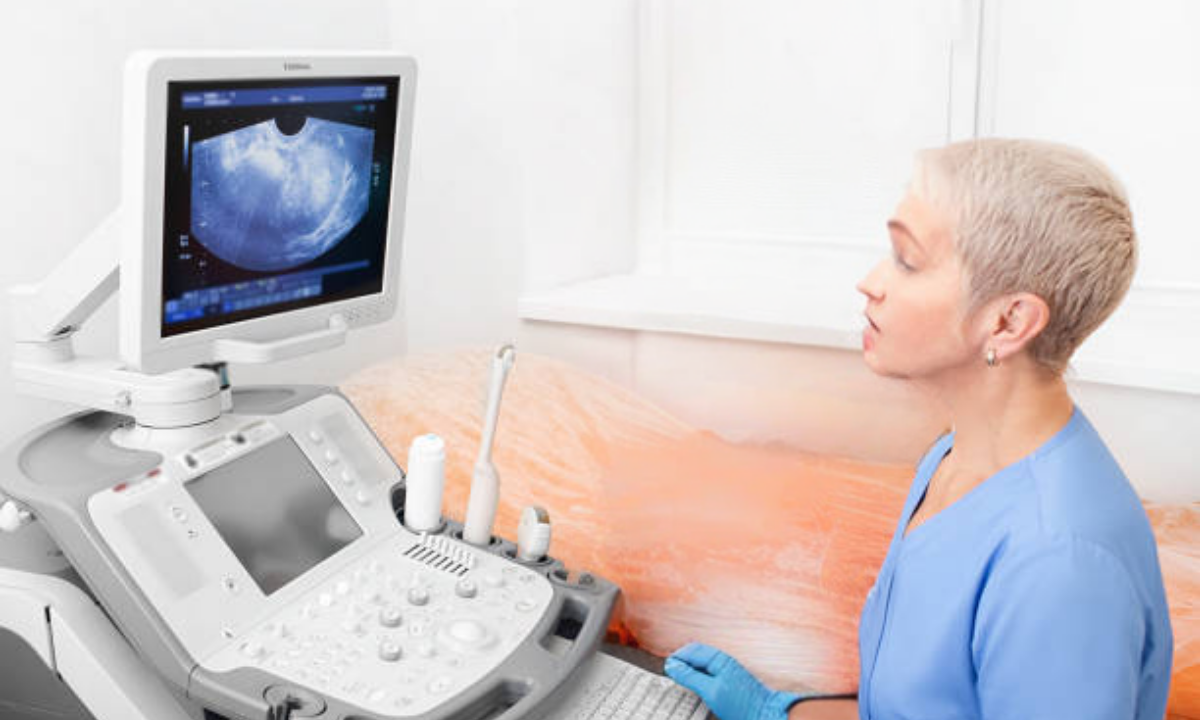
How Transvaginal Ultrasound Detects Ovarian Cysts

During a transvaginal ultrasound scan, the transvaginal probe is gently inserted into the vagina. The probe emits sound waves that create real-time images of the ovaries and surrounding tissues.

Ovarian cysts in ultrasound may appear as fluid-filled sacs or complex structures, depending on their type. The scan allows doctors to:

  • Identify the size, shape, and number of cysts
  • Determine if cysts are simple (usually harmless) or complex (may need further evaluation)
  • Track changes over time to monitor natural resolution or response to treatment

Advantages of Transvaginal Ultrasound

Some key benefits of transvaginal ultrasound include:

  • Early detection: Small cysts can be identified before symptoms appear.
  • Non-invasive and safe: No radiation exposure or surgical procedures required.
  • Accurate imaging: Offers detailed views that abdominal scans may miss.
  • Guided treatment planning: Helps in deciding whether ovarian cysts treatment is necessary.
  • Comfortable and quick: Minimal preparation is needed, and results are fast.

Ovarian Cysts Treatment Options

Treatment depends on the cyst type, size, and symptoms. Common approaches include:

Treatment TypeWhen Used
Watchful waitingSmall, simple cysts without symptoms
MedicationHormonal therapy to prevent new cysts
SurgeryLarge, persistent, or complex cysts
Fertility monitoringWhen cysts affect reproductive planning

Regular ovarian cysts ultrasound images help doctors monitor changes and decide the best approach.

Preparing for a Transvaginal Ultrasound

Preparation is simple:

  • Empty your bladder before the scan for comfort.
  • Wear comfortable clothing.
  • Remove jewelry near the pelvic area.
  • Follow any specific instructions given by your doctor or imaging centre.

No fasting is required. The scan is brief, painless, and usually well-tolerated.

Tips for Maintaining Ovarian Health

  • Track menstrual cycles and note unusual symptoms.
  • Maintain a healthy diet and exercise routine.
  • Schedule a routine ultrasound scan in Bangalore at trusted centres.
  • Consult your doctor promptly if you notice pelvic pain or irregular periods.
  • Keep all past ultrasound reports for reference during follow-ups.

Routine monitoring at a health screening centre in Bangalore ensures early detection and timely care.

Key Takeaways

A transvaginal ultrasound is a reliable, safe, and effective tool for detecting ovarian cysts early. By providing accurate images, it helps in timely diagnosis and management.

Regular check-ups at a health screening centre in Bangalore can prevent complications, support fertility planning, and maintain overall reproductive health.

Monitoring ovarian cysts with ultrasound ensures that you and your doctor can make informed decisions about treatment or observation.

Final Thoughts

Your reproductive and ovarian health deserve attention even when there are no obvious symptoms. Early detection through transvaginal ultrasound empowers women to take control of their health.

At Koshikaa, we combine advanced technology, experienced radiologists, and a patient-friendly approach to deliver precise scans. If you are looking for a reliable health screening centre in Bangalore, our team is here to guide you with care, comfort, and accurate results.

Prioritize your health today, because early detection of ovarian cysts makes a significant difference in long-term wellness.

FAQs

1. What is the best ultrasound for ovarian cysts?

A transvaginal ultrasound is the most accurate for detecting small ovarian cysts, providing detailed views of the ovaries and surrounding tissues.

2. Is a transvaginal ultrasound painful?

No, it is usually painless. You may feel slight pressure during the probe insertion, but the procedure is generally comfortable.

3. How to prepare for a transvaginal ultrasound?

Empty your bladder, wear comfortable clothing, and follow any instructions from the imaging centre. No fasting or special prep is needed.

4. Can ovarian cysts affect fertility?

Some cysts may interfere with ovulation, but most resolve naturally. Regular monitoring helps manage fertility-related concerns.

5. How often should I get an ovarian cyst ultrasound?

Follow-up depends on cyst type and symptoms. Doctors may recommend scans every 6–12 months or sooner if abnormalities persist.

MRI of the uterus is primarily used for diagnosing and evaluating conditions such as uterine fibroids, adenomyosis, and malignancies. It is particularly useful in the planning of surgical procedures and assessing the extent of gynecological diseases. This imaging test is valuable for its ability to deliver detailed cross-sectional images of the uterus and surrounding structures.
An MRI of the spleen can help diagnose various disorders affecting the spleen including splenomegaly (enlarged spleen), tumors, and other abnormalities. It provides clear, detailed images that are crucial for assessing the health of the spleen and planning potential surgical interventions.
Prostate MRI is a non-invasive imaging technique that provides detailed pictures of the prostate gland and surrounding tissues. It helps in the detection, staging, and monitoring of prostate cancer. Prostate MRIs are increasingly used to guide biopsy procedures and assist in the planning of treatment strategies for prostate health.
An MRI of the pancreas is used to identify issues such as pancreatitis, pancreatic cancer, and other pancreatic disorders. It offers high-resolution images that help in detecting small lesions and assessing the pancreatic ducts. This imaging technique is valuable for guiding biopsies and planning surgeries involving the pancreas.
MRI scans of the liver, kidney, and bile ducts provide detailed images that help diagnose various conditions, such as tumors, cysts, and blockages in the bile ducts. They are crucial for assessing organ damage, staging cancer, and planning liver or kidney-related surgeries. This type of MRI can also evaluate the health and function of these organs comprehensively.
Breast MRI is highly sensitive for detecting breast cancer, especially in individuals with dense breast tissue or a high risk of cancer. It is often used in conjunction with mammography to provide a more comprehensive evaluation. Breast MRIs can also monitor the response to breast cancer treatments and provide detailed pre-surgical mapping.
Brain and spine MRIs are critical tools for diagnosing and monitoring neurological conditions, including tumors, spinal cord injuries, and multiple sclerosis. These MRIs can reveal abnormalities in brain tissue and spinal cord structure that might not be visible with other imaging methods. They are essential for planning surgeries and assessing the effectiveness of treatments in neurology.
PET-CT scans of transplant organs like the liver, kidney, or pancreas are performed to assess metabolic activity, detect complications, and monitor post-transplant function. These scans play a vital role in the early detection of issues such as organ rejection, recurrence of disease, or infection, guiding timely interventions and optimizing transplant outcomes for patients.
PET-CT examinations of organs such as the liver, kidney, or prostate are crucial for assessing metabolic activity, detecting abnormalities, and monitoring disease progression. These scans help diagnose conditions such as liver metastases, renal cell carcinoma, or prostate cancer, guiding treatment decisions and monitoring response to therapy over time.
PET-CT scans of soft-tissue masses are utilized to assess metabolic activity and detect malignant tumors or other abnormalities. By combining PET imaging with CT imaging, these scans provide detailed information about tumor location, size, and metabolic characteristics, aiding in accurate diagnosis and treatment planning for soft-tissue cancers.
PET-CT scans are valuable for detecting cysts or fluid collections within the body, including cysts in the liver, kidneys, or other organs. These scans help characterize the nature and metabolic activity of cysts, guiding diagnosis and treatment planning for conditions such as cystic liver disease, renal cysts, or pancreatic cysts.
PET-CT scans of muscles are performed to assess metabolic activity and detect abnormalities such as inflammation, infection, or tumors. These scans provide valuable information about muscle function and pathology, aiding in the diagnosis and management of conditions like myositis, muscular dystrophy, or soft-tissue sarcomas.
PET-CT scans of the brain are utilized to evaluate brain metabolism and detect abnormalities such as tumors, epilepsy, or neurodegenerative diseases. By combining functional PET imaging with anatomical CT imaging, these scans offer comprehensive information about brain function and structure, guiding neurosurgical interventions and treatment decisions for neurological disorders
PET-CT scans of bones are commonly used to detect bone metastases in cancer patients, assess bone health in conditions like osteoporosis, and evaluate response to treatment. By combining PET and CT imaging, these scans provide detailed information about metabolic activity and anatomical changes in bone structures, aiding in accurate diagnosis and treatment planning for various bone-related conditions.
CT scans of the spine are conducted to assess the vertebrae, spinal cord, and surrounding soft tissues for conditions like herniated discs, spinal fractures, or spinal stenosis, aiding in the diagnosis and management of spinal disorders with detailed imaging essential for surgical planning and intervention.
CT scans of the pelvis are utilized to visualize the pelvic bones, reproductive organs, bladder, and surrounding structures for conditions such as fractures, tumors, or pelvic inflammatory disease, guiding treatment plans and surgical interventions with precise anatomical information.
CT scans of the neck are performed to evaluate structures such as the throat, thyroid gland, lymph nodes, and blood vessels for abnormalities like tumors, infections, or injuries, assisting in diagnosis and management of head and neck disorders with detailed imaging.
CT scans of the extremities are employed to assess bones, joints, and soft tissues in the arms or legs, aiding in the diagnosis of fractures, arthritis, or other musculoskeletal conditions, guiding orthopedic treatment and rehabilitation plans for optimal recovery.
CT scans of the chest are utilized to examine the lungs, heart, blood vessels, and surrounding structures for conditions such as lung nodules, pneumonia, or pulmonary embolism, facilitating accurate diagnosis and management of respiratory and cardiovascular diseases.
CT scans of the brain are conducted to assess brain structures, identify abnormalities like tumors, hemorrhages, or strokes, and aid in diagnosing neurological conditions, guiding neurosurgical interventions and treatment strategies with precise imaging of the brain’s anatomy.
CT scans of the abdomen are performed to visualize and evaluate the internal organs such as the liver, kidneys, pancreas, and intestines for conditions like tumors, infections, or injuries, providing detailed images essential for diagnosis and treatment planning.
Transplant organ ultrasound scans, including those of the liver, kidney, or pancreas, are performed to assess organ viability, detect complications, and monitor post-transplant function. These scans play a vital role in the early detection of issues such as organ rejection, vascular complications, or fluid collections, guiding timely interventions and optimizing transplant outcomes for patients.
Ultrasound examinations of organs such as the liver, kidney, or prostate are crucial for assessing organ structure, detecting abnormalities, and monitoring disease progression. These scans help diagnose conditions such as liver cirrhosis, kidney stones, or prostate enlargement, guiding treatment decisions and monitoring response to therapy over time.
Soft-tissue masses can be evaluated using ultrasound to determine their size, location, and characteristics. Ultrasound imaging helps differentiate between benign and malignant masses, guiding further diagnostic evaluation and treatment planning. By providing real-time imaging, ultrasound assists in the precise localization of soft-tissue masses for biopsy or surgical removal if needed.
Ultrasound scans are valuable for detecting cysts or fluid collections within the body, including cysts in the liver, kidneys, or other organs. These scans help characterize the nature and size of cysts, aiding in diagnosis and treatment planning for conditions such as cystic liver disease or renal cysts, and guiding interventions such as cyst drainage or sclerotherapy.
Musculoskeletal ultrasound scans are used to assess the health and integrity of muscles, identifying issues such as strains, tears, or muscle inflammation. These scans provide detailed images of muscle structures, allowing healthcare providers to diagnose muscle injuries accurately and develop personalized rehabilitation plans to promote recovery and restore function.
Ultrasound examinations of joints are essential for evaluating joint health, detecting abnormalities such as inflammation, fluid accumulation, or cartilage damage. By visualizing the joint structures in real-time, ultrasound helps diagnose conditions like arthritis, bursitis, or ligament injuries, guiding treatment decisions and monitoring disease progression over time.
Ultrasound scans of tendons are performed to assess for injuries, inflammation, or tears, aiding in the diagnosis and management of conditions such as tendonitis or tendon tears. These scans provide detailed images of tendon structures, helping healthcare providers determine the extent of damage and plan appropriate treatment strategies, including physical therapy or surgical intervention if necessary.
Pelvis X-rays play a crucial role in diagnosing injuries, diseases, and developmental conditions affecting the pelvic region. They provide detailed images of the hip bones, sacrum, and coccyx, aiding in the assessment of fractures, dislocations, or signs of arthritis. Pelvic X-rays are instrumental in guiding surgical interventions, monitoring healing progress, and evaluating conditions such as osteoporosis or tumors. By providing precise anatomical information, these imaging studies enable healthcare professionals to deliver personalized care and improve patient outcomes.
Teeth X-rays, known as dental radiographs, are indispensable tools for dentists to assess oral health. They aid in diagnosing dental decay, gum disease, and abnormalities in tooth structure or alignment. These images provide valuable insights into hidden dental issues, allowing for early intervention and prevention of complications. Additionally, dental X-rays are essential for planning treatments such as orthodontics, root canals, or dental implants, ensuring optimal outcomes and patient satisfaction.
Spine X-rays are pivotal in diagnosing spinal fractures, degenerative conditions, and spinal misalignments, providing critical information for treatment planning and monitoring spinal health. They offer detailed images of the vertebrae and surrounding structures, enabling healthcare professionals to identify issues such as scoliosis, disc herniation, or spinal stenosis
Skull X-rays are valuable for detecting fractures, assessing head trauma, and evaluating the alignment of facial bones, crucial in diagnosing conditions like fractures, tumors, or developmental abnormalities in the skull. They provide detailed views of the cranial structure, aiding clinicians in determining appropriate treatment strategies and ensuring patient safety.
For the heart, X-ray solutions can aid in the detection of various cardiac problems, ranging from mild to severe, including – Heart failure, Cardiac congestion and other heart-related conditions. In addition to cardiac assessments, X-ray solutions are also used for breast examinations to identify tumors or signs of breast cancer.

Bone X-rays are essential for diagnosing fractures and assessing bone alignment, aiding doctors in developing treatment plans and monitoring healing progress. They provide detailed images of the skeletal system, helping identify abnormalities or injuries that may not be visible through other imaging techniques

Abdominal X-rays are performed when patients experience symptoms such as significant nausea, ongoing vomiting, abdominal pain, flank or back pain, or abdominal swelling. These X-rays can help diagnose various conditions.

Frequently Booked Tests

Frequently Booked Checkups