X Ray Scan Centre in Bangalore

With Koshikaa X Ray scan Centre in Bangalore , Experience the benefits of advanced digital X-ray technology. Contact us today for a full body x ray. Our experienced staff is ready to assist you and answer any questions.

What is Digital X ray?

Digital x ray is an advanced imaging technique using electromagnetic radiation that penetrates the body and generate images of internal structures by a non- invasive process. This imaging technique helps in diagnosing various diseases and health conditions.

At our x ray centre in bangalore, we provide comprehensive x ray services for a variety of medical illnesses as well as for any type of recurring checkup, ensuring that patients receive proper treatment and the best healthcare results.

Request a Call Back

X Ray Cost in Bangalore

Abdominal X ray₹1200

Bone X ray₹1200

Chest X ray₹1200

Spine X ray₹1200

Teeth X ray₹1200

Pelvis X ray₹1200

Skull X ray₹1200

Note: Price may varies

Types Of X Ray in Bangalore

Utilizing X ray in Bangalore enables effective visualization of various structures and conditions within the body,

Abdomen
799/-

Bones
599/-

Chest
499/-

Skull
699/-

Spine
1199/-

Teeth
499/-

Pelvis
899/-

Why Choose Koshikaa For X ray?

Advanced X-Ray Technology in Bangalore

Advanced X-ray Technology

Accessible and affordable

Accessible and affordable

X Ray in Bangalore with Patient Focused Approach

Patient-Focused Approach

100% Reliable and accurate X Ray reports in Bangalore

100% Reliable and accurate reports

Easy and convenient X Ray booking in Bangalore

Easy and convenient booking

Comprehensive Screening using X Ray in Bangalore

Comprehensive Screening

How An X Ray Happen

X Ray Bangalore

How To Prepare For An X Ray ?

Dress in Comfortable Clothing

Wear loose and comfortable clothing on the day of your X-ray. This allows ease of movement and ensures your comfort during the process.

Remove Metal Items

Before your X-ray, kindly remove any jewelry or clothes containing metal, such as zippers or metal buttons. These items may interfere with the X-ray images and need to be taken off.

Normal Eating and Drinking

You can eat and drink as usual before your X-ray. There is no need to fast or avoid any specific food or beverages

Take Regular Medications

 Continue taking your regular medications as prescribed. It is essential to maintain your usual medication schedule unless advised otherwise by your healthcare provider.

ATTENTION, TO-BE-MOMMYS!!

If you are pregnant or suspect you might be, please inform the staff prior to the digital X-ray. While X-rays are generally safe, special precautions may be necessary for pregnant individuals, and in non-urgent cases, X-rays may be deferred until after pregnancy.

How does X Ray help in early cancer detection?

Diagnostic Precision

X-rays provide diagnostic precision, to capture detailed images of your bones, lungs, and other internal structures. It help s to identify fractures, tumors, infections, and other medical conditions with remarkable accuracy.

Fast and Non-Invasive

X-ray health screening at Koshikaa Screening Center is quick and non-invasive, allowing for efficient evaluations without the need for surgery or invasive procedures. This ensures your comfort and convenience during the screening process.

Comprehensive Health Insights

Our X-ray technology provides comprehensive insights into your health, allowing our medical professionals to tailor personalized treatment plans and preventive measures based on accurate and detailed imaging.

Dental Health Assessment

X-rays are also employed in dental health screening, enabling us to visualize your teeth, gums, and jaw. Our dental X-rays help diagnose cavities, assess tooth root health, and plan appropriate orthodontic treatments.

Trusted Expertise

At Koshikaa Screening Center, our radiographers are highly trained specialists who ensure the highest standard of care during your X-ray health screening. Your safety and well-being are our top priorities.

Early Detection of Health Issues

By utilizing X-rays for health screening, we can identify potential health issues at their earliest stages. Early detection is crucial for timely intervention and improved treatment outcomes.

Reviews

Our Recent Blogs

Our Location

Get your X-ray done at Koshikaa. Take the next step toward accurate diagnosis and timely treatment with confidence, supported by reliable imaging and expert care.

Visit our centre for professional X-ray services using advanced technology and precise imaging protocols, ensuring clear results, minimal exposure, and dependable diagnostic insights.

Frequently Asked Questions

What Is An X-ray Test?

An X-ray test is a safe, painless and non-invasive imaging procedure that uses a small amount of radiation to capture images of the inside of your body. It is mainly used to view:

  • Bones and joints
  • Chest and lungs
  • Teeth and jaw
  • Abdomen and digestive tract
  • Injuries, fractures and infections

An X-ray helps doctors diagnose medical conditions quickly and accurately without surgery.

What Is A Digital X-ray? Is This Available In Bangalore?

A Digital X-ray is an advanced form of X-ray that uses digital sensors instead of traditional film to capture images.

Key benefits of digital X-ray:

  • Faster results
  • Higher image clarity
  • Up to 80% less radiation exposure
  • Easy digital storage & sharing
  • More accurate diagnosis

Yes, Digital X-ray is available in Bangalore, and at Koshikaa Screening Centre, you get access to modern, high-resolution digital X-ray technology for safe and precise imaging.

Why And Where Is An X-ray Test Needed?

Doctors usually recommend an X-ray test to diagnose or monitor:

  • Bone fractures and dislocations
  • Chest infections (like pneumonia / TB)
  • Arthritis and joint problems
  • Dental issues
  • Kidney stones
  • Intestinal blockages
  • Tumours or abnormal growths

An X-ray can be done at a certified diagnostic centre in Bangalore like Koshikaa, where trained radiographers and advanced machines ensure accurate results.

Why Are X-ray Tests Important?

X-ray tests play a crucial role in medical diagnosis because they:

  • Detect injuries and diseases early
  • Help doctors plan treatment or surgery
  • Monitor recovery and progress
  • Prevent complications
  • Provide a quick and painless diagnosis

It is one of the most commonly used imaging techniques in the world due to its speed, low cost and reliability.

What Are The Types Of X-ray Tests?

At Koshikaa in Bangalore, multiple types of X-rays are performed, including:

  • Chest X-ray
  • Hand X-ray
  • Spine (Cervical, Thoracic, Lumbar) X-ray
  • Pelvis / Hip X-ray
  • Abdominal X-ray
  • Dental X-ray
  • Skull X-ray

Each type is used to examine a specific part of the body based on your symptoms and the doctor’s recommendation.

When And Who Needs To Take A Hand X-ray?

A hand X-ray is usually recommended when:

  • There is pain or swelling in the hand
  • You experience an injury or fracture
  • There is restricted movement
  • Arthritis or bone infection is suspected
  • A child’s bone age needs assessment

Doctors, orthopaedic specialists and emergency physicians commonly advise a hand X-ray for quick and accurate diagnosis.

Do I Need A Doctor’s Prescription For An X-ray Test?

In most cases, yes, a doctor’s prescription is advised before taking an X-ray test. This ensures that:

  • The right body part is scanned
  • The correct type of X-ray is done
  • Unnecessary radiation exposure is avoided

However, in case of injury or emergency, you can walk into Koshikaa and our team will guide you.

What Is The Expected Time Duration For An X-ray Test?

From entry to exit, an X-ray visit usually takes:

ProcessTime
Registration5 minutes
X-ray scan5–10 minutes
Total time15–20 minutes

It is a quick, smooth and hassle-free process at Koshikaa.

How Long Does An X-ray Take?

An X-ray test is one of the fastest imaging procedures.

  • The actual scan takes 5-10 minutes
  • You may spend 15-20 minutes total at the centre, including positioning and preparation

At Koshikaa, most reports are available on the same day.

What Is The Expected Time Duration For An X-ray Test?

The average cost of an X-ray test in Bangalore ranges between: ₹200 – ₹1,500, depending on:

  • Body part scanned
  • Number of views required
  • Technology used (digital/advanced)


Compared to major cities in India, Bangalore offers very affordable X-ray services.

What X-ray Test Packages Are Available At Koshikaa?

Koshikaa offers individual and multi-part X-ray packages, such as:

  • Chest + Spine X-ray
  • Hand + Wrist X-ray
  • Full spine series
  • Trauma/fracture packages
  • Custom packages recommended by doctors


These packages help patients save money while receiving complete diagnostic support.

Why Is Koshikaa Considered One Of The Best X-ray Test Centres In Bangalore?

Koshikaa stands out because of:

  • Advanced Digital X-ray machines
  • Highly trained radiology staff
  • Accurate & fast reports
  • Low radiation exposure
  • Affordable pricing
  • Clean and hygienic environment
  • Excellent patient reviews

We focus on accuracy, safety and patient comfort at every step.

Why Choose Koshikaa In Bangalore For Your X-ray Test?

People across Bangalore choose Koshikaa because we provide:

  • Trusted diagnostic services
  • High-resolution imaging
  • Same-day reports
  • Easy booking system
  • Convenient location
  • Consistent quality & care


If you’re looking for a reliable X-ray centre in Bangalore, Koshikaa is the right choice.

How To Book An X-ray Test At Koshikaa In Bangalore?

Booking an X-ray at Koshikaa is very simple:


Our team ensures minimum wait time and fast processing.

Is there an affordable X-ray centre near me that doesn't require a long wait for appointments?

Yes, Koshikaa Screening Centre is always a one-stop destination offering affordable X-ray services with minimal wait times. We prioritize patient convenience and have flexible appointment slots to ensure you receive timely care. Feel free to contact us to check availability and schedule your visit!

What are the uses of X-rays for different organs?

Radiography plays a crucial role in diagnostic procedures, providing essential X-ray images to identify various illnesses.

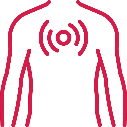
In the chest, X-rays help detect pulmonary diseases such as asthma, pneumonia, and lung cancer. They also reveal heart issues, including heart failure and congestive heart failure. Breast examinations use X-rays to identify tumors, aiding early detection and treatment of breast cancer.

Abdominal X-rays assist in diagnosing conditions involving vomiting, abdominal pain, and swelling, such as pancreatitis, appendicitis, and G.I. perforation.

In summary, X-rays help doctors visualize and understand numerous medical conditions, making them invaluable tools in healthcare. Whenever you need a diagnostic procedure, you prefer to visit the Koshikaa’s X-ray centre for quick and accurate results.

Who all needs X-Ray?

X-rays are often needed for various individuals based on their health conditions.

  • Common scenarios include assessing bone injuries from trauma, evaluating joint issues in arthritis patients, and examining lungs in those with respiratory symptoms.
  • Dental visits frequently require X-rays for diagnosis, and individuals with abdominal pain might need them to identify problems.
  • Cancer patients and those preparing for surgery also benefit from X-rays to guide treatment.

Ultimately, healthcare professionals decide the necessity based on each patient’s specific symptoms and medical history.

What are the advantages of X-Rays?

Cancer screening has several advantages:

  • It can be done before the onset of signs and symptoms, treatment procedures are easier and overall survival rates are high.
  • The improvement of the treatment for early-diagnosed patients includes a higher number of choices which might be non-invasive and more efficient treatments.
  • Last but not least, screening results have better five-year survival rates by early detection and adequate management.

Koshikaa an x-ray centre near you is suggested that offers quick service and a friendly staff.

Abdominal X-rays are performed when patients experience symptoms such as significant nausea, ongoing vomiting, abdominal pain, flank or back pain, or abdominal swelling. These X-rays can help diagnose various conditions.

Bone X-rays are essential for diagnosing fractures and assessing bone alignment, aiding doctors in developing treatment plans and monitoring healing progress. They provide detailed images of the skeletal system, helping identify abnormalities or injuries that may not be visible through other imaging techniques

For the heart, X-ray solutions can aid in the detection of various cardiac problems, ranging from mild to severe, including – Heart failure, Cardiac congestion and other heart-related conditions. In addition to cardiac assessments, X-ray solutions are also used for breast examinations to identify tumors or signs of breast cancer.
Skull X-rays are valuable for detecting fractures, assessing head trauma, and evaluating the alignment of facial bones, crucial in diagnosing conditions like fractures, tumors, or developmental abnormalities in the skull. They provide detailed views of the cranial structure, aiding clinicians in determining appropriate treatment strategies and ensuring patient safety.
Spine X-rays are pivotal in diagnosing spinal fractures, degenerative conditions, and spinal misalignments, providing critical information for treatment planning and monitoring spinal health. They offer detailed images of the vertebrae and surrounding structures, enabling healthcare professionals to identify issues such as scoliosis, disc herniation, or spinal stenosis
Teeth X-rays, known as dental radiographs, are indispensable tools for dentists to assess oral health. They aid in diagnosing dental decay, gum disease, and abnormalities in tooth structure or alignment. These images provide valuable insights into hidden dental issues, allowing for early intervention and prevention of complications. Additionally, dental X-rays are essential for planning treatments such as orthodontics, root canals, or dental implants, ensuring optimal outcomes and patient satisfaction.
Pelvis X-rays play a crucial role in diagnosing injuries, diseases, and developmental conditions affecting the pelvic region. They provide detailed images of the hip bones, sacrum, and coccyx, aiding in the assessment of fractures, dislocations, or signs of arthritis. Pelvic X-rays are instrumental in guiding surgical interventions, monitoring healing progress, and evaluating conditions such as osteoporosis or tumors. By providing precise anatomical information, these imaging studies enable healthcare professionals to deliver personalized care and improve patient outcomes.
MRI of the uterus is primarily used for diagnosing and evaluating conditions such as uterine fibroids, adenomyosis, and malignancies. It is particularly useful in the planning of surgical procedures and assessing the extent of gynecological diseases. This imaging test is valuable for its ability to deliver detailed cross-sectional images of the uterus and surrounding structures.
An MRI of the spleen can help diagnose various disorders affecting the spleen including splenomegaly (enlarged spleen), tumors, and other abnormalities. It provides clear, detailed images that are crucial for assessing the health of the spleen and planning potential surgical interventions.
Prostate MRI is a non-invasive imaging technique that provides detailed pictures of the prostate gland and surrounding tissues. It helps in the detection, staging, and monitoring of prostate cancer. Prostate MRIs are increasingly used to guide biopsy procedures and assist in the planning of treatment strategies for prostate health.
An MRI of the pancreas is used to identify issues such as pancreatitis, pancreatic cancer, and other pancreatic disorders. It offers high-resolution images that help in detecting small lesions and assessing the pancreatic ducts. This imaging technique is valuable for guiding biopsies and planning surgeries involving the pancreas.
MRI scans of the liver, kidney, and bile ducts provide detailed images that help diagnose various conditions, such as tumors, cysts, and blockages in the bile ducts. They are crucial for assessing organ damage, staging cancer, and planning liver or kidney-related surgeries. This type of MRI can also evaluate the health and function of these organs comprehensively.
Breast MRI is highly sensitive for detecting breast cancer, especially in individuals with dense breast tissue or a high risk of cancer. It is often used in conjunction with mammography to provide a more comprehensive evaluation. Breast MRIs can also monitor the response to breast cancer treatments and provide detailed pre-surgical mapping.
Brain and spine MRIs are critical tools for diagnosing and monitoring neurological conditions, including tumors, spinal cord injuries, and multiple sclerosis. These MRIs can reveal abnormalities in brain tissue and spinal cord structure that might not be visible with other imaging methods. They are essential for planning surgeries and assessing the effectiveness of treatments in neurology.
PET-CT scans of transplant organs like the liver, kidney, or pancreas are performed to assess metabolic activity, detect complications, and monitor post-transplant function. These scans play a vital role in the early detection of issues such as organ rejection, recurrence of disease, or infection, guiding timely interventions and optimizing transplant outcomes for patients.
PET-CT examinations of organs such as the liver, kidney, or prostate are crucial for assessing metabolic activity, detecting abnormalities, and monitoring disease progression. These scans help diagnose conditions such as liver metastases, renal cell carcinoma, or prostate cancer, guiding treatment decisions and monitoring response to therapy over time.
PET-CT scans of soft-tissue masses are utilized to assess metabolic activity and detect malignant tumors or other abnormalities. By combining PET imaging with CT imaging, these scans provide detailed information about tumor location, size, and metabolic characteristics, aiding in accurate diagnosis and treatment planning for soft-tissue cancers.
PET-CT scans are valuable for detecting cysts or fluid collections within the body, including cysts in the liver, kidneys, or other organs. These scans help characterize the nature and metabolic activity of cysts, guiding diagnosis and treatment planning for conditions such as cystic liver disease, renal cysts, or pancreatic cysts.
PET-CT scans of muscles are performed to assess metabolic activity and detect abnormalities such as inflammation, infection, or tumors. These scans provide valuable information about muscle function and pathology, aiding in the diagnosis and management of conditions like myositis, muscular dystrophy, or soft-tissue sarcomas.
PET-CT scans of the brain are utilized to evaluate brain metabolism and detect abnormalities such as tumors, epilepsy, or neurodegenerative diseases. By combining functional PET imaging with anatomical CT imaging, these scans offer comprehensive information about brain function and structure, guiding neurosurgical interventions and treatment decisions for neurological disorders
PET-CT scans of bones are commonly used to detect bone metastases in cancer patients, assess bone health in conditions like osteoporosis, and evaluate response to treatment. By combining PET and CT imaging, these scans provide detailed information about metabolic activity and anatomical changes in bone structures, aiding in accurate diagnosis and treatment planning for various bone-related conditions.
CT scans of the spine are conducted to assess the vertebrae, spinal cord, and surrounding soft tissues for conditions like herniated discs, spinal fractures, or spinal stenosis, aiding in the diagnosis and management of spinal disorders with detailed imaging essential for surgical planning and intervention.
CT scans of the pelvis are utilized to visualize the pelvic bones, reproductive organs, bladder, and surrounding structures for conditions such as fractures, tumors, or pelvic inflammatory disease, guiding treatment plans and surgical interventions with precise anatomical information.
CT scans of the neck are performed to evaluate structures such as the throat, thyroid gland, lymph nodes, and blood vessels for abnormalities like tumors, infections, or injuries, assisting in diagnosis and management of head and neck disorders with detailed imaging.
CT scans of the extremities are employed to assess bones, joints, and soft tissues in the arms or legs, aiding in the diagnosis of fractures, arthritis, or other musculoskeletal conditions, guiding orthopedic treatment and rehabilitation plans for optimal recovery.
CT scans of the chest are utilized to examine the lungs, heart, blood vessels, and surrounding structures for conditions such as lung nodules, pneumonia, or pulmonary embolism, facilitating accurate diagnosis and management of respiratory and cardiovascular diseases.
CT scans of the brain are conducted to assess brain structures, identify abnormalities like tumors, hemorrhages, or strokes, and aid in diagnosing neurological conditions, guiding neurosurgical interventions and treatment strategies with precise imaging of the brain’s anatomy.
CT scans of the abdomen are performed to visualize and evaluate the internal organs such as the liver, kidneys, pancreas, and intestines for conditions like tumors, infections, or injuries, providing detailed images essential for diagnosis and treatment planning.
Transplant organ ultrasound scans, including those of the liver, kidney, or pancreas, are performed to assess organ viability, detect complications, and monitor post-transplant function. These scans play a vital role in the early detection of issues such as organ rejection, vascular complications, or fluid collections, guiding timely interventions and optimizing transplant outcomes for patients.
Ultrasound examinations of organs such as the liver, kidney, or prostate are crucial for assessing organ structure, detecting abnormalities, and monitoring disease progression. These scans help diagnose conditions such as liver cirrhosis, kidney stones, or prostate enlargement, guiding treatment decisions and monitoring response to therapy over time.
Soft-tissue masses can be evaluated using ultrasound to determine their size, location, and characteristics. Ultrasound imaging helps differentiate between benign and malignant masses, guiding further diagnostic evaluation and treatment planning. By providing real-time imaging, ultrasound assists in the precise localization of soft-tissue masses for biopsy or surgical removal if needed.
Ultrasound scans are valuable for detecting cysts or fluid collections within the body, including cysts in the liver, kidneys, or other organs. These scans help characterize the nature and size of cysts, aiding in diagnosis and treatment planning for conditions such as cystic liver disease or renal cysts, and guiding interventions such as cyst drainage or sclerotherapy.
Musculoskeletal ultrasound scans are used to assess the health and integrity of muscles, identifying issues such as strains, tears, or muscle inflammation. These scans provide detailed images of muscle structures, allowing healthcare providers to diagnose muscle injuries accurately and develop personalized rehabilitation plans to promote recovery and restore function.
Ultrasound examinations of joints are essential for evaluating joint health, detecting abnormalities such as inflammation, fluid accumulation, or cartilage damage. By visualizing the joint structures in real-time, ultrasound helps diagnose conditions like arthritis, bursitis, or ligament injuries, guiding treatment decisions and monitoring disease progression over time.
Ultrasound scans of tendons are performed to assess for injuries, inflammation, or tears, aiding in the diagnosis and management of conditions such as tendonitis or tendon tears. These scans provide detailed images of tendon structures, helping healthcare providers determine the extent of damage and plan appropriate treatment strategies, including physical therapy or surgical intervention if necessary.

Frequently Booked Tests

Frequently Booked Checkups