Advanced CT Scan Technology
Koshikaa uses high-resolution CT scanners that provide detailed imaging with minimal radiation exposure. This makes every CT scan in Bangalore at our centre precise and reliable. Advanced software converts data into accurate 3D images for thorough diagnosis.

Experienced Radiology Team
Skilled radiologists analyse each CT scan centre across Bangalore with precision, providing your doctor with dependable diagnostic insights and personalised care recommendations.

Patient-Centric Approach
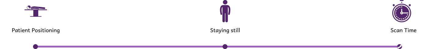
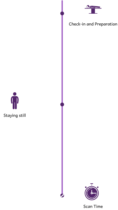
From registration to post-scan guidance, Koshikaa’s staff ensures a smooth, stress-free CT scan in Bangalore experience. Comfort and clear communication are prioritised at every step.

Quick & Reliable Reports
Most scans are completed within 10–20 minutes, and reports from your CT scan are delivered promptly to enable timely treatment decisions.
Affordable & Transparent Pricing
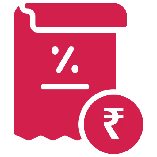
Koshikaa offers competitive costs for a CT scan in Bangalore with no hidden charges, making quality diagnostic care accessible to all.