PET CT Scan vs. X Ray vs. MRI: Which One is Right for You?

pet ct scan vs. x ray

Table of Contents

Medical imaging technology is vital in medical diagnosis and treatment procedures for different illnesses. Today’s medical imaging market includes several options including PET CT scan along with X-rays and MRI scans which makes patients struggle to determine which test is appropriate for their needs. This article analyzes the unique aspects of these procedures while discussing their clinical applications and necessary indications.  

Users who seek PET CT scan, x-ray, or MRI scan in Bangalore will benefit from information about these imaging techniques before they make their choice.  

What Is an X-ray?  

X-ray imaging represents the most frequently employed diagnostic medical tool currently used in healthcare centres. A low dose of radiation enables medical professionals to generate pictures that show your bones and body tissues together with other inside structures. Medical practitioners use two-dimensional X-ray images to examine patients and determine the existence of broken bones and other dental or pulmonary conditions.  

How Does It Work?  

X-ray machines transmit minimal doses of radiation into your body while tissue materials block the radiation at different speeds. X-rays project dense materials such as bones to appear white when they stop the rays thus producing contrasting images that darker areas show softer material types such as muscle.  

Applications of X-Rays  

The main utilization of X-ray technology in medical diagnosis leads to identifying fractures as well as treating lung infections and detecting cavities in teeth. The medical procedure takes minimal time while staying comfortable for all patients undergoing it. Doctors often start their imaging strategy for typical medical problems with examinations of X-ray in Bangalore.  

Pros and Cons of X-Rays 

Pros:  

  • Fast and readily available  
  • This imaging technique costs less than other available imaging tests.  
  • The technique proves valuable when doctors need to detect bone fractures as well as assess correct bone alignment.  

Cons: 

  • Limited detail, especially for soft tissues  
  • Radiation exposure (though usually minimal)  

The medical facilities operating in Bangalore city offer efficient X-ray services for diagnosing patients.  

What Is an MRI Scan? 

mri scan in bangalore

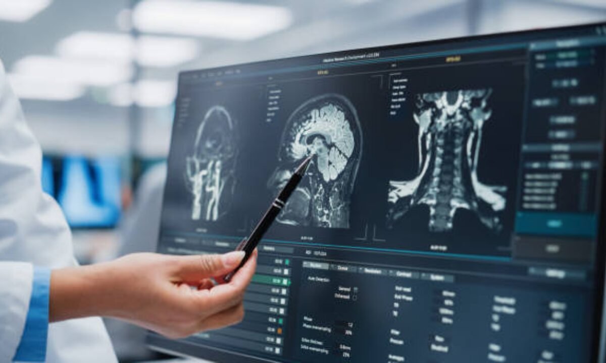
Using Magnetomic Resonance Imaging technology medical professionals can create comprehensive sectional images of human body structures. An MRI generates images through a magnetic field and radio wave technology rather than the radiation that powers X-ray machines.  

Why Is MRI Different?  

Brain tissue along with muscles organs and ligaments appear as highly precise images when scanned by MRI imaging techniques. Medical professionals use this diagnostic procedure to spot tumours together with ligament tears and spinal cord problems.   

How Is an MRI Performed?  

MRI scans usually take longer than X-rays (ranging from 30 minutes to an hour). During the scan, patients are required to lie completely still inside a large MRI machine, which has a tunnel-like structure. Some people may feel claustrophobic, but in most cases, there is no physical discomfort involved. 

Advantages and Limitations of MRI Scans  

Pros:  

  • The technique delivers detailed visualizations of all bodily soft materials.  
  • No radiation exposure  
  • This scan method serves as an effective tool to find tumours and nerve-related abnormalities in the body.  

Cons: 

  • Time-consuming  
  • The costs associated with MRI scanning exceed what X-rays typically cost.  
  • The procedure is unsuitable for patients who possess metallic implants in their bodies.  

People who need deep imaging studies for problems affecting organs and soft tissues should obtain an MRI scan in Bangalore facilities. Advanced MRI facilities are available at leading hospital institutions throughout Bangalore.  

What Is a PET CT Scan?  

PET CT Scan procedures distinguish themselves from other scanning methods because of fundamental reasons.  

The medical imaging device PET CT scan unites two technologies called Positron Emission Tomography and Computed Tomography (CT scan) to produce single imaging results. PET identifies metabolic patterns through radioactive substance use while CT generates detailed anatomical images of the body. PET CT imaging produces combined data about both molecular activity and structural features of body organs and tissues.  

How Does It Work?  

A radioactive tracer is provided to patients during hospital PET CT scan procedures. The tracer detects metabolic issues in the different processes that occur in your body. New technology adapted to this process enables physicians to detect abnormalities more easily by providing both metabolic and structural information from CT scans.  

When Is This Scan Used? 

PET CT scanning becomes necessary to obtain very precise medical information when doctors need to detect cancer, assess heart health or evaluate brain activity conditions. PET CT stands as one of the most advanced diagnostic methods that healthcare professionals recommend as an extension to existing imaging procedures.  

Strengths and Drawbacks of PET CT Scans  

Pros: 

  • During cancer diagnosis and disease monitoring PET CT scan provides excellent diagnostic capabilities.  
  • Combines metabolic and anatomical details  
  • PET CT examination helps healthcare providers detect diseases before they advance to late stages.  

Cons:  

  • PET CT scans require the patient to encounter radioactive substances during the examination.  
  • The costs of this procedure remain higher than what other imaging techniques need to spend.  
  • The medical procedure necessitates patients to empty their stomachs before undergoing testing.  

Modern healthcare developments enable numerous hospitals to implement this service. People requiring particular diagnostic images for cancer testing should consider making an appointment for a pet CT scan in Bangalore.   

Key Factors to Consider for Selecting the Suitable Imaging Test

The choice of medical imaging depends entirely on your medical condition together with your symptoms and the evaluation requirements that your doctor sets. Medical scenarios include the following breakdown:  

  • Individuals dealing with bone injuries or dental concerns should get an X-ray as the most suitable imaging procedure. The process delivers valuable information about dental problems while revealing bone fracture details at reasonable costs and through quick completion.  
  • An MRI scan stands out as the optimal tool for heart and brain examinations and muscle tissue assessments due to its exceptional diagnostic capability.   
  • Oncology or Detailed Assessments: If you or your doctor need specific metabolic data, such as for detecting cancer or its progression, a PET CT scan combines function and structure in a single test.  

When to Choose Each Scan? 

It is important to understand when each imaging test—PET CT scan, X-ray, or MRI scan—is chosen by doctors. The selection of diagnostic tests depends on the medical condition together with the examined tissue and the required clarity level.

Imaging MethodBest Chosen For
X-RayBone fractures, chest/lung conditions, dental imaging
MRI ScanBrain tumours, spinal cord issues, soft tissue injuries, detailed organ imaging
PET CT ScanCancer detection, heart function, neurological disorders

The time of scan selection provides valuable information to determine which test matches your symptoms the most.  

Final Thoughts

There is no standard evaluation procedure for choosing between a PET CT scan, X-ray or MRI scan since each medical evaluation has its unique advantages and limitations. Your medical condition together with the needed detail level and doctor recommendations determine the suitable imaging test.  If you need immediate results for a simple issue like a bone fracture, an X-ray is perfect. For complex issues involving soft tissues, an MRI may be more appropriate. A PET CT scan should be reserved for more detailed diagnostics, particularly in cases of cancer or metabolic disorders. 

People seeking examinations for X-ray in Bangalore can access world-class diagnostic facilities and patients also have access to superior MRI and PET CT diagnostic services. You need to speak with your doctor to select the right imaging test according to your needs.   

Koshikaa stands as the best health screening centre in Bangalore dedicated to providing precise diagnostic services through PET CT along with MRI and X-ray scans for enhanced patient experiences. Directly connect with the team for your queries. 

MRI of the uterus is primarily used for diagnosing and evaluating conditions such as uterine fibroids, adenomyosis, and malignancies. It is particularly useful in the planning of surgical procedures and assessing the extent of gynecological diseases. This imaging test is valuable for its ability to deliver detailed cross-sectional images of the uterus and surrounding structures.
An MRI of the spleen can help diagnose various disorders affecting the spleen including splenomegaly (enlarged spleen), tumors, and other abnormalities. It provides clear, detailed images that are crucial for assessing the health of the spleen and planning potential surgical interventions.
Prostate MRI is a non-invasive imaging technique that provides detailed pictures of the prostate gland and surrounding tissues. It helps in the detection, staging, and monitoring of prostate cancer. Prostate MRIs are increasingly used to guide biopsy procedures and assist in the planning of treatment strategies for prostate health.
An MRI of the pancreas is used to identify issues such as pancreatitis, pancreatic cancer, and other pancreatic disorders. It offers high-resolution images that help in detecting small lesions and assessing the pancreatic ducts. This imaging technique is valuable for guiding biopsies and planning surgeries involving the pancreas.
MRI scans of the liver, kidney, and bile ducts provide detailed images that help diagnose various conditions, such as tumors, cysts, and blockages in the bile ducts. They are crucial for assessing organ damage, staging cancer, and planning liver or kidney-related surgeries. This type of MRI can also evaluate the health and function of these organs comprehensively.
Breast MRI is highly sensitive for detecting breast cancer, especially in individuals with dense breast tissue or a high risk of cancer. It is often used in conjunction with mammography to provide a more comprehensive evaluation. Breast MRIs can also monitor the response to breast cancer treatments and provide detailed pre-surgical mapping.
Brain and spine MRIs are critical tools for diagnosing and monitoring neurological conditions, including tumors, spinal cord injuries, and multiple sclerosis. These MRIs can reveal abnormalities in brain tissue and spinal cord structure that might not be visible with other imaging methods. They are essential for planning surgeries and assessing the effectiveness of treatments in neurology.
PET-CT scans of transplant organs like the liver, kidney, or pancreas are performed to assess metabolic activity, detect complications, and monitor post-transplant function. These scans play a vital role in the early detection of issues such as organ rejection, recurrence of disease, or infection, guiding timely interventions and optimizing transplant outcomes for patients.
PET-CT examinations of organs such as the liver, kidney, or prostate are crucial for assessing metabolic activity, detecting abnormalities, and monitoring disease progression. These scans help diagnose conditions such as liver metastases, renal cell carcinoma, or prostate cancer, guiding treatment decisions and monitoring response to therapy over time.
PET-CT scans of soft-tissue masses are utilized to assess metabolic activity and detect malignant tumors or other abnormalities. By combining PET imaging with CT imaging, these scans provide detailed information about tumor location, size, and metabolic characteristics, aiding in accurate diagnosis and treatment planning for soft-tissue cancers.
PET-CT scans are valuable for detecting cysts or fluid collections within the body, including cysts in the liver, kidneys, or other organs. These scans help characterize the nature and metabolic activity of cysts, guiding diagnosis and treatment planning for conditions such as cystic liver disease, renal cysts, or pancreatic cysts.
PET-CT scans of muscles are performed to assess metabolic activity and detect abnormalities such as inflammation, infection, or tumors. These scans provide valuable information about muscle function and pathology, aiding in the diagnosis and management of conditions like myositis, muscular dystrophy, or soft-tissue sarcomas.
PET-CT scans of the brain are utilized to evaluate brain metabolism and detect abnormalities such as tumors, epilepsy, or neurodegenerative diseases. By combining functional PET imaging with anatomical CT imaging, these scans offer comprehensive information about brain function and structure, guiding neurosurgical interventions and treatment decisions for neurological disorders
PET-CT scans of bones are commonly used to detect bone metastases in cancer patients, assess bone health in conditions like osteoporosis, and evaluate response to treatment. By combining PET and CT imaging, these scans provide detailed information about metabolic activity and anatomical changes in bone structures, aiding in accurate diagnosis and treatment planning for various bone-related conditions.
CT scans of the spine are conducted to assess the vertebrae, spinal cord, and surrounding soft tissues for conditions like herniated discs, spinal fractures, or spinal stenosis, aiding in the diagnosis and management of spinal disorders with detailed imaging essential for surgical planning and intervention.
CT scans of the pelvis are utilized to visualize the pelvic bones, reproductive organs, bladder, and surrounding structures for conditions such as fractures, tumors, or pelvic inflammatory disease, guiding treatment plans and surgical interventions with precise anatomical information.
CT scans of the neck are performed to evaluate structures such as the throat, thyroid gland, lymph nodes, and blood vessels for abnormalities like tumors, infections, or injuries, assisting in diagnosis and management of head and neck disorders with detailed imaging.
CT scans of the extremities are employed to assess bones, joints, and soft tissues in the arms or legs, aiding in the diagnosis of fractures, arthritis, or other musculoskeletal conditions, guiding orthopedic treatment and rehabilitation plans for optimal recovery.
CT scans of the chest are utilized to examine the lungs, heart, blood vessels, and surrounding structures for conditions such as lung nodules, pneumonia, or pulmonary embolism, facilitating accurate diagnosis and management of respiratory and cardiovascular diseases.
CT scans of the brain are conducted to assess brain structures, identify abnormalities like tumors, hemorrhages, or strokes, and aid in diagnosing neurological conditions, guiding neurosurgical interventions and treatment strategies with precise imaging of the brain’s anatomy.
CT scans of the abdomen are performed to visualize and evaluate the internal organs such as the liver, kidneys, pancreas, and intestines for conditions like tumors, infections, or injuries, providing detailed images essential for diagnosis and treatment planning.
Transplant organ ultrasound scans, including those of the liver, kidney, or pancreas, are performed to assess organ viability, detect complications, and monitor post-transplant function. These scans play a vital role in the early detection of issues such as organ rejection, vascular complications, or fluid collections, guiding timely interventions and optimizing transplant outcomes for patients.
Ultrasound examinations of organs such as the liver, kidney, or prostate are crucial for assessing organ structure, detecting abnormalities, and monitoring disease progression. These scans help diagnose conditions such as liver cirrhosis, kidney stones, or prostate enlargement, guiding treatment decisions and monitoring response to therapy over time.
Soft-tissue masses can be evaluated using ultrasound to determine their size, location, and characteristics. Ultrasound imaging helps differentiate between benign and malignant masses, guiding further diagnostic evaluation and treatment planning. By providing real-time imaging, ultrasound assists in the precise localization of soft-tissue masses for biopsy or surgical removal if needed.
Ultrasound scans are valuable for detecting cysts or fluid collections within the body, including cysts in the liver, kidneys, or other organs. These scans help characterize the nature and size of cysts, aiding in diagnosis and treatment planning for conditions such as cystic liver disease or renal cysts, and guiding interventions such as cyst drainage or sclerotherapy.
Musculoskeletal ultrasound scans are used to assess the health and integrity of muscles, identifying issues such as strains, tears, or muscle inflammation. These scans provide detailed images of muscle structures, allowing healthcare providers to diagnose muscle injuries accurately and develop personalized rehabilitation plans to promote recovery and restore function.
Ultrasound examinations of joints are essential for evaluating joint health, detecting abnormalities such as inflammation, fluid accumulation, or cartilage damage. By visualizing the joint structures in real-time, ultrasound helps diagnose conditions like arthritis, bursitis, or ligament injuries, guiding treatment decisions and monitoring disease progression over time.
Ultrasound scans of tendons are performed to assess for injuries, inflammation, or tears, aiding in the diagnosis and management of conditions such as tendonitis or tendon tears. These scans provide detailed images of tendon structures, helping healthcare providers determine the extent of damage and plan appropriate treatment strategies, including physical therapy or surgical intervention if necessary.
Pelvis X-rays play a crucial role in diagnosing injuries, diseases, and developmental conditions affecting the pelvic region. They provide detailed images of the hip bones, sacrum, and coccyx, aiding in the assessment of fractures, dislocations, or signs of arthritis. Pelvic X-rays are instrumental in guiding surgical interventions, monitoring healing progress, and evaluating conditions such as osteoporosis or tumors. By providing precise anatomical information, these imaging studies enable healthcare professionals to deliver personalized care and improve patient outcomes.
Teeth X-rays, known as dental radiographs, are indispensable tools for dentists to assess oral health. They aid in diagnosing dental decay, gum disease, and abnormalities in tooth structure or alignment. These images provide valuable insights into hidden dental issues, allowing for early intervention and prevention of complications. Additionally, dental X-rays are essential for planning treatments such as orthodontics, root canals, or dental implants, ensuring optimal outcomes and patient satisfaction.
Spine X-rays are pivotal in diagnosing spinal fractures, degenerative conditions, and spinal misalignments, providing critical information for treatment planning and monitoring spinal health. They offer detailed images of the vertebrae and surrounding structures, enabling healthcare professionals to identify issues such as scoliosis, disc herniation, or spinal stenosis
Skull X-rays are valuable for detecting fractures, assessing head trauma, and evaluating the alignment of facial bones, crucial in diagnosing conditions like fractures, tumors, or developmental abnormalities in the skull. They provide detailed views of the cranial structure, aiding clinicians in determining appropriate treatment strategies and ensuring patient safety.
For the heart, X-ray solutions can aid in the detection of various cardiac problems, ranging from mild to severe, including – Heart failure, Cardiac congestion and other heart-related conditions. In addition to cardiac assessments, X-ray solutions are also used for breast examinations to identify tumors or signs of breast cancer.

Bone X-rays are essential for diagnosing fractures and assessing bone alignment, aiding doctors in developing treatment plans and monitoring healing progress. They provide detailed images of the skeletal system, helping identify abnormalities or injuries that may not be visible through other imaging techniques

Abdominal X-rays are performed when patients experience symptoms such as significant nausea, ongoing vomiting, abdominal pain, flank or back pain, or abdominal swelling. These X-rays can help diagnose various conditions.
Search Bar

Frequently Booked Tests

Frequently Booked Checkups