Pelvis USG Scan Explained: Purpose, Process & What to Expect

Table of Contents

The USG pelvis scan is a harmless examination that poses no threat to the patient, as it involves the use of sound waves to display the organs in the pelvis. Physicians frequently use it to detect issues in the uterus, ovaries, bladder, prostate, and surrounding tissue. It uses no X-rays or radiation, making it safe for all ages, including pregnant women. This guide explains the test’s purpose, preparation, procedure, cost, and how to find quality service. These are simple steps to follow, and they will make you feel prepared to face your appointment.

What is a pelvic ultrasound?

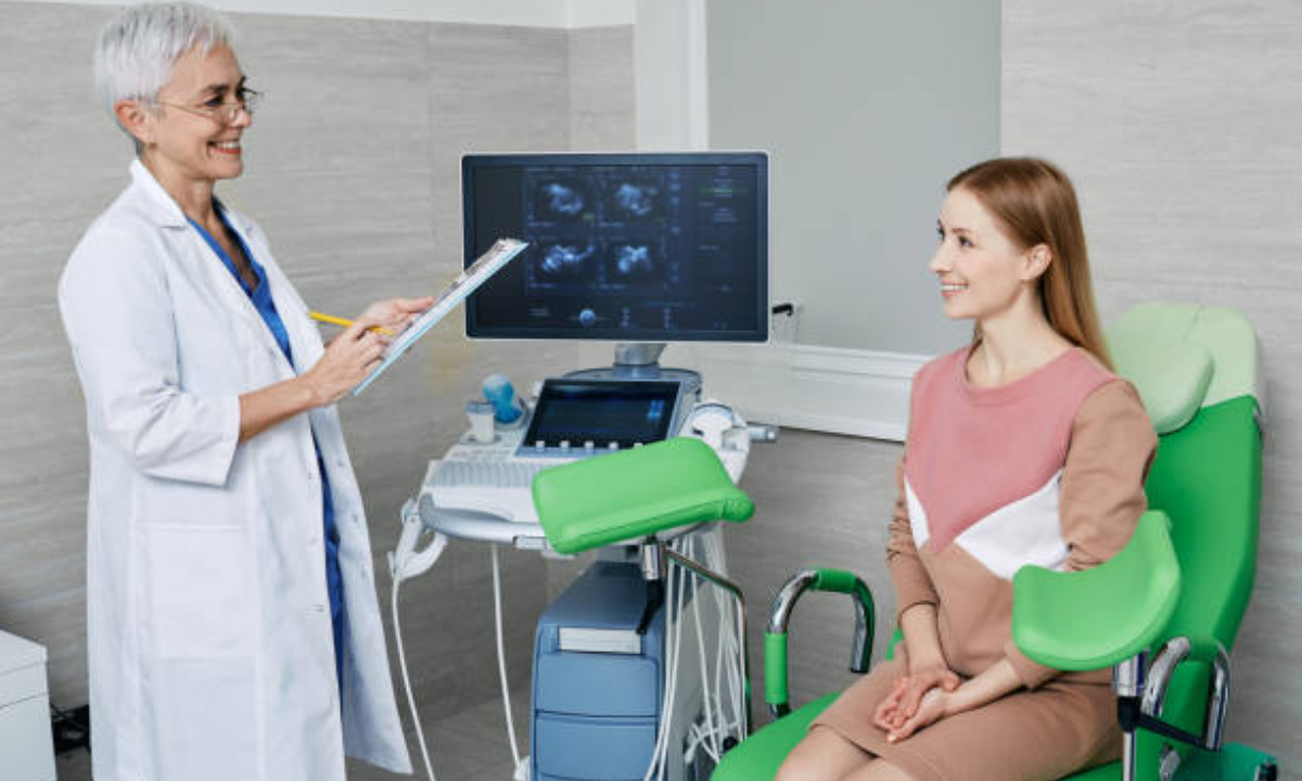
In a pelvic ultrasound, the images of the lower abdomen structures are developed in real-time. A sonographer will apply a sonographic gel to your bare skin during the exam and hold a thin, wand-like device called a transducer. A transducer sends sound waves with high frequency to bounce off the organs and produce a reflection. These replicates are displayed in the form of pictorial details on a monitor. There are a lot of healthcare providers who refer to this test as merely a USG pelvis scan since USG refers to ultrasonography. It is painless, will take 15-30 minutes, and needs no recovery period–so you can resume normal activities immediately.

What is the purpose of a USG pelvis scan?

Ultrasound scan in Bangalore

The objectives of the test will be the following:

• Identify abnormalities: Diagnosing cysts, fibroids, tumors, or a collection of fluid tissue inside the pelvic cavity.  

• Determine pain or bleeding: Find the causes of pelvic pain, abnormal periods, or post menopausal bleeding.  

• Interventional guidance: Aid in other procedures like drainage of fluids, biopsies, or IUD inserts, which are being done with real-time visualization imaging.  

• Treatment monitoring: Detect the change of known conditions, time-dependent, like the size of a cyst on the ovary, to be able to gauge the success of the therapy.

The scan helps in making fast clinical judgments in both the outpatient and emergency rooms by providing information pertaining to structures without the involvement of invasive surgery.

What should you do before taking a USG pelvis?

  • Consume water and make sure that you do not pee before the test.  
  • Walk in loose, comfortable clothes.
  • You need to drink and eat as usual before the exam.

What happens in the USG scan of the pelvis?

  • You lie on a table, put the back of your pelvis on a pillow.  
  • The sonographer will apply warmed gel on your skin and move the transducer over the region.  
  • The transducer will send high-frequency sounds and reflect the sounds back depending on the organs. These reverberations become highly pictorial images on the screen.  
  • The whole operation is without pain and takes approximately 15-30 minutes.  
  • This is the time that you are returning to normal activities.

Where will you get good-quality USG pelvis scan services?

  • Refer yourself to a good clinic by asking your doctor to advise a good clinic. 
  • Talk to one of your good friends.  
  • Find out your covered facilities. 

Pelvis USG Preparation

Plan ahead of your visit to get your scan to pass more smoothly:

  1. Water wherever and whenever you are instructed. A micturating bladder separates bowel loops from the pelvis region, making it possible for the doctor to efficiently.
  2. Wear comfortable clothes that fit loosely. A medical gown will also be asked to be worn by you.
  3. Tell staff about your existing operation plans, allergies, or medication you take.
  4. Get there early to set up paperwork and place your queries that require an urgent answer.
  5. Proper preparation will minimize delays, and there will be clear pictures to assist the sonographer in making a proper report.

Sequential steps of USG pelvis scan

1. Positioning: You are lying down on an exam table most of the time in a supine position with your lower abdominal area open.

2. Gel application: This is done by covering your skin with a transparent water-based gel to fully exclude air pockets.

3. Transducer movement: The sonographer moves the transducer around your pelvis and changes angles to see each of your organs.

4. Optional probes: Depending on greater detail, a transvaginal or a transrectal probe can be placed in the vaginal canal or the rectum, tenderly.

5. Monitoring image: The live pictures are captured, measurements are determined, and major observations are noted.

6. Finishing: The Gel is stripped, and it is possible to take off the clothes immediately.

Good communication during the scan makes you relaxed and aware. 

What are the factors affecting the USG pelvis scan cost?

Facility type: Hospitals are determined to be very expensive as opposed to individual clinics.

Extra imaging- There is an extra cost of either Doppler studies or contrast agents.

Reporting services: In some centers, radiologist interpretation is covered in the basic fee, and in others, they charge extra.

Geographic location: More expensive prices are likely to be met in cities where different taxes and costs of operation are higher.

An average basic scan varies in price; it is mostly affordable to moderate. In any call, it is always good to ask for an itemized quote so you know what services are included and possible add-ons.

Intimation and The Access: Ultrasound scan in Bangalore

These are some points to be considered when you make your choice of an Ultrasound scan in Bangalore:

Accreditation: Clinics that have been nationally accredited by an imaging body with sonographers who are trained on how to handle their work.

Quality of equipment: The generation of machines allows for obtaining clearer images, which helps to diagnose more precisely.

Patient reviews: Actually, reality reviews can be presented on professionalism, waiting periods, and report turnaround.

Comfort amenities: Your experience is easier due to such amenities as online booking, report delivery (it is digital), and flexible hours.

Researching ahead of time will help you choose a facility that is a balance of all three: experience, comfort, and affordability.

Expectations During Your Scan

The environment is characterized by most patients as calm and professional. The sonographer describes the process, one by one, before beginning, considering possible questions. The gel feels cold at first, then warms, with slight pressure from the moving transducer. When internal probes are involved, you will be shown a smooth insertion procedure that does not take long and is rather comfortable. The overall time takes around half an hour, and in case any gel is left, you simply wipe it off, and you are free to go without any limitations.

Interpretation of the Results

Once the images are acquired, a radiologist conducts a review and outlines the main findings, measurements, the look of an organ, and any anomalies that have been detected. You might get instant verbal feedback and later get a documented report of your assessment within 24 to 48 hours. Suppose there are no abnormal findings; otherwise, the patient is recommended to have follow-up examinations, take drugs, or visit a specialist. Always talk to the referring doctor about your report to put imaging into the clinical assessment and map the patient’s course of care.

Conclusion

The pelvic ultrasound is a required measure to examine the condition of the pelvis in terms of safety, affordability, and efficiency. Understanding the purpose of the USG pelvis scan, preparing accordingly, being aware of every step of the USG pelvis scan process, and preparing a budget regarding the USG pelvis scan cost would make the process smooth. When researching Ultrasound scans in Bangalore, it is good to conduct thorough research and discuss matters clearly with your representative so that you capture the most correct and comfortable images. 

Armed with this information, you can make it to your appointment confidently while being aware of the state of progress at each step of the journey.

Koshikaa Sceening Centre is a topmost center that provides Ultrasound scans in Bangalore. It has highly advanced imaging technology, and its sonographers are highly skilled; therefore, the exams are accurate and comfortable.

MRI of the uterus is primarily used for diagnosing and evaluating conditions such as uterine fibroids, adenomyosis, and malignancies. It is particularly useful in the planning of surgical procedures and assessing the extent of gynecological diseases. This imaging test is valuable for its ability to deliver detailed cross-sectional images of the uterus and surrounding structures.
An MRI of the spleen can help diagnose various disorders affecting the spleen including splenomegaly (enlarged spleen), tumors, and other abnormalities. It provides clear, detailed images that are crucial for assessing the health of the spleen and planning potential surgical interventions.
Prostate MRI is a non-invasive imaging technique that provides detailed pictures of the prostate gland and surrounding tissues. It helps in the detection, staging, and monitoring of prostate cancer. Prostate MRIs are increasingly used to guide biopsy procedures and assist in the planning of treatment strategies for prostate health.
An MRI of the pancreas is used to identify issues such as pancreatitis, pancreatic cancer, and other pancreatic disorders. It offers high-resolution images that help in detecting small lesions and assessing the pancreatic ducts. This imaging technique is valuable for guiding biopsies and planning surgeries involving the pancreas.
MRI scans of the liver, kidney, and bile ducts provide detailed images that help diagnose various conditions, such as tumors, cysts, and blockages in the bile ducts. They are crucial for assessing organ damage, staging cancer, and planning liver or kidney-related surgeries. This type of MRI can also evaluate the health and function of these organs comprehensively.
Breast MRI is highly sensitive for detecting breast cancer, especially in individuals with dense breast tissue or a high risk of cancer. It is often used in conjunction with mammography to provide a more comprehensive evaluation. Breast MRIs can also monitor the response to breast cancer treatments and provide detailed pre-surgical mapping.
Brain and spine MRIs are critical tools for diagnosing and monitoring neurological conditions, including tumors, spinal cord injuries, and multiple sclerosis. These MRIs can reveal abnormalities in brain tissue and spinal cord structure that might not be visible with other imaging methods. They are essential for planning surgeries and assessing the effectiveness of treatments in neurology.
PET-CT scans of transplant organs like the liver, kidney, or pancreas are performed to assess metabolic activity, detect complications, and monitor post-transplant function. These scans play a vital role in the early detection of issues such as organ rejection, recurrence of disease, or infection, guiding timely interventions and optimizing transplant outcomes for patients.
PET-CT examinations of organs such as the liver, kidney, or prostate are crucial for assessing metabolic activity, detecting abnormalities, and monitoring disease progression. These scans help diagnose conditions such as liver metastases, renal cell carcinoma, or prostate cancer, guiding treatment decisions and monitoring response to therapy over time.
PET-CT scans of soft-tissue masses are utilized to assess metabolic activity and detect malignant tumors or other abnormalities. By combining PET imaging with CT imaging, these scans provide detailed information about tumor location, size, and metabolic characteristics, aiding in accurate diagnosis and treatment planning for soft-tissue cancers.
PET-CT scans are valuable for detecting cysts or fluid collections within the body, including cysts in the liver, kidneys, or other organs. These scans help characterize the nature and metabolic activity of cysts, guiding diagnosis and treatment planning for conditions such as cystic liver disease, renal cysts, or pancreatic cysts.
PET-CT scans of muscles are performed to assess metabolic activity and detect abnormalities such as inflammation, infection, or tumors. These scans provide valuable information about muscle function and pathology, aiding in the diagnosis and management of conditions like myositis, muscular dystrophy, or soft-tissue sarcomas.
PET-CT scans of the brain are utilized to evaluate brain metabolism and detect abnormalities such as tumors, epilepsy, or neurodegenerative diseases. By combining functional PET imaging with anatomical CT imaging, these scans offer comprehensive information about brain function and structure, guiding neurosurgical interventions and treatment decisions for neurological disorders
PET-CT scans of bones are commonly used to detect bone metastases in cancer patients, assess bone health in conditions like osteoporosis, and evaluate response to treatment. By combining PET and CT imaging, these scans provide detailed information about metabolic activity and anatomical changes in bone structures, aiding in accurate diagnosis and treatment planning for various bone-related conditions.
CT scans of the spine are conducted to assess the vertebrae, spinal cord, and surrounding soft tissues for conditions like herniated discs, spinal fractures, or spinal stenosis, aiding in the diagnosis and management of spinal disorders with detailed imaging essential for surgical planning and intervention.
CT scans of the pelvis are utilized to visualize the pelvic bones, reproductive organs, bladder, and surrounding structures for conditions such as fractures, tumors, or pelvic inflammatory disease, guiding treatment plans and surgical interventions with precise anatomical information.
CT scans of the neck are performed to evaluate structures such as the throat, thyroid gland, lymph nodes, and blood vessels for abnormalities like tumors, infections, or injuries, assisting in diagnosis and management of head and neck disorders with detailed imaging.
CT scans of the extremities are employed to assess bones, joints, and soft tissues in the arms or legs, aiding in the diagnosis of fractures, arthritis, or other musculoskeletal conditions, guiding orthopedic treatment and rehabilitation plans for optimal recovery.
CT scans of the chest are utilized to examine the lungs, heart, blood vessels, and surrounding structures for conditions such as lung nodules, pneumonia, or pulmonary embolism, facilitating accurate diagnosis and management of respiratory and cardiovascular diseases.
CT scans of the brain are conducted to assess brain structures, identify abnormalities like tumors, hemorrhages, or strokes, and aid in diagnosing neurological conditions, guiding neurosurgical interventions and treatment strategies with precise imaging of the brain’s anatomy.
CT scans of the abdomen are performed to visualize and evaluate the internal organs such as the liver, kidneys, pancreas, and intestines for conditions like tumors, infections, or injuries, providing detailed images essential for diagnosis and treatment planning.
Transplant organ ultrasound scans, including those of the liver, kidney, or pancreas, are performed to assess organ viability, detect complications, and monitor post-transplant function. These scans play a vital role in the early detection of issues such as organ rejection, vascular complications, or fluid collections, guiding timely interventions and optimizing transplant outcomes for patients.
Ultrasound examinations of organs such as the liver, kidney, or prostate are crucial for assessing organ structure, detecting abnormalities, and monitoring disease progression. These scans help diagnose conditions such as liver cirrhosis, kidney stones, or prostate enlargement, guiding treatment decisions and monitoring response to therapy over time.
Soft-tissue masses can be evaluated using ultrasound to determine their size, location, and characteristics. Ultrasound imaging helps differentiate between benign and malignant masses, guiding further diagnostic evaluation and treatment planning. By providing real-time imaging, ultrasound assists in the precise localization of soft-tissue masses for biopsy or surgical removal if needed.
Ultrasound scans are valuable for detecting cysts or fluid collections within the body, including cysts in the liver, kidneys, or other organs. These scans help characterize the nature and size of cysts, aiding in diagnosis and treatment planning for conditions such as cystic liver disease or renal cysts, and guiding interventions such as cyst drainage or sclerotherapy.
Musculoskeletal ultrasound scans are used to assess the health and integrity of muscles, identifying issues such as strains, tears, or muscle inflammation. These scans provide detailed images of muscle structures, allowing healthcare providers to diagnose muscle injuries accurately and develop personalized rehabilitation plans to promote recovery and restore function.
Ultrasound examinations of joints are essential for evaluating joint health, detecting abnormalities such as inflammation, fluid accumulation, or cartilage damage. By visualizing the joint structures in real-time, ultrasound helps diagnose conditions like arthritis, bursitis, or ligament injuries, guiding treatment decisions and monitoring disease progression over time.
Ultrasound scans of tendons are performed to assess for injuries, inflammation, or tears, aiding in the diagnosis and management of conditions such as tendonitis or tendon tears. These scans provide detailed images of tendon structures, helping healthcare providers determine the extent of damage and plan appropriate treatment strategies, including physical therapy or surgical intervention if necessary.
Pelvis X-rays play a crucial role in diagnosing injuries, diseases, and developmental conditions affecting the pelvic region. They provide detailed images of the hip bones, sacrum, and coccyx, aiding in the assessment of fractures, dislocations, or signs of arthritis. Pelvic X-rays are instrumental in guiding surgical interventions, monitoring healing progress, and evaluating conditions such as osteoporosis or tumors. By providing precise anatomical information, these imaging studies enable healthcare professionals to deliver personalized care and improve patient outcomes.
Teeth X-rays, known as dental radiographs, are indispensable tools for dentists to assess oral health. They aid in diagnosing dental decay, gum disease, and abnormalities in tooth structure or alignment. These images provide valuable insights into hidden dental issues, allowing for early intervention and prevention of complications. Additionally, dental X-rays are essential for planning treatments such as orthodontics, root canals, or dental implants, ensuring optimal outcomes and patient satisfaction.
Spine X-rays are pivotal in diagnosing spinal fractures, degenerative conditions, and spinal misalignments, providing critical information for treatment planning and monitoring spinal health. They offer detailed images of the vertebrae and surrounding structures, enabling healthcare professionals to identify issues such as scoliosis, disc herniation, or spinal stenosis
Skull X-rays are valuable for detecting fractures, assessing head trauma, and evaluating the alignment of facial bones, crucial in diagnosing conditions like fractures, tumors, or developmental abnormalities in the skull. They provide detailed views of the cranial structure, aiding clinicians in determining appropriate treatment strategies and ensuring patient safety.
For the heart, X-ray solutions can aid in the detection of various cardiac problems, ranging from mild to severe, including – Heart failure, Cardiac congestion and other heart-related conditions. In addition to cardiac assessments, X-ray solutions are also used for breast examinations to identify tumors or signs of breast cancer.

Bone X-rays are essential for diagnosing fractures and assessing bone alignment, aiding doctors in developing treatment plans and monitoring healing progress. They provide detailed images of the skeletal system, helping identify abnormalities or injuries that may not be visible through other imaging techniques

Abdominal X-rays are performed when patients experience symptoms such as significant nausea, ongoing vomiting, abdominal pain, flank or back pain, or abdominal swelling. These X-rays can help diagnose various conditions.

Frequently Booked Tests

Frequently Booked Checkups