
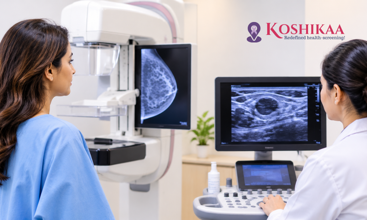
Breast imaging plays a vital role in finding breast changes early and investigating symptoms such as a lump or pain. Breast cancer is the most common cancer among women in India, and it is often diagnosed at a younger age than in many Western countries, which makes awareness of the right tests especially important.
Two of the most widely used breast imaging tests are mammography and breast ultrasound. Many women wonder which test they need and whether one is better than the other. The short answer is that they are different tools with different strengths. A mammogram uses low-dose X-rays and is the main test for routine breast cancer screening, while ultrasound uses sound waves and is especially useful for examining a specific lump, dense breast tissue and younger women.
This guide explains how each test works, the key differences, when each is recommended and why doctors often use both together.
What Is a Mammogram?
A mammogram is an X-ray of the breast. During the test, each breast is placed between two flat plates and gently compressed for a few seconds while images are taken from different angles. Compression spreads the breast tissue so that small changes can be seen clearly and the radiation dose is kept low.
Mammograms are very good at detecting tiny calcium deposits, called microcalcifications, which can be an early sign of some breast cancers, including early non-invasive cancers. They can often find changes years before a lump can be felt.
There are two main types:
- Screening mammography: used for women without symptoms to look for early signs of breast cancer
- Diagnostic mammography: used to investigate a symptom, such as a lump, or to look more closely at an area found on a screening mammogram
Some centres also offer 3D mammography, known as tomosynthesis, which takes multiple images to create a layered view of the breast. Learn more about our mammography in Bangalore services.
What Is a Breast Ultrasound?
A breast ultrasound uses high-frequency sound waves to create real-time images of the breast tissue. A sonographer or radiologist applies gel to the skin and moves a small handheld device, called a transducer, over the breast and armpit.
Ultrasound is painless, involves no compression and does not use any radiation. It is particularly good at showing whether a lump is a fluid-filled cyst, which is usually harmless, or a solid mass that may need further assessment. It can also guide a needle precisely during a biopsy.
Find out more about our ultrasound scan in Bangalore services.
Mammogram vs Breast Ultrasound: Key Differences
| Feature | Mammogram | Breast Ultrasound |
| Technology | Low-dose X-rays | High-frequency sound waves |
| Radiation | Yes, a very low dose | None |
| Main use | Routine screening and investigating symptoms | Investigating a specific lump or area; supplementary imaging |
| Best at detecting | Microcalcifications and early changes across the whole breast | Cysts vs solid lumps; changes in dense breast tissue |
| Comfort | Brief compression, which may feel uncomfortable | Painless, no compression |
| Dense breasts | Less sensitive, as dense tissue can hide abnormalities | May be added after mammography; does not replace it |
| Age and risk | Established screening test for eligible women; test choice also depends on symptoms, density and family history | Added after mammography or used to assess a specific concern; often first for symptoms in women under 30 or in pregnancy |
| Guiding biopsies | Can be used with specialised equipment | Commonly used |
Neither test is better in every situation. Each has strengths that the other lacks. Understanding these strengths helps explain why your doctor may recommend one test, the other or both, depending on your age, symptoms and breast tissue.
When Is Mammography Recommended?
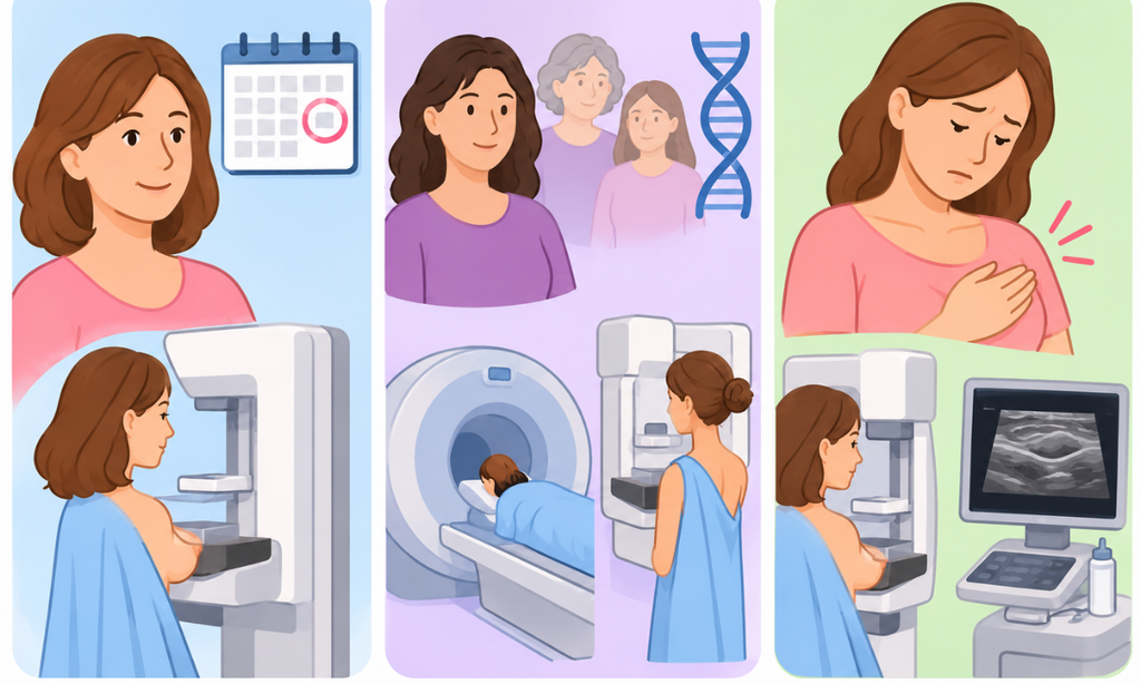
Routine Breast Cancer Screening
Mammography is the standard test for routine breast cancer screening. Many guidelines recommend that women at average risk begin regular screening mammograms from around the age of 40, although some start at 45 or 50, with screening repeated every one to two years. The best age to start and how often to repeat the test depend on your risk and your doctor’s advice.
Higher-Risk Women
Women with a strong family history of breast or ovarian cancer, a known inherited gene change such as BRCA1 or BRCA2, or previous radiotherapy to the chest may be advised to start screening earlier. For many of these women, annual breast MRI is recommended alongside mammography, typically beginning around age 25 for MRI and around age 30 for mammography, depending on individual risk and specialist advice. Ultrasound may be used in selected situations but is not usually the main additional test.
Investigating Breast Symptoms
When a woman notices a lump, nipple discharge, skin changes or other breast symptoms, the first test depends on her age and the type of symptom. For women aged around 30 and over, a diagnostic mammogram is often recommended, usually together with an ultrasound. This is different from routine screening, which is for women without symptoms.
When Is Breast Ultrasound Used?
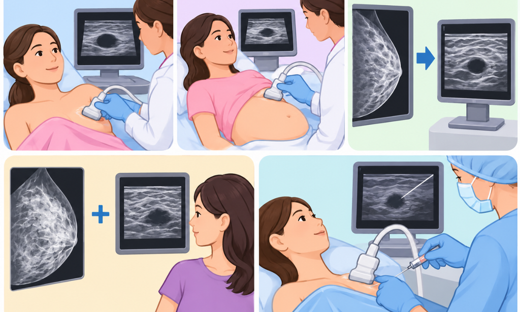
Investigating a Lump in Younger Women
In women under 30, breast tissue is usually dense, and breast cancer is less common. Ultrasound is therefore often the first test for a breast lump or pain in this age group.
During Pregnancy and Breastfeeding
Ultrasound is the preferred first test for breast symptoms during pregnancy and breastfeeding, as it does not use radiation and is not affected by the changes in breast tissue at these times.
Checking a Finding on a Mammogram
If a mammogram shows an area that needs a closer look, ultrasound can help determine whether it is a cyst or a solid mass.
Dense Breasts
Dense breasts contain more glandular and fibrous tissue and less fat. On a mammogram, dense tissue appears white, as do many cancers, which can make abnormalities harder to see. Breast density is common, particularly in younger women and many Asian women. Ultrasound may be recommended as an additional test after mammography for women with dense breasts, depending on their overall risk. It adds information but does not replace the mammogram.
Guiding Biopsies and Procedures
Ultrasound is widely used to guide needles during biopsies or to drain cysts, as the doctor can watch the needle in real time.
Can Both Tests Be Needed?
Yes. Mammography and ultrasound often work best together. A mammogram gives an overview of the whole breast and is excellent at detecting calcifications, while ultrasound provides a closer look at specific areas and dense tissue.
For example, a woman in her 40s with a new lump may have a diagnostic mammogram to check both breasts, followed by an ultrasound to examine the lump in detail. If either test shows a suspicious area, a biopsy may be recommended to confirm the diagnosis.
It is important to remember that ultrasound is not a replacement for screening mammography in women who are due for routine screening. Ultrasound alone may miss early changes, such as microcalcifications, that a mammogram can detect.
Breast Imaging and Screening: What Should Patients Know?
How Results Are Reported
Both mammogram and ultrasound reports often use a standard scoring system called BI-RADS, which ranges from 0 to 6. Lower scores usually mean a normal or benign finding, while higher scores indicate a greater need for further tests. Your doctor will explain what your score means and what to do next.
An Abnormal Result Does Not Always Mean Cancer
Many abnormal findings turn out to be harmless, such as cysts, fibroadenomas or benign calcifications. Further imaging or a biopsy is usually needed to confirm the cause.
Do Not Ignore Symptoms
A normal screening result does not rule out every problem. If you notice a new lump, nipple changes, skin dimpling or any other breast change, see a doctor promptly, even if your last mammogram was normal.
Choosing the Right Test
The right test depends on your age, symptoms, breast density, family history and your doctor’s assessment. If you are unsure, discuss your options with your doctor. Our guide to cancer screening and diagnostic tests explains how breast imaging fits into wider cancer screening.
How to Prepare for Each Test
Preparing for a Mammogram
- Avoid deodorant, powder, lotion or perfume on your breasts and underarms on the day of the test, as they can show up on the images
- Wear a two-piece outfit, so you only need to undress from the waist up
- If your breasts are tender before your period, try to book the test for a week or so after your period starts
- Tell the team if you have breast implants, are pregnant or breastfeeding
- Bring previous mammogram images and reports for comparison
Preparing for a Breast Ultrasound
No special preparation is usually needed. Wear a two-piece outfit and bring any previous breast imaging reports. Let the team know about any lump or area of concern so they can examine it carefully.
Breast Awareness: Know What Is Normal for You
Imaging tests are important, but being familiar with how your breasts normally look and feel is just as valuable. Breast tissue can change naturally with your menstrual cycle, pregnancy and age, so knowing what is normal for you makes it easier to notice anything new. See a doctor if you notice:
- A new lump or thickening in the breast or armpit
- A change in the size or shape of a breast
- Dimpling, puckering or redness of the skin
- Nipple changes, such as inversion, a rash or discharge
- Persistent pain in one area of the breast
Most breast changes are not cancer, but they should always be checked.
Mammogram and Breast Ultrasound Costs at Koshikaa
At Koshikaa, a screening mammogram is currently listed from ₹1,499, with digital and 3D mammography priced higher. Breast ultrasound can be performed on its own or alongside a mammogram when needed, and its price depends on the type of scan, so please confirm with the centre when you book.
Frequently Asked Questions
Is a breast ultrasound better than a mammogram?
Not in general. Mammography is the main screening test and is better at detecting microcalcifications, while ultrasound is better for examining specific lumps, dense tissue and younger women. They are often used together.
Can a breast ultrasound replace a mammogram?
No, not for routine screening in women who are due for mammograms. Ultrasound is usually used alongside mammography or for specific situations, such as younger women or pregnancy.
At what age should I start having mammograms?
Many guidelines suggest starting regular screening mammograms from around the age of 40 for women at average risk, though some recommend 45 or 50. Women at higher risk may need to start earlier, often with breast MRI as well. Your doctor can advise you.
Is a mammogram painful?
The compression can feel uncomfortable for a few seconds, but it is usually brief and well tolerated. Booking the test when your breasts are less tender can help.
Why do I need an ultrasound after my mammogram?
Your doctor may recommend ultrasound to take a closer look at an area seen on the mammogram, to check whether a lump is a cyst or solid, or because you have dense breast tissue.
Is the radiation from a mammogram safe?
Mammograms use a very low dose of radiation, and the benefits of finding breast cancer early generally outweigh the small risk.
Conclusion
Mammography and breast ultrasound are complementary tests, each with its own strengths. Mammography is the main tool for routine screening and detecting early changes such as microcalcifications, while ultrasound is ideal for examining specific lumps, dense breasts, younger women and pregnancy. Your doctor will recommend the test, or combination of tests, that best suits your age, symptoms and risk.
To book a mammogram or breast ultrasound, contact Koshikaa in Banashankari, Bengaluru, on +91 7996666106.