
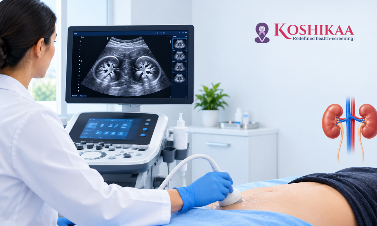
If your doctor has recommended a KUB ultrasound, you may be wondering what the scan involves and how to prepare. The good news is that it is one of the simplest and safest imaging tests available. It is painless, uses no radiation and usually takes less than half an hour.
KUB stands for kidneys, ureters and bladder, the main organs of the urinary tract. A KUB ultrasound uses sound waves to create images of these organs, helping doctors look for problems such as kidney stones, blockages, cysts, infection-related changes and difficulties with bladder emptying. In men, doctors may also suggest a PSA test alongside the scan if prostate problems are suspected.
This guide explains what a KUB ultrasound checks, why doctors recommend it, how to prepare, what happens during the scan, and what it can and cannot detect. If this is your first scan, knowing what to expect can make the experience much more comfortable, especially when you choose a trusted health screening centre in Bangalore.
What Is a KUB Ultrasound?
How Does a KUB Ultrasound Work?
A KUB ultrasound uses a small handheld device called a transducer, which sends high-frequency sound waves into the body. These sound waves bounce back from organs and tissues, and a computer turns the echoes into real-time images on a screen.
Because ultrasound does not use ionising radiation, it is considered safe for children and during pregnancy, and it can be repeated when needed for follow-up. This makes it particularly useful for monitoring people with recurring kidney stones, who may need several scans over time.
KUB Ultrasound vs KUB X-Ray
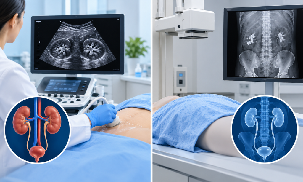
The term “KUB” is also used for a plain X-ray of the abdomen that shows the kidneys, ureters and bladder. A KUB X-ray and a KUB ultrasound are different tests. A KUB X-ray may show some calcium-containing stones, but it can miss uric acid or cystine stones and stones hidden by bowel gas. The ultrasound shows the structure of the organs and signs such as swelling or blockage. Check your prescription to confirm which test your doctor has requested.
On some reports and prescriptions, a KUB ultrasound may also be written as “USG KUB” or “USG kidney and bladder”.
What Organs Does a KUB Ultrasound Check?
Kidneys
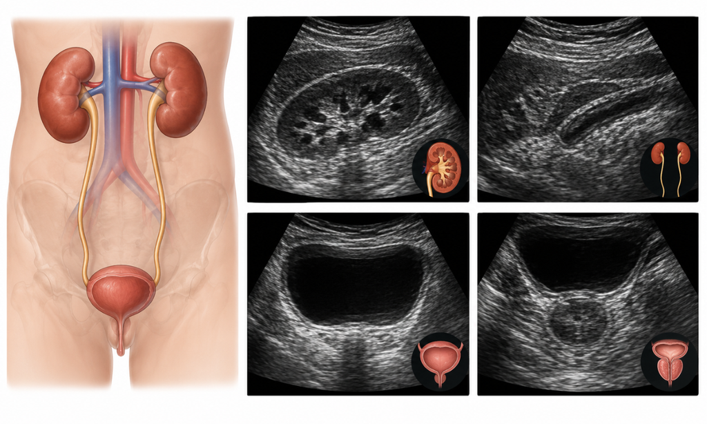
The kidneys are two bean-shaped organs at the back of the abdomen that filter waste from the blood and produce urine. The scan shows their size, shape and position, and can reveal stones, cysts, swelling caused by a blockage, scarring and some masses.
Ureters
The ureters are thin tubes that carry urine from each kidney to the bladder. Normal ureters are often difficult to see on ultrasound because they are narrow and lie deep in the body. They become easier to see when they are widened, for example because a stone is blocking the flow of urine.
Bladder
The bladder stores urine before it leaves the body. With a full bladder, the scan can show the thickness of the bladder wall, stones, growths and other abnormalities. Many centres also measure how much urine remains after you empty your bladder, known as the post-void residual volume.
Prostate in Men
In men, a KUB ultrasound often includes an assessment of the prostate gland, which sits just below the bladder. This can provide basic information about prostate size and how well the bladder empties in men with urinary symptoms. It does not replace a urological assessment or prostate cancer screening.
Why Might a Doctor Recommend a KUB Ultrasound?
A doctor may recommend a KUB ultrasound to investigate:
• Pain in the side, back or lower abdomen that may be caused by kidney stones
• Blood in the urine
• Frequent or painful urination, or recurrent urinary tract infections
• Difficulty passing urine or a feeling that the bladder is not emptying fully
• Abnormal kidney function blood tests
• Swelling of the kidneys seen on another scan
The scan is also used to monitor known conditions such as kidney stones or kidney cysts, to follow up after treatment or surgery, and to check the urinary tract in children with recurrent infections.
How to Prepare for a KUB Ultrasound

Drink Water and Arrive With a Full Bladder
A full bladder gives clearer images of the bladder and helps the sonographer see the lower urinary tract. You will usually be asked to drink several glasses of water about an hour before your appointment and to avoid passing urine until the scan is complete. The exact amount varies from person to person. Follow the exact instructions you are given when you book.
Do You Need to Fast?
Fasting is not usually required for a KUB ultrasound alone. However, if your scan is combined with an abdominal ultrasound, you may be asked to fast for six to eight hours beforehand, so that gas in the bowel does not block the view of other organs.
What to Wear and Bring
Wear loose, comfortable clothing, as you may need to uncover your abdomen. Bring your doctor’s prescription, any previous scan reports and a list of your current medicines. If you have had a previous KUB ultrasound or CT scan, bringing the images or report allows the radiologist to compare findings, which is especially useful when monitoring a known stone or cyst.
What Happens During the Procedure?
Step by Step
1. You lie on your back on an examination bed.
2. The sonographer or radiologist applies a cool, water-based gel to your abdomen. The gel helps the sound waves pass into the body.
3. The transducer is moved gently over your abdomen and sides. You may be asked to turn onto your side or take a deep breath and hold it to get clearer views of the kidneys.
4. Images of the bladder are taken while it is full.
5. You may then be asked to empty your bladder, after which a quick scan measures any urine left behind.
How Long Does It Take, and Does It Hurt?
The scan usually takes about 15 to 20 minutes. It is painless, although pressing on a very full bladder can feel uncomfortable for a short time. There is no recovery time, and you can eat, drink and return to normal activities straight away.
When Will You Get Your Report?
A radiologist reviews the images and prepares a report, which is shared with you and your doctor. At Koshikaa, ultrasound reports are usually shared digitally within a few hours.
What Can a KUB Ultrasound Detect, and What Are Its Limitations?
Conditions a KUB Ultrasound Can Help Identify
| Condition | What the Scan May Show |
| Kidney stones | Bright stones in the kidney, often with a shadow behind them |
| Hydronephrosis | Swelling of the kidney’s urine-collecting system, which may occur when urine flow is blocked |
| Kidney cysts | Fluid-filled sacs, most of which are harmless |
| Bladder stones or growths | Abnormal areas within the bladder |
| Incomplete bladder emptying | Urine remaining after passing urine |
| Enlarged prostate in men | Increased prostate size that may affect urine flow |
| Some congenital abnormalities | Differences in the shape or position of the kidneys |
Limitations of KUB Ultrasound
Ultrasound is an excellent first test, but it has some limits:
• Small stones may be missed, especially those under about 3 to 4 mm or lodged in the middle part of the ureters, which are often hidden by bowel gas. Ultrasound may also overestimate the size of stones it does detect.
• It does not measure kidney function. Blood tests such as creatinine are needed to assess how well the kidneys are working.
• Image quality can vary with body size, bowel gas and how full the bladder is.
• Some findings need further tests, such as a CT scan, MRI or cystoscopy, to confirm the cause.
KUB Ultrasound vs Other Scans
| Test | Main Strengths | Radiation |
| KUB ultrasound | Safe first test for structure, swelling, cysts and bladder emptying | None |
| Non-contrast CT KUB | Generally the most accurate test for urinary stones, particularly small ureteric stones, when clinically indicated | Yes |
| MRI | Detailed images for selected complex cases | None |
| KUB X-ray | May show some calcium-containing stones; can miss uric acid or cystine stones | Yes, low dose |
Doctors often start with ultrasound because it is safe, quick and affordable, then recommend a non-contrast CT scan if more detail is needed, particularly to locate small stones before treatment.
Understanding Your KUB Ultrasound Report
Ultrasound reports use some technical terms that can be confusing. Here are a few you may see:
• Normal size and echotexture: the kidneys look healthy in size and internal appearance.
• Corticomedullary differentiation: the normal distinction between the outer and inner parts of the kidney. “Maintained” is a reassuring finding.
• Calculus or calculi: a stone or stones. The report usually gives the size in millimetres and the location.
• Hydronephrosis: swelling of the kidney’s urine-collecting system, which may occur when urine flow is blocked. It is often graded as mild, moderate or severe.
• Simple cortical cyst: a fluid-filled sac in the kidney, which is usually harmless.
• Post-void residual (PVR): the amount of urine left in the bladder after you pass urine. A raised residual volume can have several causes and needs clinical interpretation; it does not by itself diagnose prostate disease.
• Prostatomegaly: an enlarged prostate in men.
A finding on your report is not a diagnosis on its own. For example, a small simple cyst rarely needs treatment, while a stone causing hydronephrosis may need prompt attention. Share your report with the doctor who requested the scan, who will explain what the findings mean for you and whether any further tests or treatment are needed.
KUB Ultrasound in Bangalore at Koshikaa
At Koshikaa in Banashankari, KUB ultrasound scans are performed and reported by our radiology team. A KUB ultrasound is currently listed at ₹1,250.You can learn more about our ultrasound scan in Bangalore services or call +91 7996666106 to book.
Frequently Asked Questions
Is a KUB ultrasound the same as a kidney ultrasound?
Not exactly. A kidney ultrasound focuses on the kidneys, while a KUB ultrasound also examines the ureters and bladder, and often the prostate in men. Some reports use the terms loosely, so check with your doctor which scan has been requested.
Is a KUB ultrasound painful?
No. You may feel the cool gel and some light pressure from the transducer, particularly over a full bladder, but the scan itself is painless.
Can a KUB ultrasound detect all kidney stones?
It detects most stones in the kidneys and bladder, but very small stones, especially in the middle of the ureters, can be missed. A CT scan may be recommended if a stone is suspected but not seen.
Why do I need a full bladder for a KUB ultrasound?
A full bladder gives clearer images of the bladder wall and the lower urinary tract, and it allows the team to measure how well your bladder empties.
How long does a KUB ultrasound take?
The scan usually takes about 15 to 20 minutes, though you should allow extra time for drinking water and waiting for your bladder to fill.
What does hydronephrosis on a KUB ultrasound mean?
Hydronephrosis means the kidney’s urine-collecting system is swollen. It may occur when urine flow is blocked, for example by a stone, but it can also have other causes, including mild changes during pregnancy. Your doctor will explain what it means in your case and whether further tests are needed.
Is a KUB ultrasound safe during pregnancy?
Yes. Ultrasound does not use ionising radiation and is considered safe during pregnancy. Mild swelling of the kidneys is common in pregnancy and is usually harmless, but your doctor will interpret the findings.
Do I need to fast before a KUB ultrasound?
Not usually. Fasting may be needed only if the KUB scan is combined with an abdominal ultrasound. Follow the instructions you are given when you book.
Conclusion
A KUB ultrasound is a simple, safe and painless way to examine the kidneys, ureters and bladder. It helps doctors investigate symptoms such as flank pain, blood in the urine and urinary problems, and it is often the first step before any further tests. Preparing with a full bladder and following your centre’s instructions will help ensure clear, useful images.
If your doctor has recommended a KUB ultrasound, contact Koshikaa in Banashankari, Bengaluru, on +91 7996666106 to book your scan.