Everything You Should Know About PET CT Scans

Table of Contents

PET CT scans have transformed medical diagnostics, providing healthcare staff with a strong tool for accurately detecting and monitoring a wide range of disorders. This cutting-edge technology, which combines Positron Emission Tomography (PET) and Computed Tomography (CT), allows for a precise image of tissue and organ structure. This fusion allows doctors to discover diseases like cancer at an early stage, when treatment is most successful, making it an invaluable tool in modern medicine.

PET CT scans have become crucial for diagnosing and tracking the evolution of illnesses ranging from cancer to neurological disorders in cities like Bangalore, where advanced healthcare services are becoming more accessible. This hybrid imaging technology not only aids in the detection of anomalies but also provides a more comprehensive picture of a patient’s health, allowing for more precise treatment recommendations. PET CT scan in Bangalore, whether for early detection or continuing monitoring, guarantee that patients receive prompt and precise therapy based on their unique needs.

What Is a PET CT Scan?

PET CT Scan in Bangalore

A PET CT scan is a sophisticated medical imaging method that integrates two powerful technologies, positron emission tomography (PET) and computed tomography (CT), into a single treatment. The PET scan entails injecting a tracer, or a small amount of radioactive material, into the body. This tracer identifies areas of heightened metabolic activity, which is especially useful for spotting cancer cells, which normally spend more energy than healthy cells. Meanwhile, a CT scan in Bangalore offers comprehensive X-ray images of the body’s internal components, such as organs and tissues, allowing doctors to see both healthy and diseased areas.

PET scans have emerged as a significant tool for detecting and treating diseases such as cancer, heart disease, and neurological disorders in Bangalore, where advanced healthcare is readily available. A PET scan in Bangalore gives clinicians a more complete picture of a patient’s health by integrating functional and structural imaging, allowing them to devise precise, tailored treatment plans. This dual-scan method allows for earlier discovery, more accurate monitoring, and improved treatment outcomes for patients.

Uses of PET CT Scans in Medical Diagnosis

PET CT scans are useful in identifying and monitoring a variety of medical diseases, particularly those involving aberrant cell activity. Here’s how this advanced imaging technology is applied:

Cancer Diagnosis and Monitoring: PET CT scans are useful for finding malignant tumors, determining their stage, and evaluating the efficacy of treatments like chemotherapy. This information is crucial for formulating treatment plans and monitoring disease development.

Heart Disease: These scans evaluate blood flow and identify damaged or malfunctioning cardiac components, providing important information about heart health. This aids in the accurate diagnosis and treatment of conditions such as coronary artery disease.

Brain Disorders: PET CT scans produce precise images of brain activity and structure in neurological conditions like Alzheimer’s disease, epilepsy, and brain cancer. This broadens our understanding of diseases and allows us to build more effective treatments.

PET CT scans are commonly used in Bangalore cancer screening services to give comprehensive evaluations and precise diagnoses, allowing for the most effective management and treatment options.

Types of PET CT Scans Offered

PET CT scans are classified into several categories, each with a focus on a unique diagnostic necessity. Here are a few common types:

Standard PET CT Scan: This is commonly used for general cancer diagnosis. It requires injecting a radioactive tracer to identify areas with elevated metabolic activity, which may indicate the presence of cancer cells.

Contrast PET CT Scan: This variant incorporates a contrast dye into the radioactive tracer. The dye increases the visibility of blood vessels and tissues, resulting in a more detailed and vivid depiction of the body’s interior structure.

3D PET CT Scan: A sophisticated imaging technique that produces three-dimensional images of the body. This scan provides a more thorough image of the area under examination, allowing for a very precise diagnosis.

PSMA PET CT Scan: This scan detects prostate cancer using a PSMA (Prostate-Specific Membrane Antigen) tracer. The PSMA PET CT scans tracer attaches to prostate cancer cells, enabling more accurate tumor location and evaluation.

Advantages of PET CT Scans Over Other Imaging Techniques

PET CT scans have several significant advantages over standard imaging modalities like MRIs and stand-alone CT scanners.

Early Detection: One of the key advantages of PET CT scans is their ability to diagnose diseases at an early stage, sometimes before symptoms develop. Early detection allows for more rapid responses, which improves treatment outcomes.

Functional and structural information: Unlike MRI and CT scans, which primarily examine the structural features of tissues and organs, PET CT scans provide both functional and structural information. They show how tissues work and detect metabolic abnormalities, giving a more complete picture of the body’s health.

Precise Staging: PET CT scans are very useful in oncology when determining the existence of cancer metastases. By properly staging the condition, doctors can select the most effective therapy options and monitor disease progression.

Treatment Monitoring: These scans are useful for determining the efficacy of current treatments, especially for cancer patients. They help to determine how well therapies are working, allowing adjustments to be made as necessary.

Overall, PET CT scans provide a detailed and dynamic picture of a patient’s health, making them an indispensable tool for current diagnosis and treatment planning.

PET CT scans cost and availability.

The cost of a PET CT scan varies according to several factors, including the type of scan, the location, and whether insurance coverage is available. Consult your healthcare practitioner or insurance company for more accurate information about your sickness.

PET CT scans are readily available in big cities, particularly at specialized radiology institutes and hospitals. In Bangalore, for example, numerous advanced healthcare facilities provide PET CT scanning services. Renowned scanning centers like “KOSHIKAA” offer complete PET CT scan services, giving patients access to cutting-edge diagnostic technologies. This widespread availability in metropolitan settings ensures that people may get prompt and reliable diagnostic imaging, which is critical for optimal treatment planning and disease management.

Preparing for a PET CT Scan

A PET CT scan requires proper preparation to yield precise findings. Here’s what you should know.

Pre-Scan Instructions: You may need to fast for 4-6 hours before your PET CT scan, however water is usually tolerated. If you have diabetes, you should talk to your doctor about how to manage insulin and other drugs to avoid problems.

Clothing and Accessories: Wear comfortable, loose-fitting clothing on the day of the scan. Make sure to remove any metal objects, such as jewelry, as they can interfere with the imaging process and reduce scan accuracy.

Medications: Tell your healthcare practitioner about any medications you are currently taking. Some drugs may need to be stopped or modified before the scan to obtain the best possible results.

What to Expect During the Procedure

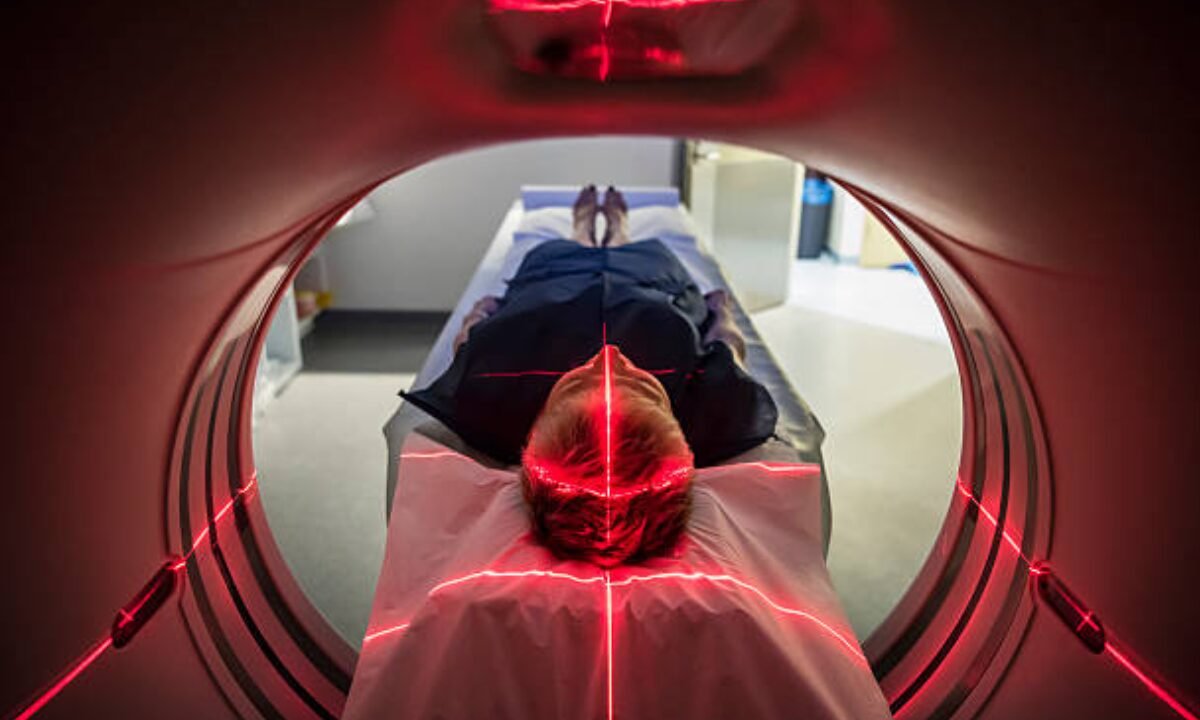
A PET CT scan is a non-invasive treatment that takes from 30 minutes to an hour. Here’s a step-by-step explanation of what to expect:

Tracer Injection: The procedure begins with a small amount of radioactive tracer being injected into a vein, usually in your arm. After the injection, you will need to wait 30-60 minutes for the tracer to be absorbed into your body.

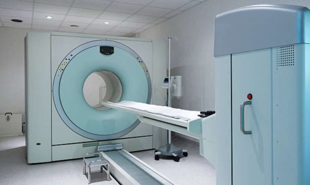
Imaging: During the scan, you will lie on a table that slowly moves into the PET CT equipment. It is critical to keep as still as possible while the scanner captures precise photos of your body. The treatment is relatively painless and rapid.

Post-Scan: Once the imaging is complete, you can resume your routine activities. Drinking plenty of water can help flush the tracer out of your system.

Risks and Side Effects of PET CT Scans

While PET CT scans are generally safe and effective, you should be aware of the following possible risks and side effects.

Radiation Exposure: PET CT scans emit relatively low quantities of radiation. Although the dose is small and typically safe for most individuals, pregnant women and young children should avoid it unless necessary. Always inform your healthcare provider if you are or may be pregnant.

Allergic Reactions: In rare situations, people may develop an allergic reaction to the contrast dye used in some PET CT scans. If you have a known allergy to contrast agents, consult your doctor first.

Side Effects: Most people have no negative side effects after a PET CT scan. However, if you become ill or notice any strange symptoms following surgery, you should contact your doctor for more information.

Conclusion and Future Developments in PET CT Scan

To summarize, PET CT scans have become an essential component of modern healthcare, allowing for the early identification and accurate diagnosis of a wide range of illnesses, including cancer, heart disease, and neurological issues. Their ability to combine functional and structural imaging enables a more comprehensive understanding of health conditions, which is extremely useful in treatment planning and illness management. PET CT scans in Bangalore have played an important role in providing these modern diagnostic capabilities to patients, ensuring that they receive the best possible care.

Moving forward, advances in PET CT technology will bring even more benefits. Future improvements will include the development of more specific tracers as well as efforts to limit radiation exposure, all of which will improve the safety and accuracy of PET CT scans. These advancements are projected to make PET CT scans more effective and accessible, reinforcing its role as an important tool in medical diagnostics and enhancing patient outcomes overall.

Koshikaa Screening Centre offers a wide range of comprehensive cancer screening services, ensuring accurate results and personalized health care. Contact us today at +91 9019800764

MRI of the uterus is primarily used for diagnosing and evaluating conditions such as uterine fibroids, adenomyosis, and malignancies. It is particularly useful in the planning of surgical procedures and assessing the extent of gynecological diseases. This imaging test is valuable for its ability to deliver detailed cross-sectional images of the uterus and surrounding structures.
An MRI of the spleen can help diagnose various disorders affecting the spleen including splenomegaly (enlarged spleen), tumors, and other abnormalities. It provides clear, detailed images that are crucial for assessing the health of the spleen and planning potential surgical interventions.
Prostate MRI is a non-invasive imaging technique that provides detailed pictures of the prostate gland and surrounding tissues. It helps in the detection, staging, and monitoring of prostate cancer. Prostate MRIs are increasingly used to guide biopsy procedures and assist in the planning of treatment strategies for prostate health.
An MRI of the pancreas is used to identify issues such as pancreatitis, pancreatic cancer, and other pancreatic disorders. It offers high-resolution images that help in detecting small lesions and assessing the pancreatic ducts. This imaging technique is valuable for guiding biopsies and planning surgeries involving the pancreas.
MRI scans of the liver, kidney, and bile ducts provide detailed images that help diagnose various conditions, such as tumors, cysts, and blockages in the bile ducts. They are crucial for assessing organ damage, staging cancer, and planning liver or kidney-related surgeries. This type of MRI can also evaluate the health and function of these organs comprehensively.
Breast MRI is highly sensitive for detecting breast cancer, especially in individuals with dense breast tissue or a high risk of cancer. It is often used in conjunction with mammography to provide a more comprehensive evaluation. Breast MRIs can also monitor the response to breast cancer treatments and provide detailed pre-surgical mapping.
Brain and spine MRIs are critical tools for diagnosing and monitoring neurological conditions, including tumors, spinal cord injuries, and multiple sclerosis. These MRIs can reveal abnormalities in brain tissue and spinal cord structure that might not be visible with other imaging methods. They are essential for planning surgeries and assessing the effectiveness of treatments in neurology.
PET-CT scans of transplant organs like the liver, kidney, or pancreas are performed to assess metabolic activity, detect complications, and monitor post-transplant function. These scans play a vital role in the early detection of issues such as organ rejection, recurrence of disease, or infection, guiding timely interventions and optimizing transplant outcomes for patients.
PET-CT examinations of organs such as the liver, kidney, or prostate are crucial for assessing metabolic activity, detecting abnormalities, and monitoring disease progression. These scans help diagnose conditions such as liver metastases, renal cell carcinoma, or prostate cancer, guiding treatment decisions and monitoring response to therapy over time.
PET-CT scans of soft-tissue masses are utilized to assess metabolic activity and detect malignant tumors or other abnormalities. By combining PET imaging with CT imaging, these scans provide detailed information about tumor location, size, and metabolic characteristics, aiding in accurate diagnosis and treatment planning for soft-tissue cancers.
PET-CT scans are valuable for detecting cysts or fluid collections within the body, including cysts in the liver, kidneys, or other organs. These scans help characterize the nature and metabolic activity of cysts, guiding diagnosis and treatment planning for conditions such as cystic liver disease, renal cysts, or pancreatic cysts.
PET-CT scans of muscles are performed to assess metabolic activity and detect abnormalities such as inflammation, infection, or tumors. These scans provide valuable information about muscle function and pathology, aiding in the diagnosis and management of conditions like myositis, muscular dystrophy, or soft-tissue sarcomas.
PET-CT scans of the brain are utilized to evaluate brain metabolism and detect abnormalities such as tumors, epilepsy, or neurodegenerative diseases. By combining functional PET imaging with anatomical CT imaging, these scans offer comprehensive information about brain function and structure, guiding neurosurgical interventions and treatment decisions for neurological disorders
PET-CT scans of bones are commonly used to detect bone metastases in cancer patients, assess bone health in conditions like osteoporosis, and evaluate response to treatment. By combining PET and CT imaging, these scans provide detailed information about metabolic activity and anatomical changes in bone structures, aiding in accurate diagnosis and treatment planning for various bone-related conditions.
CT scans of the spine are conducted to assess the vertebrae, spinal cord, and surrounding soft tissues for conditions like herniated discs, spinal fractures, or spinal stenosis, aiding in the diagnosis and management of spinal disorders with detailed imaging essential for surgical planning and intervention.
CT scans of the pelvis are utilized to visualize the pelvic bones, reproductive organs, bladder, and surrounding structures for conditions such as fractures, tumors, or pelvic inflammatory disease, guiding treatment plans and surgical interventions with precise anatomical information.
CT scans of the neck are performed to evaluate structures such as the throat, thyroid gland, lymph nodes, and blood vessels for abnormalities like tumors, infections, or injuries, assisting in diagnosis and management of head and neck disorders with detailed imaging.
CT scans of the extremities are employed to assess bones, joints, and soft tissues in the arms or legs, aiding in the diagnosis of fractures, arthritis, or other musculoskeletal conditions, guiding orthopedic treatment and rehabilitation plans for optimal recovery.
CT scans of the chest are utilized to examine the lungs, heart, blood vessels, and surrounding structures for conditions such as lung nodules, pneumonia, or pulmonary embolism, facilitating accurate diagnosis and management of respiratory and cardiovascular diseases.
CT scans of the brain are conducted to assess brain structures, identify abnormalities like tumors, hemorrhages, or strokes, and aid in diagnosing neurological conditions, guiding neurosurgical interventions and treatment strategies with precise imaging of the brain’s anatomy.
CT scans of the abdomen are performed to visualize and evaluate the internal organs such as the liver, kidneys, pancreas, and intestines for conditions like tumors, infections, or injuries, providing detailed images essential for diagnosis and treatment planning.
Transplant organ ultrasound scans, including those of the liver, kidney, or pancreas, are performed to assess organ viability, detect complications, and monitor post-transplant function. These scans play a vital role in the early detection of issues such as organ rejection, vascular complications, or fluid collections, guiding timely interventions and optimizing transplant outcomes for patients.
Ultrasound examinations of organs such as the liver, kidney, or prostate are crucial for assessing organ structure, detecting abnormalities, and monitoring disease progression. These scans help diagnose conditions such as liver cirrhosis, kidney stones, or prostate enlargement, guiding treatment decisions and monitoring response to therapy over time.
Soft-tissue masses can be evaluated using ultrasound to determine their size, location, and characteristics. Ultrasound imaging helps differentiate between benign and malignant masses, guiding further diagnostic evaluation and treatment planning. By providing real-time imaging, ultrasound assists in the precise localization of soft-tissue masses for biopsy or surgical removal if needed.
Ultrasound scans are valuable for detecting cysts or fluid collections within the body, including cysts in the liver, kidneys, or other organs. These scans help characterize the nature and size of cysts, aiding in diagnosis and treatment planning for conditions such as cystic liver disease or renal cysts, and guiding interventions such as cyst drainage or sclerotherapy.
Musculoskeletal ultrasound scans are used to assess the health and integrity of muscles, identifying issues such as strains, tears, or muscle inflammation. These scans provide detailed images of muscle structures, allowing healthcare providers to diagnose muscle injuries accurately and develop personalized rehabilitation plans to promote recovery and restore function.
Ultrasound examinations of joints are essential for evaluating joint health, detecting abnormalities such as inflammation, fluid accumulation, or cartilage damage. By visualizing the joint structures in real-time, ultrasound helps diagnose conditions like arthritis, bursitis, or ligament injuries, guiding treatment decisions and monitoring disease progression over time.
Ultrasound scans of tendons are performed to assess for injuries, inflammation, or tears, aiding in the diagnosis and management of conditions such as tendonitis or tendon tears. These scans provide detailed images of tendon structures, helping healthcare providers determine the extent of damage and plan appropriate treatment strategies, including physical therapy or surgical intervention if necessary.
Pelvis X-rays play a crucial role in diagnosing injuries, diseases, and developmental conditions affecting the pelvic region. They provide detailed images of the hip bones, sacrum, and coccyx, aiding in the assessment of fractures, dislocations, or signs of arthritis. Pelvic X-rays are instrumental in guiding surgical interventions, monitoring healing progress, and evaluating conditions such as osteoporosis or tumors. By providing precise anatomical information, these imaging studies enable healthcare professionals to deliver personalized care and improve patient outcomes.
Teeth X-rays, known as dental radiographs, are indispensable tools for dentists to assess oral health. They aid in diagnosing dental decay, gum disease, and abnormalities in tooth structure or alignment. These images provide valuable insights into hidden dental issues, allowing for early intervention and prevention of complications. Additionally, dental X-rays are essential for planning treatments such as orthodontics, root canals, or dental implants, ensuring optimal outcomes and patient satisfaction.
Spine X-rays are pivotal in diagnosing spinal fractures, degenerative conditions, and spinal misalignments, providing critical information for treatment planning and monitoring spinal health. They offer detailed images of the vertebrae and surrounding structures, enabling healthcare professionals to identify issues such as scoliosis, disc herniation, or spinal stenosis
Skull X-rays are valuable for detecting fractures, assessing head trauma, and evaluating the alignment of facial bones, crucial in diagnosing conditions like fractures, tumors, or developmental abnormalities in the skull. They provide detailed views of the cranial structure, aiding clinicians in determining appropriate treatment strategies and ensuring patient safety.
For the heart, X-ray solutions can aid in the detection of various cardiac problems, ranging from mild to severe, including – Heart failure, Cardiac congestion and other heart-related conditions. In addition to cardiac assessments, X-ray solutions are also used for breast examinations to identify tumors or signs of breast cancer.
Bone X-rays are essential for diagnosing fractures and assessing bone alignment, aiding doctors in developing treatment plans and monitoring healing progress. They provide detailed images of the skeletal system, helping identify abnormalities or injuries that may not be visible through other imaging techniques
Abdominal X-rays are performed when patients experience symptoms such as significant nausea, ongoing vomiting, abdominal pain, flank or back pain, or abdominal swelling. These X-rays can help diagnose various conditions.
Search Bar

Frequently Booked Tests

Frequently Booked Checkups