CT Scan: What It Is, Types, Purpose, Procedure and Results

Table of Contents

Medical imaging has revolutionized how doctors diagnose and treat various health conditions, and the CT scan is among the most powerful diagnostic tools available today. Whether you have been recommended this test or are simply curious about what it involves, understanding the basics can help ease any concerns.

If you are looking for a CT scan in Bangalore, knowing what to expect from this procedure is essential for your peace of mind and preparation.

Let’s explore everything you need to know about CT scans, from what they are to how they work and what your results might mean.

Understanding the CT Scan Definition

A CT scan, or computed tomography scan, is an advanced medical imaging technique that uses X-rays and computer technology to create detailed cross-sectional images of your body.

Unlike regular X-rays that produce flat, two-dimensional images, CT scans generate multiple images from different angles.

These images are then processed by a computer to create comprehensive 3D views of your bones, blood vessels, and soft tissues.

The technology allows doctors to see inside your body without making a single incision, making it an invaluable diagnostic tool.

What Is a CT Scan Used For?

CT scans serve numerous diagnostic purposes across various medical specialities.

Doctors commonly order CT scans to detect cancers, monitor tumors, and plan cancer treatments effectively.

They’re essential for diagnosing internal injuries after accidents or trauma, especially to identify internal bleeding or bone fractures.

CT scans help detect blood clots, heart disease, and stroke by providing clear images of blood vessels and the heart.

They’re also used to diagnose infections, kidney stones, appendicitis, and other abdominal conditions with remarkable accuracy.

Additionally, CT scans guide biopsies and other medical procedures by showing doctors exactly where to target.

Common Types of CT Scans

Different types of CT scans target specific areas of your body based on your symptoms and medical needs.

CT Scan of Lungs

This scan examines your chest area to detect lung cancer, infections, pulmonary embolism, or chronic lung diseases. It’s particularly useful for smokers or those with persistent respiratory symptoms.

CT Scan of Heart

Also called cardiac CT, this specialized scan evaluates your heart’s structure, coronary arteries, and surrounding blood vessels. It helps detect blockages, heart disease, and calcium buildup in arteries.

CT Scan Abdomen

This comprehensive Abdominal CT scan examines your liver, kidneys, pancreas, spleen, and intestines. Doctors order it to investigate abdominal pain, digestive issues, or suspected organ problems.

CT Scan for Kidney Stones

This is one of the most accurate methods for detecting kidney stones, showing their exact size, location, and number. It helps doctors plan the most appropriate treatment approach.

The CT Scan Procedure: What to Expect

Understanding the CT scan procedure helps reduce anxiety and ensures you’re properly prepared.

Before the Scan:

  • You may be asked to avoid eating or drinking for a few hours before your scan, especially if contrast dye will be used.
  • Inform your doctor about any allergies, especially to iodine or contrast materials, and if you’re pregnant or might be pregnant.
  • Remove all metal objects, including jewellery, glasses, and dentures, as they can interfere with the imaging.

During the Scan:

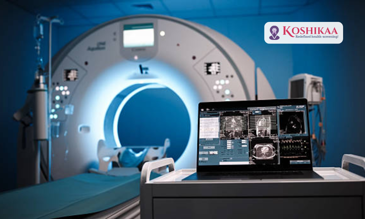
  • You will lie on a motorized table that slides into the CT scanner, which looks like a large doughnut.
  • The technician will position you correctly and may use pillows or straps to help you stay still during the procedure.
  • You’ll need to remain very still and may be asked to hold your breath briefly for clearer images.
  • The machine rotates around you, taking multiple X-ray images from different angles. You might hear whirring or clicking sounds.
  • Some scans require contrast dye, which may be injected through an IV or taken orally. This helps certain tissues show up more clearly.

After the Scan:

  • The entire process is painless and non-invasive. You can typically resume normal activities immediately after the scan.
  • If contrast dye was used, drink plenty of water to help flush it out of your system.

How Long Do CT Scans Take?

The duration of a CT scan depends on which part of your body is being examined.

Most CT scans take between 10 and 30 minutes from start to finish, including preparation time.

The actual scanning time is often just 5 to 10 minutes, though more complex scans may take longer.

Scans requiring contrast dye may add a few extra minutes to the procedure.

We always recommend arriving 15 minutes early to complete any necessary paperwork and preparation.

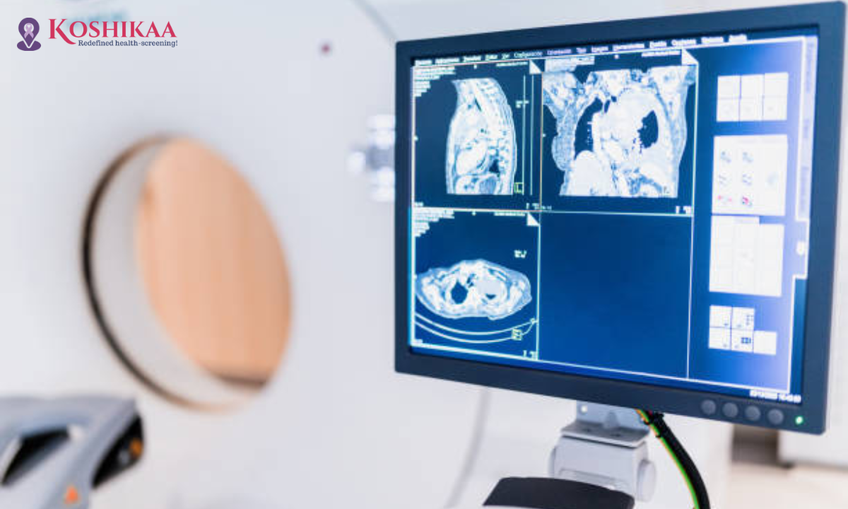
Understanding Your CT Scan Report

Your CT scan report is a detailed document prepared by a radiologist who specializes in interpreting medical images.

The report describes what was observed in each area of your body that was scanned.

It includes measurements of any abnormalities, their location, and their appearance compared to normal tissue.

Most centres provide reports within 24 to 48 hours, though urgent cases may receive faster turnaround times.

Your referring doctor will review the report with you and explain what the findings mean for your health and treatment plan.

Never try to interpret your CT scan report alone, as medical terminology can be confusing without proper context.

Understanding CT Scan Price in Bangalore

Various factors affect the overall cost, including the body area being scanned and whether contrast dye is required.

More specialized scans, like a cardiac CT or a full-body CT scan, typically involve different considerations.

Choosing a CT Scan Centre

When considering a CT scan in Bangalore, several factors influence your choice of diagnostic centre.

  • Look for facilities with modern, well-maintained CT scan equipment that ensures accurate imaging.
  • Check if the centre has experienced radiologists who specialize in interpreting CT scan images.
  • The health screening centre in Bangalore offers comprehensive diagnostic services under one roof for your convenience.
  • Always enquire about the turnaround time for reports and available payment options before booking your appointment.
  • Verify if your health insurance covers the scan and whether the centre offers cashless claim facilities to make the process smoother.

Tips for a Successful CT Scan

Here are some practical tips to ensure your CT scan goes smoothly:

  • Wear comfortable, loose-fitting clothing without metal zippers or buttons
  • Leave valuables at home to avoid any loss during the procedure
  • Inform the technician if you feel claustrophobic or anxious in enclosed spaces
  • Ask all your questions before the scan begins to feel more comfortable
  • Follow all pre-scan instructions carefully, especially regarding fasting
  • Bring your previous medical reports and images for comparison, if available

Final Thoughts

A CT scan has become an indispensable tool in modern medicine, offering detailed insights that help doctors make accurate diagnoses and create effective treatment plans.

Understanding what the procedure involves, how to prepare, and what to expect from your results can significantly reduce any anxiety you might feel.

If you need a CT scan in Bangalore, choosing a reliable health screening centre in Bangalore with modern equipment and experienced radiologists is crucial for accurate results.

At Koshikaa, we are committed to providing comprehensive diagnostic services with advanced CT scan technology and expert radiologists who ensure accurate, timely reports.

Remember, a CT scan is a safe, quick, and painless procedure that could provide critical information about your health.

Frequently Asked Questions

Q: Is a CT scan safe?

Yes, CT scans are generally safe. While they use X-rays, the radiation exposure is minimal, and the diagnostic benefits usually far outweigh the risks.

Q: Can I eat before a CT scan?

It depends on the type of scan. For some CT scans, especially those using contrast dye, you may need to fast for 4-6 hours beforehand.

Q: Will I feel anything during the CT scan?

The scan itself is painless. If contrast dye is used, you might feel a warm sensation or metallic taste temporarily.

Q: How soon will I get my CT scan report?

Most centres provide reports within 24-48 hours. Urgent cases may receive results on the same day.

Q: Can pregnant women have CT scans?

CT scans are generally avoided during pregnancy unless necessary, as radiation may affect the developing baby. Always inform your doctor if you’re pregnant.

MRI of the uterus is primarily used for diagnosing and evaluating conditions such as uterine fibroids, adenomyosis, and malignancies. It is particularly useful in the planning of surgical procedures and assessing the extent of gynecological diseases. This imaging test is valuable for its ability to deliver detailed cross-sectional images of the uterus and surrounding structures.
An MRI of the spleen can help diagnose various disorders affecting the spleen including splenomegaly (enlarged spleen), tumors, and other abnormalities. It provides clear, detailed images that are crucial for assessing the health of the spleen and planning potential surgical interventions.
Prostate MRI is a non-invasive imaging technique that provides detailed pictures of the prostate gland and surrounding tissues. It helps in the detection, staging, and monitoring of prostate cancer. Prostate MRIs are increasingly used to guide biopsy procedures and assist in the planning of treatment strategies for prostate health.
An MRI of the pancreas is used to identify issues such as pancreatitis, pancreatic cancer, and other pancreatic disorders. It offers high-resolution images that help in detecting small lesions and assessing the pancreatic ducts. This imaging technique is valuable for guiding biopsies and planning surgeries involving the pancreas.
MRI scans of the liver, kidney, and bile ducts provide detailed images that help diagnose various conditions, such as tumors, cysts, and blockages in the bile ducts. They are crucial for assessing organ damage, staging cancer, and planning liver or kidney-related surgeries. This type of MRI can also evaluate the health and function of these organs comprehensively.
Breast MRI is highly sensitive for detecting breast cancer, especially in individuals with dense breast tissue or a high risk of cancer. It is often used in conjunction with mammography to provide a more comprehensive evaluation. Breast MRIs can also monitor the response to breast cancer treatments and provide detailed pre-surgical mapping.
Brain and spine MRIs are critical tools for diagnosing and monitoring neurological conditions, including tumors, spinal cord injuries, and multiple sclerosis. These MRIs can reveal abnormalities in brain tissue and spinal cord structure that might not be visible with other imaging methods. They are essential for planning surgeries and assessing the effectiveness of treatments in neurology.
PET-CT scans of transplant organs like the liver, kidney, or pancreas are performed to assess metabolic activity, detect complications, and monitor post-transplant function. These scans play a vital role in the early detection of issues such as organ rejection, recurrence of disease, or infection, guiding timely interventions and optimizing transplant outcomes for patients.
PET-CT examinations of organs such as the liver, kidney, or prostate are crucial for assessing metabolic activity, detecting abnormalities, and monitoring disease progression. These scans help diagnose conditions such as liver metastases, renal cell carcinoma, or prostate cancer, guiding treatment decisions and monitoring response to therapy over time.
PET-CT scans of soft-tissue masses are utilized to assess metabolic activity and detect malignant tumors or other abnormalities. By combining PET imaging with CT imaging, these scans provide detailed information about tumor location, size, and metabolic characteristics, aiding in accurate diagnosis and treatment planning for soft-tissue cancers.
PET-CT scans are valuable for detecting cysts or fluid collections within the body, including cysts in the liver, kidneys, or other organs. These scans help characterize the nature and metabolic activity of cysts, guiding diagnosis and treatment planning for conditions such as cystic liver disease, renal cysts, or pancreatic cysts.
PET-CT scans of muscles are performed to assess metabolic activity and detect abnormalities such as inflammation, infection, or tumors. These scans provide valuable information about muscle function and pathology, aiding in the diagnosis and management of conditions like myositis, muscular dystrophy, or soft-tissue sarcomas.
PET-CT scans of the brain are utilized to evaluate brain metabolism and detect abnormalities such as tumors, epilepsy, or neurodegenerative diseases. By combining functional PET imaging with anatomical CT imaging, these scans offer comprehensive information about brain function and structure, guiding neurosurgical interventions and treatment decisions for neurological disorders
PET-CT scans of bones are commonly used to detect bone metastases in cancer patients, assess bone health in conditions like osteoporosis, and evaluate response to treatment. By combining PET and CT imaging, these scans provide detailed information about metabolic activity and anatomical changes in bone structures, aiding in accurate diagnosis and treatment planning for various bone-related conditions.
CT scans of the spine are conducted to assess the vertebrae, spinal cord, and surrounding soft tissues for conditions like herniated discs, spinal fractures, or spinal stenosis, aiding in the diagnosis and management of spinal disorders with detailed imaging essential for surgical planning and intervention.
CT scans of the pelvis are utilized to visualize the pelvic bones, reproductive organs, bladder, and surrounding structures for conditions such as fractures, tumors, or pelvic inflammatory disease, guiding treatment plans and surgical interventions with precise anatomical information.
CT scans of the neck are performed to evaluate structures such as the throat, thyroid gland, lymph nodes, and blood vessels for abnormalities like tumors, infections, or injuries, assisting in diagnosis and management of head and neck disorders with detailed imaging.
CT scans of the extremities are employed to assess bones, joints, and soft tissues in the arms or legs, aiding in the diagnosis of fractures, arthritis, or other musculoskeletal conditions, guiding orthopedic treatment and rehabilitation plans for optimal recovery.
CT scans of the chest are utilized to examine the lungs, heart, blood vessels, and surrounding structures for conditions such as lung nodules, pneumonia, or pulmonary embolism, facilitating accurate diagnosis and management of respiratory and cardiovascular diseases.
CT scans of the brain are conducted to assess brain structures, identify abnormalities like tumors, hemorrhages, or strokes, and aid in diagnosing neurological conditions, guiding neurosurgical interventions and treatment strategies with precise imaging of the brain’s anatomy.
CT scans of the abdomen are performed to visualize and evaluate the internal organs such as the liver, kidneys, pancreas, and intestines for conditions like tumors, infections, or injuries, providing detailed images essential for diagnosis and treatment planning.
Transplant organ ultrasound scans, including those of the liver, kidney, or pancreas, are performed to assess organ viability, detect complications, and monitor post-transplant function. These scans play a vital role in the early detection of issues such as organ rejection, vascular complications, or fluid collections, guiding timely interventions and optimizing transplant outcomes for patients.
Ultrasound examinations of organs such as the liver, kidney, or prostate are crucial for assessing organ structure, detecting abnormalities, and monitoring disease progression. These scans help diagnose conditions such as liver cirrhosis, kidney stones, or prostate enlargement, guiding treatment decisions and monitoring response to therapy over time.
Soft-tissue masses can be evaluated using ultrasound to determine their size, location, and characteristics. Ultrasound imaging helps differentiate between benign and malignant masses, guiding further diagnostic evaluation and treatment planning. By providing real-time imaging, ultrasound assists in the precise localization of soft-tissue masses for biopsy or surgical removal if needed.
Ultrasound scans are valuable for detecting cysts or fluid collections within the body, including cysts in the liver, kidneys, or other organs. These scans help characterize the nature and size of cysts, aiding in diagnosis and treatment planning for conditions such as cystic liver disease or renal cysts, and guiding interventions such as cyst drainage or sclerotherapy.
Musculoskeletal ultrasound scans are used to assess the health and integrity of muscles, identifying issues such as strains, tears, or muscle inflammation. These scans provide detailed images of muscle structures, allowing healthcare providers to diagnose muscle injuries accurately and develop personalized rehabilitation plans to promote recovery and restore function.
Ultrasound examinations of joints are essential for evaluating joint health, detecting abnormalities such as inflammation, fluid accumulation, or cartilage damage. By visualizing the joint structures in real-time, ultrasound helps diagnose conditions like arthritis, bursitis, or ligament injuries, guiding treatment decisions and monitoring disease progression over time.
Ultrasound scans of tendons are performed to assess for injuries, inflammation, or tears, aiding in the diagnosis and management of conditions such as tendonitis or tendon tears. These scans provide detailed images of tendon structures, helping healthcare providers determine the extent of damage and plan appropriate treatment strategies, including physical therapy or surgical intervention if necessary.
Pelvis X-rays play a crucial role in diagnosing injuries, diseases, and developmental conditions affecting the pelvic region. They provide detailed images of the hip bones, sacrum, and coccyx, aiding in the assessment of fractures, dislocations, or signs of arthritis. Pelvic X-rays are instrumental in guiding surgical interventions, monitoring healing progress, and evaluating conditions such as osteoporosis or tumors. By providing precise anatomical information, these imaging studies enable healthcare professionals to deliver personalized care and improve patient outcomes.
Teeth X-rays, known as dental radiographs, are indispensable tools for dentists to assess oral health. They aid in diagnosing dental decay, gum disease, and abnormalities in tooth structure or alignment. These images provide valuable insights into hidden dental issues, allowing for early intervention and prevention of complications. Additionally, dental X-rays are essential for planning treatments such as orthodontics, root canals, or dental implants, ensuring optimal outcomes and patient satisfaction.
Spine X-rays are pivotal in diagnosing spinal fractures, degenerative conditions, and spinal misalignments, providing critical information for treatment planning and monitoring spinal health. They offer detailed images of the vertebrae and surrounding structures, enabling healthcare professionals to identify issues such as scoliosis, disc herniation, or spinal stenosis
Skull X-rays are valuable for detecting fractures, assessing head trauma, and evaluating the alignment of facial bones, crucial in diagnosing conditions like fractures, tumors, or developmental abnormalities in the skull. They provide detailed views of the cranial structure, aiding clinicians in determining appropriate treatment strategies and ensuring patient safety.
For the heart, X-ray solutions can aid in the detection of various cardiac problems, ranging from mild to severe, including – Heart failure, Cardiac congestion and other heart-related conditions. In addition to cardiac assessments, X-ray solutions are also used for breast examinations to identify tumors or signs of breast cancer.

Bone X-rays are essential for diagnosing fractures and assessing bone alignment, aiding doctors in developing treatment plans and monitoring healing progress. They provide detailed images of the skeletal system, helping identify abnormalities or injuries that may not be visible through other imaging techniques

Abdominal X-rays are performed when patients experience symptoms such as significant nausea, ongoing vomiting, abdominal pain, flank or back pain, or abdominal swelling. These X-rays can help diagnose various conditions.

Frequently Booked Tests

Frequently Booked Checkups