
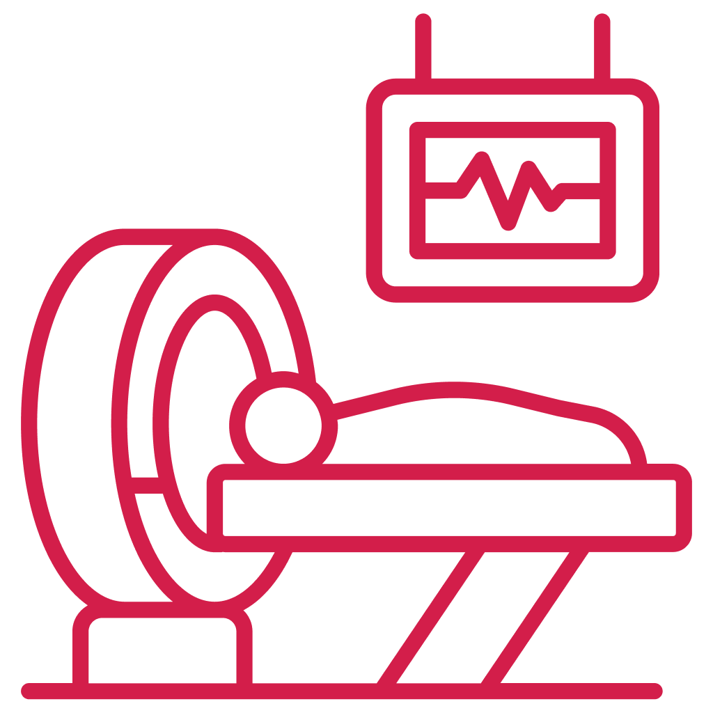
We use a head and neck CT scan to quickly detect tumors, infections, or fractures with high clarity for early decision-making.

Our CT scan of the head and neck helps visualize deep-lying nerves, vessels, and tissues that regular X-rays cannot capture accurately.

Many choose our CT scanning services because it delivers fast results, reducing delays in starting the right medical treatment or surgery.

With a CT scan, we minimize exploratory procedures by giving doctors a clear internal map before planning further interventions.
We use a head and neck CT scan to quickly detect tumors, infections, or fractures with high clarity for early decision-making.
Our CT scan of the head and neck helps visualize deep-lying nerves, vessels, and tissues that regular X-rays cannot capture accurately.
Many choose our CT scanning services because it delivers fast results, reducing delays in starting the right medical treatment or surgery.
With a CT scan, we minimize exploratory procedures by giving doctors a clear internal map before planning further interventions.
We perform every head and neck CT scan on advanced scanners, ensuring sharp image quality that helps doctors make confident and early decisions.

We perform every head and neck CT scan on advanced scanners, ensuring sharp image quality that helps doctors make confident and early decisions.

Many patients choose us for CT scanning services because we maintain strict safety, hygiene, and radiation protocols throughout the procedure.

We provide a head and neck CT scan with fast appointments, same-day reports, and smooth patient handling to reduce stress and waiting time.

With transparent CT scan cost in Bangalore, we make premium CT scan of the head and neck services affordable without compromising on quality.
We perform every head and neck CT scan on advanced scanners, ensuring sharp image quality that helps doctors make confident and early decisions.
Our CT scan reports are interpreted by skilled radiologists who focus on accuracy, clarity, and clinical relevance for patients.
Many patients choose us for CT scanning services because we maintain strict safety, hygiene, and radiation protocols throughout the procedure.
We provide a head and neck CT scan with fast appointments, same-day reports, and smooth patient handling to reduce stress and waiting time.
With transparent CT scan cost in Bangalore, we make premium CT scan of the head and neck services affordable without compromising on quality.
“Koshikaa’s Digital Mammography in Bangalore was quick, accurate, and comfortable. The staff made me feel confident and well cared for.”
“I trust Koshikaa for all screenings. The digital mammography provided precise results and peace of mind throughout the process.”
This mammography centre in Bangalore truly cares. The procedure was painless, and the results were fast and easy to understand.”


