Comparison of CT Scan and MRI Scan

CT scan vs MRI scan

Table of Contents

Modern medical diagnostics heavily rely on imaging for doctors during the diagnosis and treatment of patients. Among various scanning methods, CT scans and MRI scans are the two most commonly used types. While both are essential for diagnosing and detecting numerous diseases, they differ significantly based on technology, application, and accuracy. We will explore the advantages, limitations, and differences between CT scans and MRI scans.

What is a CT scan?

CT scan in Bangalore

A CT scan is a process in which X-rays create images of the body’s internal structures. It is highly effective and quick in diagnosing internal injuries or abnormalities like fractures, tumors, and infections. The procedure involves taking multiple X-ray images from various angles and combined by a computer to produce a cross-sectional view of the body’s internal structures.

Advantages of CT Scans

1. Speed: One of the Key advantages of CT scans is their speed. CT scans are fast and provide quick results, making them particularly useful in emergencies, where rapid diagnosis is necessary. It can be crucial in situations where timely medical intervention is critical.

2. Bone Imaging: CT scans are excellent at imaging bones and are particularly effective at detecting fractures and diagnosing bone damage. Making them an indispensable tool in orthopedics medicine, since they can provide detailed images of bone structures and aid in accurate diagnoses.

3. Cost-Effective: In general, CT scans are more cost-effective compared to MRI scans, making them a more commonly available imaging option. This cost-effectiveness can make CT scan in Bangalore a preferred choice for both patients and healthcare providers, especially when comprehensive imaging is required.

Disadvantages of CT Scans

1. Radiation Exposure: Of course, as they utilize X-rays, CT scans do expose patients to some small amount of ionizing radiation, which can prove harmful in case of repeated exposure.

2. Imaging of Soft Tissues: CT scans might not provide detailed images of soft tissues such as the brain, spinal cord, or muscles.

A person interested in getting a CT scan should find a diagnostic center that uses modern technology and professional expertise to provide accurate results. MRI, or Magnetic Resonance Imaging, is a cutting-edge medical imaging technique that utilizes powerful magnets and radio waves to generate incredibly detailed images of the body’s internal organs and tissues. Unlike CT scans, MRI scans are especially effective in producing high-resolution images of soft tissues such as the brain, spinal cord, joints, and internal organs. MRI scan is an essential tool for accurate diagnosis and treatment planning for various medical conditions.

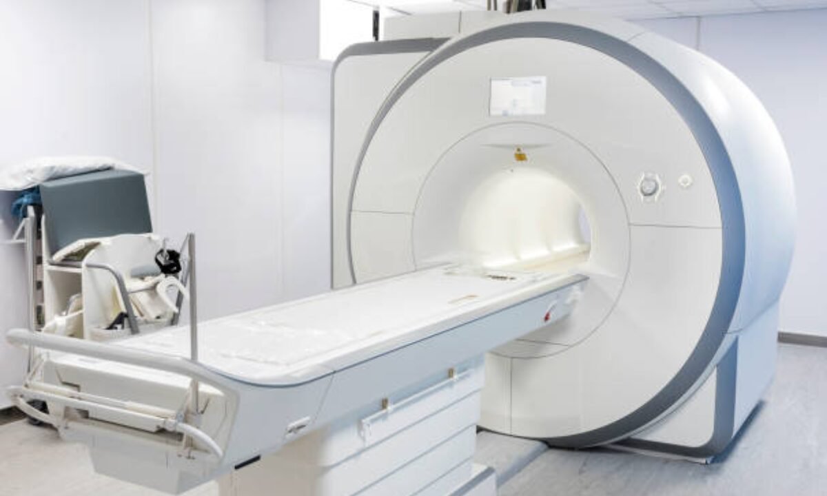
What is an MRI scan?

Magnetic Resonance Imaging (MRI) Scan is a medical imaging technique used to visualize internal structures of the body in detail. It uses strong magnetic fields and radio waves to generate images of the organs and tissues. It is commonly used to diagnose various conditions and is extremely useful for imaging the brain, spine, joints, and soft tissues.

Advantages associated with MRI Scans

1. No radiation: A major benefit of an MRI scan is that it does not use ionizing radiation, so almost all patients coming to the MRI center are at no risk, especially children and pregnant women.

2. Superior Soft Tissue Imaging: MRI is unique in its ability to provide high-resolution images of soft tissues, making it primarily used for tumor detection, brain disorders, and musculoskeletal-related issues.

3. Accuracy: MRI provides a more detailed and accurate diagnosis than CT scans in cases where close observation of soft tissue is necessary.

Limitations of MRI Scans

When considering the limitations of MRI scans, it’s important to note that they generally take longer time, when compared to CT scans. An MRI typically takes about 30 minutes to over an hour, which can be a significant factor for individuals with busy schedules or those who may find it challenging to remain still for an extended period.

In addition, the cost of an MRI scan is usually higher than that of a CT scan, making it important to consider budget and other financial requirements when opting for this type of imaging.

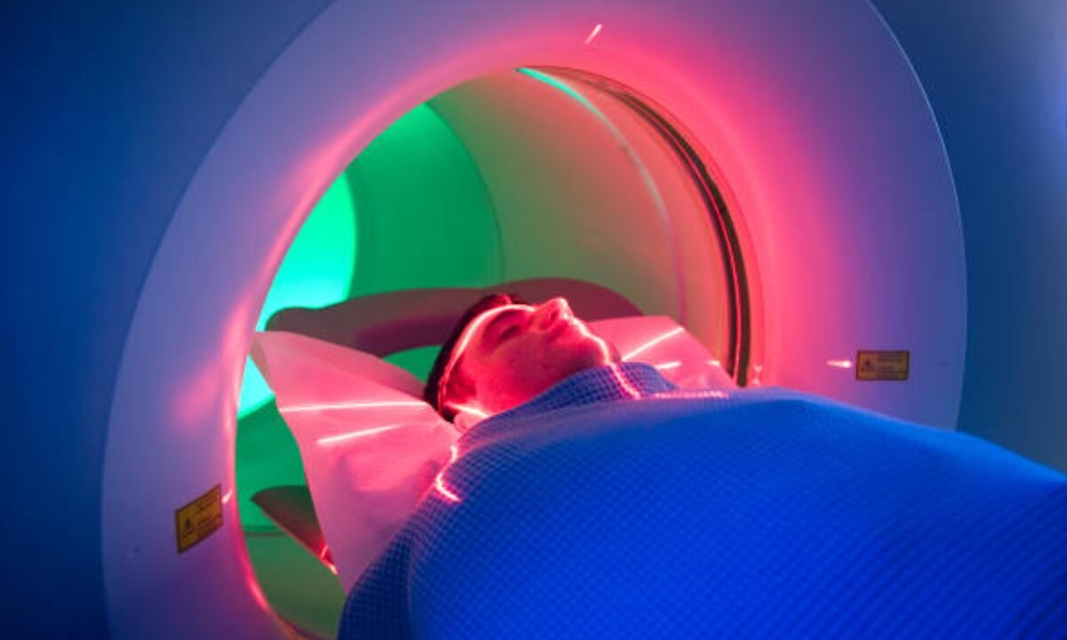
Furthermore, the confined space of the MRI machine may contribute to feelings of claustrophobia for some patients, which can be a source of anxiety or discomfort during the scan.

If you’re in search of an MRI scan in Bangalore, it’s advisable to seek out a reputable diagnostic center known for offering high-end MRI facilities and employing experienced radiologists who can ensure a smooth and reliable imaging experience.

Differences Between CT Scans And MRI Scans

When it comes to the differences between CT scans and MRI scans, there are several key points to consider.

1. Technology: CT scans utilize X-rays, providing detailed images of the body’s internal structures. On the other hand, MRI scans use a combination of magnetic fields and radio waves to generate detailed images, especially for soft tissues like the brain and muscles.

2. CT scans are particularly effective for imaging bones and the lungs, making them useful for detecting cancer or internal injuries. On the other hand, MRI scans excel in visualizing soft tissues such as the brain, spinal cord, and muscles.

3. Duration: CT scans are relatively quick, typically taking only a few minutes to complete. Conversely, MRI scans tend to take longer due to the detailed nature of the images they produce.

4. Cost and Availability: CT scans are generally more affordable and widely available compared to MRI scans, making them more accessible for many patients.

5. Radiation: CT scans involve minimal radiation exposure, while MRI scans do not expose the patient to any radiation, making them a safer option in that regard.

There is often a choice between a CT scan and an MRI scan, as one may be more suitable for the specific condition being diagnosed. For patients requiring a CT scan or an MRI scan in Bangalore, Koshikaa Screening Centre provides advanced imaging technology and precise expert analysis to ensure accurate diagnosis.

MRI of the uterus is primarily used for diagnosing and evaluating conditions such as uterine fibroids, adenomyosis, and malignancies. It is particularly useful in the planning of surgical procedures and assessing the extent of gynecological diseases. This imaging test is valuable for its ability to deliver detailed cross-sectional images of the uterus and surrounding structures.
An MRI of the spleen can help diagnose various disorders affecting the spleen including splenomegaly (enlarged spleen), tumors, and other abnormalities. It provides clear, detailed images that are crucial for assessing the health of the spleen and planning potential surgical interventions.
Prostate MRI is a non-invasive imaging technique that provides detailed pictures of the prostate gland and surrounding tissues. It helps in the detection, staging, and monitoring of prostate cancer. Prostate MRIs are increasingly used to guide biopsy procedures and assist in the planning of treatment strategies for prostate health.
An MRI of the pancreas is used to identify issues such as pancreatitis, pancreatic cancer, and other pancreatic disorders. It offers high-resolution images that help in detecting small lesions and assessing the pancreatic ducts. This imaging technique is valuable for guiding biopsies and planning surgeries involving the pancreas.
MRI scans of the liver, kidney, and bile ducts provide detailed images that help diagnose various conditions, such as tumors, cysts, and blockages in the bile ducts. They are crucial for assessing organ damage, staging cancer, and planning liver or kidney-related surgeries. This type of MRI can also evaluate the health and function of these organs comprehensively.
Breast MRI is highly sensitive for detecting breast cancer, especially in individuals with dense breast tissue or a high risk of cancer. It is often used in conjunction with mammography to provide a more comprehensive evaluation. Breast MRIs can also monitor the response to breast cancer treatments and provide detailed pre-surgical mapping.
Brain and spine MRIs are critical tools for diagnosing and monitoring neurological conditions, including tumors, spinal cord injuries, and multiple sclerosis. These MRIs can reveal abnormalities in brain tissue and spinal cord structure that might not be visible with other imaging methods. They are essential for planning surgeries and assessing the effectiveness of treatments in neurology.
PET-CT scans of transplant organs like the liver, kidney, or pancreas are performed to assess metabolic activity, detect complications, and monitor post-transplant function. These scans play a vital role in the early detection of issues such as organ rejection, recurrence of disease, or infection, guiding timely interventions and optimizing transplant outcomes for patients.
PET-CT examinations of organs such as the liver, kidney, or prostate are crucial for assessing metabolic activity, detecting abnormalities, and monitoring disease progression. These scans help diagnose conditions such as liver metastases, renal cell carcinoma, or prostate cancer, guiding treatment decisions and monitoring response to therapy over time.
PET-CT scans of soft-tissue masses are utilized to assess metabolic activity and detect malignant tumors or other abnormalities. By combining PET imaging with CT imaging, these scans provide detailed information about tumor location, size, and metabolic characteristics, aiding in accurate diagnosis and treatment planning for soft-tissue cancers.
PET-CT scans are valuable for detecting cysts or fluid collections within the body, including cysts in the liver, kidneys, or other organs. These scans help characterize the nature and metabolic activity of cysts, guiding diagnosis and treatment planning for conditions such as cystic liver disease, renal cysts, or pancreatic cysts.
PET-CT scans of muscles are performed to assess metabolic activity and detect abnormalities such as inflammation, infection, or tumors. These scans provide valuable information about muscle function and pathology, aiding in the diagnosis and management of conditions like myositis, muscular dystrophy, or soft-tissue sarcomas.
PET-CT scans of the brain are utilized to evaluate brain metabolism and detect abnormalities such as tumors, epilepsy, or neurodegenerative diseases. By combining functional PET imaging with anatomical CT imaging, these scans offer comprehensive information about brain function and structure, guiding neurosurgical interventions and treatment decisions for neurological disorders
PET-CT scans of bones are commonly used to detect bone metastases in cancer patients, assess bone health in conditions like osteoporosis, and evaluate response to treatment. By combining PET and CT imaging, these scans provide detailed information about metabolic activity and anatomical changes in bone structures, aiding in accurate diagnosis and treatment planning for various bone-related conditions.
CT scans of the spine are conducted to assess the vertebrae, spinal cord, and surrounding soft tissues for conditions like herniated discs, spinal fractures, or spinal stenosis, aiding in the diagnosis and management of spinal disorders with detailed imaging essential for surgical planning and intervention.
CT scans of the pelvis are utilized to visualize the pelvic bones, reproductive organs, bladder, and surrounding structures for conditions such as fractures, tumors, or pelvic inflammatory disease, guiding treatment plans and surgical interventions with precise anatomical information.
CT scans of the neck are performed to evaluate structures such as the throat, thyroid gland, lymph nodes, and blood vessels for abnormalities like tumors, infections, or injuries, assisting in diagnosis and management of head and neck disorders with detailed imaging.
CT scans of the extremities are employed to assess bones, joints, and soft tissues in the arms or legs, aiding in the diagnosis of fractures, arthritis, or other musculoskeletal conditions, guiding orthopedic treatment and rehabilitation plans for optimal recovery.
CT scans of the chest are utilized to examine the lungs, heart, blood vessels, and surrounding structures for conditions such as lung nodules, pneumonia, or pulmonary embolism, facilitating accurate diagnosis and management of respiratory and cardiovascular diseases.
CT scans of the brain are conducted to assess brain structures, identify abnormalities like tumors, hemorrhages, or strokes, and aid in diagnosing neurological conditions, guiding neurosurgical interventions and treatment strategies with precise imaging of the brain’s anatomy.
CT scans of the abdomen are performed to visualize and evaluate the internal organs such as the liver, kidneys, pancreas, and intestines for conditions like tumors, infections, or injuries, providing detailed images essential for diagnosis and treatment planning.
Transplant organ ultrasound scans, including those of the liver, kidney, or pancreas, are performed to assess organ viability, detect complications, and monitor post-transplant function. These scans play a vital role in the early detection of issues such as organ rejection, vascular complications, or fluid collections, guiding timely interventions and optimizing transplant outcomes for patients.
Ultrasound examinations of organs such as the liver, kidney, or prostate are crucial for assessing organ structure, detecting abnormalities, and monitoring disease progression. These scans help diagnose conditions such as liver cirrhosis, kidney stones, or prostate enlargement, guiding treatment decisions and monitoring response to therapy over time.
Soft-tissue masses can be evaluated using ultrasound to determine their size, location, and characteristics. Ultrasound imaging helps differentiate between benign and malignant masses, guiding further diagnostic evaluation and treatment planning. By providing real-time imaging, ultrasound assists in the precise localization of soft-tissue masses for biopsy or surgical removal if needed.
Ultrasound scans are valuable for detecting cysts or fluid collections within the body, including cysts in the liver, kidneys, or other organs. These scans help characterize the nature and size of cysts, aiding in diagnosis and treatment planning for conditions such as cystic liver disease or renal cysts, and guiding interventions such as cyst drainage or sclerotherapy.
Musculoskeletal ultrasound scans are used to assess the health and integrity of muscles, identifying issues such as strains, tears, or muscle inflammation. These scans provide detailed images of muscle structures, allowing healthcare providers to diagnose muscle injuries accurately and develop personalized rehabilitation plans to promote recovery and restore function.
Ultrasound examinations of joints are essential for evaluating joint health, detecting abnormalities such as inflammation, fluid accumulation, or cartilage damage. By visualizing the joint structures in real-time, ultrasound helps diagnose conditions like arthritis, bursitis, or ligament injuries, guiding treatment decisions and monitoring disease progression over time.
Ultrasound scans of tendons are performed to assess for injuries, inflammation, or tears, aiding in the diagnosis and management of conditions such as tendonitis or tendon tears. These scans provide detailed images of tendon structures, helping healthcare providers determine the extent of damage and plan appropriate treatment strategies, including physical therapy or surgical intervention if necessary.
Pelvis X-rays play a crucial role in diagnosing injuries, diseases, and developmental conditions affecting the pelvic region. They provide detailed images of the hip bones, sacrum, and coccyx, aiding in the assessment of fractures, dislocations, or signs of arthritis. Pelvic X-rays are instrumental in guiding surgical interventions, monitoring healing progress, and evaluating conditions such as osteoporosis or tumors. By providing precise anatomical information, these imaging studies enable healthcare professionals to deliver personalized care and improve patient outcomes.
Teeth X-rays, known as dental radiographs, are indispensable tools for dentists to assess oral health. They aid in diagnosing dental decay, gum disease, and abnormalities in tooth structure or alignment. These images provide valuable insights into hidden dental issues, allowing for early intervention and prevention of complications. Additionally, dental X-rays are essential for planning treatments such as orthodontics, root canals, or dental implants, ensuring optimal outcomes and patient satisfaction.
Spine X-rays are pivotal in diagnosing spinal fractures, degenerative conditions, and spinal misalignments, providing critical information for treatment planning and monitoring spinal health. They offer detailed images of the vertebrae and surrounding structures, enabling healthcare professionals to identify issues such as scoliosis, disc herniation, or spinal stenosis
Skull X-rays are valuable for detecting fractures, assessing head trauma, and evaluating the alignment of facial bones, crucial in diagnosing conditions like fractures, tumors, or developmental abnormalities in the skull. They provide detailed views of the cranial structure, aiding clinicians in determining appropriate treatment strategies and ensuring patient safety.
For the heart, X-ray solutions can aid in the detection of various cardiac problems, ranging from mild to severe, including – Heart failure, Cardiac congestion and other heart-related conditions. In addition to cardiac assessments, X-ray solutions are also used for breast examinations to identify tumors or signs of breast cancer.
Bone X-rays are essential for diagnosing fractures and assessing bone alignment, aiding doctors in developing treatment plans and monitoring healing progress. They provide detailed images of the skeletal system, helping identify abnormalities or injuries that may not be visible through other imaging techniques
Abdominal X-rays are performed when patients experience symptoms such as significant nausea, ongoing vomiting, abdominal pain, flank or back pain, or abdominal swelling. These X-rays can help diagnose various conditions.
Search Bar

Frequently Booked Tests

Frequently Booked Checkups