Koshikaa’s Advanced Diagnostics Detects Illnesses Early in Bangalore

MRI scan in Bangalore

Introduction In today’s fast-paced, high-pressure world—especially in a tech hub like Bangalore—health problems often go unnoticed until it’s too late. Fatigue is brushed off as overwork, weight gain as aging, and stress as a side effect of success. But what if these were early warning signs of something more serious? Koshikaa’s Advanced Diagnostics is revolutionizing […]

Top 5 Essential Tests in Koshikaa’s Full Body Checkup Package

CT scan in Bangalore

Introduction Health isn’t just about feeling good—it’s about staying one step ahead of problems that haven’t even surfaced yet. In Bangalore’s high-octane environment, where professional stress and sedentary habits dominate, investing in preventive care has never been more critical. Koshikaa’s Comprehensive Full Body Checkup Package is designed to be your health’s early warning system. But […]

Why You Can’t Afford to Skip a Full Body Checkup in Bangalore

Blood test in Bangalore

Introduction In today’s fast-paced urban life, health often takes a backseat to hustle. With Bangalore’s ever-evolving tech scene, long commutes, and hectic schedules, it’s easy to ignore subtle health warnings. Neglecting your well-being can lead to severe consequences that cost more time, energy, and money down the line. That’s where a Full Body Checkup in […]

Book Full Body Checkup at Home in Minutes: Quick, Easy &  Affordable!

CT scan in Bangalore

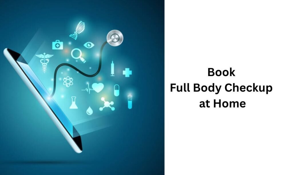
Introduction: Your Health, Your Comfort—Right at Home! Imagine getting a complete health checkup without leaving your home—no traffic, no waiting rooms, no stress. Sounds like a dream, right? Well, it’s now a reality for Bangaloreans! Thanks to modern healthcare solutions, you can book a full body checkup at home in Bangalore in just a few […]

Full Body Checkup for Senior Citizens: Special Offers

Blood test in Bangalore

Ageing is beautiful, but it also demands attention, especially regarding health. As we grow older, our bodies require more care, and early detection can make all the difference. If you’re searching for an affordable and convenient Full Body Checkup in Bangalore residents trust, especially tailored for seniors, you’ve landed in the right place. Let’s explore […]

Who Needs a Full Body Check-Up? 7 Vital Reasons in 2025

Blood test in Bangalore

Introduction: In today’s high-paced world, health often takes a back seat—until something goes wrong. But what if you could detect health issues early and take proactive steps before they turn into costly medical emergencies? That’s the power of a Full Body Check-up. But the big question is: Who should consider getting a full body check-up? […]

Same-Day Full Body Checkup in Bangalore – Book Now!

Blood test in Bangalore

Introduction: No More Waiting – Your Health Can’t Wait! In today’s fast-paced world, waiting days to get a health checkup is a luxury most people can’t afford. Thankfully, Same-Day Full Body Checkup in Bangalore is now a reality. Whether you’re preparing for a big trip, managing a health scare, or just being proactive, you can […]

Top 5 Cancers Detectable by Preventive Health Checkup

Full Body checkup in Bangalore

The world faces cancer as a prevailing health issue among its most significant medical concerns. The early identification of cancer leads to life-saving outcomes while generating superior therapy outcomes. Most people seek answers regarding the nature of screening examinations as well as their role in identifying cancers early. Regular checkups conducted at proper times provide […]

Understanding the Importance of Routine Health Screenings

Full body checkup in Bangalore

Regimen-based disease screening methods serve two essential functions which include disease prevention through early detection at treatable stages. Preventative medical attention stands equally important with sick patient care although most individuals wait until sickness to seek medical attention. The combination of regular health screenings protects lives while offering an improved quality of life to people. […]

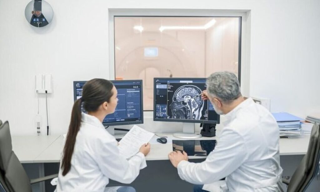
PET CT Scan vs. X Ray vs. MRI: Which One is Right for You?

pet ct scan vs. x ray

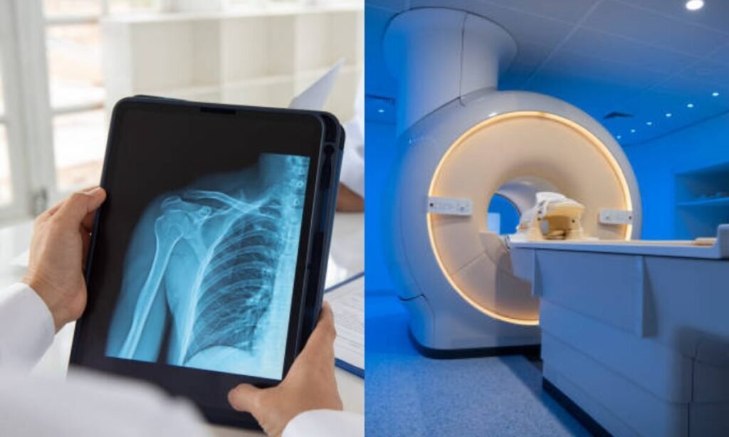
Medical imaging technology is vital in medical diagnosis and treatment procedures for different illnesses. Today’s medical imaging market includes several options including PET CT scan along with X-rays and MRI scans which makes patients struggle to determine which test is appropriate for their needs. This article analyzes the unique aspects of these procedures while discussing […]

MRI of the uterus is primarily used for diagnosing and evaluating conditions such as uterine fibroids, adenomyosis, and malignancies. It is particularly useful in the planning of surgical procedures and assessing the extent of gynecological diseases. This imaging test is valuable for its ability to deliver detailed cross-sectional images of the uterus and surrounding structures.
An MRI of the spleen can help diagnose various disorders affecting the spleen including splenomegaly (enlarged spleen), tumors, and other abnormalities. It provides clear, detailed images that are crucial for assessing the health of the spleen and planning potential surgical interventions.
Prostate MRI is a non-invasive imaging technique that provides detailed pictures of the prostate gland and surrounding tissues. It helps in the detection, staging, and monitoring of prostate cancer. Prostate MRIs are increasingly used to guide biopsy procedures and assist in the planning of treatment strategies for prostate health.
An MRI of the pancreas is used to identify issues such as pancreatitis, pancreatic cancer, and other pancreatic disorders. It offers high-resolution images that help in detecting small lesions and assessing the pancreatic ducts. This imaging technique is valuable for guiding biopsies and planning surgeries involving the pancreas.
MRI scans of the liver, kidney, and bile ducts provide detailed images that help diagnose various conditions, such as tumors, cysts, and blockages in the bile ducts. They are crucial for assessing organ damage, staging cancer, and planning liver or kidney-related surgeries. This type of MRI can also evaluate the health and function of these organs comprehensively.
Breast MRI is highly sensitive for detecting breast cancer, especially in individuals with dense breast tissue or a high risk of cancer. It is often used in conjunction with mammography to provide a more comprehensive evaluation. Breast MRIs can also monitor the response to breast cancer treatments and provide detailed pre-surgical mapping.
Brain and spine MRIs are critical tools for diagnosing and monitoring neurological conditions, including tumors, spinal cord injuries, and multiple sclerosis. These MRIs can reveal abnormalities in brain tissue and spinal cord structure that might not be visible with other imaging methods. They are essential for planning surgeries and assessing the effectiveness of treatments in neurology.
PET-CT scans of transplant organs like the liver, kidney, or pancreas are performed to assess metabolic activity, detect complications, and monitor post-transplant function. These scans play a vital role in the early detection of issues such as organ rejection, recurrence of disease, or infection, guiding timely interventions and optimizing transplant outcomes for patients.
PET-CT examinations of organs such as the liver, kidney, or prostate are crucial for assessing metabolic activity, detecting abnormalities, and monitoring disease progression. These scans help diagnose conditions such as liver metastases, renal cell carcinoma, or prostate cancer, guiding treatment decisions and monitoring response to therapy over time.
PET-CT scans of soft-tissue masses are utilized to assess metabolic activity and detect malignant tumors or other abnormalities. By combining PET imaging with CT imaging, these scans provide detailed information about tumor location, size, and metabolic characteristics, aiding in accurate diagnosis and treatment planning for soft-tissue cancers.
PET-CT scans are valuable for detecting cysts or fluid collections within the body, including cysts in the liver, kidneys, or other organs. These scans help characterize the nature and metabolic activity of cysts, guiding diagnosis and treatment planning for conditions such as cystic liver disease, renal cysts, or pancreatic cysts.
PET-CT scans of muscles are performed to assess metabolic activity and detect abnormalities such as inflammation, infection, or tumors. These scans provide valuable information about muscle function and pathology, aiding in the diagnosis and management of conditions like myositis, muscular dystrophy, or soft-tissue sarcomas.
PET-CT scans of the brain are utilized to evaluate brain metabolism and detect abnormalities such as tumors, epilepsy, or neurodegenerative diseases. By combining functional PET imaging with anatomical CT imaging, these scans offer comprehensive information about brain function and structure, guiding neurosurgical interventions and treatment decisions for neurological disorders
PET-CT scans of bones are commonly used to detect bone metastases in cancer patients, assess bone health in conditions like osteoporosis, and evaluate response to treatment. By combining PET and CT imaging, these scans provide detailed information about metabolic activity and anatomical changes in bone structures, aiding in accurate diagnosis and treatment planning for various bone-related conditions.
CT scans of the spine are conducted to assess the vertebrae, spinal cord, and surrounding soft tissues for conditions like herniated discs, spinal fractures, or spinal stenosis, aiding in the diagnosis and management of spinal disorders with detailed imaging essential for surgical planning and intervention.
CT scans of the pelvis are utilized to visualize the pelvic bones, reproductive organs, bladder, and surrounding structures for conditions such as fractures, tumors, or pelvic inflammatory disease, guiding treatment plans and surgical interventions with precise anatomical information.
CT scans of the neck are performed to evaluate structures such as the throat, thyroid gland, lymph nodes, and blood vessels for abnormalities like tumors, infections, or injuries, assisting in diagnosis and management of head and neck disorders with detailed imaging.
CT scans of the extremities are employed to assess bones, joints, and soft tissues in the arms or legs, aiding in the diagnosis of fractures, arthritis, or other musculoskeletal conditions, guiding orthopedic treatment and rehabilitation plans for optimal recovery.
CT scans of the chest are utilized to examine the lungs, heart, blood vessels, and surrounding structures for conditions such as lung nodules, pneumonia, or pulmonary embolism, facilitating accurate diagnosis and management of respiratory and cardiovascular diseases.
CT scans of the brain are conducted to assess brain structures, identify abnormalities like tumors, hemorrhages, or strokes, and aid in diagnosing neurological conditions, guiding neurosurgical interventions and treatment strategies with precise imaging of the brain’s anatomy.
CT scans of the abdomen are performed to visualize and evaluate the internal organs such as the liver, kidneys, pancreas, and intestines for conditions like tumors, infections, or injuries, providing detailed images essential for diagnosis and treatment planning.
Transplant organ ultrasound scans, including those of the liver, kidney, or pancreas, are performed to assess organ viability, detect complications, and monitor post-transplant function. These scans play a vital role in the early detection of issues such as organ rejection, vascular complications, or fluid collections, guiding timely interventions and optimizing transplant outcomes for patients.
Ultrasound examinations of organs such as the liver, kidney, or prostate are crucial for assessing organ structure, detecting abnormalities, and monitoring disease progression. These scans help diagnose conditions such as liver cirrhosis, kidney stones, or prostate enlargement, guiding treatment decisions and monitoring response to therapy over time.
Soft-tissue masses can be evaluated using ultrasound to determine their size, location, and characteristics. Ultrasound imaging helps differentiate between benign and malignant masses, guiding further diagnostic evaluation and treatment planning. By providing real-time imaging, ultrasound assists in the precise localization of soft-tissue masses for biopsy or surgical removal if needed.
Ultrasound scans are valuable for detecting cysts or fluid collections within the body, including cysts in the liver, kidneys, or other organs. These scans help characterize the nature and size of cysts, aiding in diagnosis and treatment planning for conditions such as cystic liver disease or renal cysts, and guiding interventions such as cyst drainage or sclerotherapy.
Musculoskeletal ultrasound scans are used to assess the health and integrity of muscles, identifying issues such as strains, tears, or muscle inflammation. These scans provide detailed images of muscle structures, allowing healthcare providers to diagnose muscle injuries accurately and develop personalized rehabilitation plans to promote recovery and restore function.
Ultrasound examinations of joints are essential for evaluating joint health, detecting abnormalities such as inflammation, fluid accumulation, or cartilage damage. By visualizing the joint structures in real-time, ultrasound helps diagnose conditions like arthritis, bursitis, or ligament injuries, guiding treatment decisions and monitoring disease progression over time.
Ultrasound scans of tendons are performed to assess for injuries, inflammation, or tears, aiding in the diagnosis and management of conditions such as tendonitis or tendon tears. These scans provide detailed images of tendon structures, helping healthcare providers determine the extent of damage and plan appropriate treatment strategies, including physical therapy or surgical intervention if necessary.
Pelvis X-rays play a crucial role in diagnosing injuries, diseases, and developmental conditions affecting the pelvic region. They provide detailed images of the hip bones, sacrum, and coccyx, aiding in the assessment of fractures, dislocations, or signs of arthritis. Pelvic X-rays are instrumental in guiding surgical interventions, monitoring healing progress, and evaluating conditions such as osteoporosis or tumors. By providing precise anatomical information, these imaging studies enable healthcare professionals to deliver personalized care and improve patient outcomes.
Teeth X-rays, known as dental radiographs, are indispensable tools for dentists to assess oral health. They aid in diagnosing dental decay, gum disease, and abnormalities in tooth structure or alignment. These images provide valuable insights into hidden dental issues, allowing for early intervention and prevention of complications. Additionally, dental X-rays are essential for planning treatments such as orthodontics, root canals, or dental implants, ensuring optimal outcomes and patient satisfaction.
Spine X-rays are pivotal in diagnosing spinal fractures, degenerative conditions, and spinal misalignments, providing critical information for treatment planning and monitoring spinal health. They offer detailed images of the vertebrae and surrounding structures, enabling healthcare professionals to identify issues such as scoliosis, disc herniation, or spinal stenosis
Skull X-rays are valuable for detecting fractures, assessing head trauma, and evaluating the alignment of facial bones, crucial in diagnosing conditions like fractures, tumors, or developmental abnormalities in the skull. They provide detailed views of the cranial structure, aiding clinicians in determining appropriate treatment strategies and ensuring patient safety.
For the heart, X-ray solutions can aid in the detection of various cardiac problems, ranging from mild to severe, including – Heart failure, Cardiac congestion and other heart-related conditions. In addition to cardiac assessments, X-ray solutions are also used for breast examinations to identify tumors or signs of breast cancer.

Bone X-rays are essential for diagnosing fractures and assessing bone alignment, aiding doctors in developing treatment plans and monitoring healing progress. They provide detailed images of the skeletal system, helping identify abnormalities or injuries that may not be visible through other imaging techniques

Abdominal X-rays are performed when patients experience symptoms such as significant nausea, ongoing vomiting, abdominal pain, flank or back pain, or abdominal swelling. These X-rays can help diagnose various conditions.

Frequently Booked Tests

Frequently Booked Checkups